Estimation of Cellulolytic Bacteria from Cowdung in Awka Central Abattoir, Anambra State, Nigeria.
- Chijioke Christian Aniekwu
- Ugochukwu Chukwuma Okafor
- Vitus Ikechi Onyeneho
- 228-236
- Feb 19, 2024
- Education
Estimation of Cellulolytic Bacteria from Cowdung in Awka Central Abattoir, Anambra State, Nigeria.
Chijioke Christian Aniekwu1, Ugochukwu Chukwuma Okafor2, Vitus Ikechi Onyeneho3
1,2Department of Applied Microbiology and Brewing, Faculty of Biosciences, Nnamdi Azikiwe University, Awka, Nigeria.
3Public Health Department, National Open University of Nigeria.
DOI: https://doi.org/10.51584/IJRIAS.2024.90121
Received: 15 January 2024; Accepted: 20 January 2024; Published: 19 February 2024
ABSTRACT
Bacteria obtained from cow-dungs from selected slaughtering points in Awka central abattoir were assessed for cellulose degradation. The qualitative screening of the isolates was determined by culturing the isolates on a cellulose selective medium. The isolates were obtained and identified as Serratia marcescens, Streptococcus mutans, Pseudomonas aeruginosa, Escherichia coli, Salmonella typhi, Bacillus subtilis and Lactobacillus spp. Three of the isolates with the highest cellulolytic activity are Pseudomonas aeruginosa, Bacillus subtilis and Streptococcus mutans. Pseudomonas aeruginosa gave the highest diameter clear zone value of 12mm+0.25mm Streptococcus spp. follows with clear zone of 11mm+1.02mm and Bacillus subtilis had the least diameter clear zone of 10mm+1.04mm. The three isolates were selected for optimization because they had a clear zone up to 10mm and above. These isolates were subjected to qualitative screening by culturing the isolates in a liquid medium with different incubation periods (12, 24, 48, 60, 72, 84 and 96hrs) at shack and static conditions. Potential activities of Fpase and CMC-ase were found in Pseudomonas aeruginosa (0.94u/m), Bacillus subtilis (0.88u/m) and Streptococcus mutans (0.6u/m) all at 48hrs incubation respectively. Microbes in cow dung have a potential to be Cellulolytic as the degradation of cellulose is an environmental friendly approach that can reduce the accumulation of agricultural wastes.
Key Words: Abattoir; Cellulolytic Bacteria; Cowdung; Hydrolysis Qualitative and Quantitative screening.
INTRODUCTION
Cellulose is considered the most abundant biological polymer on aquatic and terrestrial ecosystems. It is also the main component of plant biomass [1]. It is the dominant waste material generated from agricultural industry in the form of stems, stalks, and husks. Lots of interests have been generated concerning the utilization of cellulose as an energy resource and feed [2]. Cellulose can be defined as a polymer made up of D-glucose units linked together to form linear chain through ß-1, 4-glycosidic linkages. It is the substance that makes up most of the plant’s cell wall. Due to the molecular linkages of cellulose, it is insoluble or does not dissolve easily in water. These molecules are formed into a criss-cross mesh which gives the strength and shape to the cell wall. Cellulose is commonly degraded by cellulolytic enzymes known as cellulases. Cellulases have a huge potential for application in biotechnology and other industries; such applications include malting and brewing, starch processing, clarification of juice, alcoholic beverage, pulp bleaching, animal feed production and textile processing [3]. Cellulases are the group of enzymes involved in the conversion of cellulosic substrates to fermentable sugars. Main members of this group include endoglucanase (EC 3.2.1.4), exoglucanase or cellobiohydrolase (EC 3.2.1.91) and β-glucosidase (EC 3.2.1.21). The endoglucanase hydrolyzes β-1,4 bonds in cellulose molecule, whereas exoglucanase cleaves the ends to release cellobiose and β-glucosidase converts cellulobiose to glucose. However, the enzymes capable of degrading cellulose, known as cellulases are mainly produced by Bacteria, Fungi and Protozoan that hydrolyzes cellulose. Cellulolytic microbes have important role in environment for degradation of cellulose and convert it into useful products. Cellulases are mainly classified into 5 types on the basis of types of reaction catalyzed- [4].
Cellulases have attracted much interest because of the diversity of their application. The major industrial applications of cellulases are in textile industry for ‘bio-polishing’ of fabrics and producing stonewashed look of denims, as well as in household laundry detergents for improving fabric softness and brightness. Besides, they are used in animal feeds for improving the nutritional quality and digestibility, in processing of fruit juices, and in baking, while de-inking of paper is yet another emerging application. A potential challenging area where cellulases would have a central role is the bioconversion of renewable cellulosic biomass to commodity chemicals, insect repellant, thermal insulator, oil and grease removal and biofuel [5].
Screening for microbial cellulose activity is typically performed as pure cultures of bacterial isolates were individually transferred in Carboxy-methylcellulose Agar (CMC) agar plates. After incubation for 48 hrs, CMC agar plates is flooded with 1 % congored and allowed to stand for 15 mins at room temperature. One molar NaCl is thoroughly used for counterstaining the plates. Clear zones appeared around growing bacterial colonies indicating cellulose hydrolysis ability and cellulase production.
Cow dung is a waste product from bovine animals, which includes cattle, buffalo etc. Cow dung is the undigested residue of plant matter which is passed through the animal’s gut.Being a mixture of feces and urine in the ratio of 3:1, it mainly consists of lignin, celluloseand hemicelluloses. Fresh Cow dung is greenish black in colour, but when exposed to air turnd dark greenish brown. It also contains different minerals like nitrogen, potassium, along with trace amount of sulphur, iron, magnesium, copper, cobalt and manganese. Indigenous cow also contain higher amount of calcium, phosphorus, zinc and copper than the cross-breed cow [6]:[7]. Cow dung harbours a rich microbial diversity, containing different species of bacteria (Bacillus spp., Corynebacterium spp. and Lactobacillus spp.), protozoa and yeast (Saccharomyces and Candida) [7].[8] have isolated many different bacterial genera such as Citrobacter koseri, Enterobacter aerogenes, Escherichia coli, Klebsiella oxytoca, Klebsiella pneumoniae, Kluyvera spp., Morgarella morganii, Pasteurella spp., Providencia alcaligenes, Providencia stuartii and Pseudomonas spp. from cow dung.
Microbes in cow dung have a potential to be Cellulolytic as the degradation of cellulose in biomass wastes is an environmental friendly approach that can reduce the accumulation of agricultural waste and reduce the emission of greenhouse gases. This is considered suitable to improve environmental sanitation
MATERIALS AND METHODS
Study Area: The study was carried out in Awka metropolis. Awka is the capital of Anambra state. The city has an estimated population of 301,667 as of the 2006 Nigerian census, and over 2.5 milion as at 2018 estimate. Awka is located at 199.1 kilometers by road, directly north of port Harcourt in the center of the densely populated Igbo heartland in South East Nigeria. Awka central abattoir is located 3kilometers away from Appeal court, along court road in Awka south Local Government Area with a population of 189,049 people in the area [9].
Sample Collection: The cow dung used in this study was collected from the Awka central Abattoir and Amansea Abattoir located in Awka metropolis of Anambra State. Seven (7) Cow dung samples were collected from seven slaughtering points in the two abattoirs used for the study. The samples were hygienically placed in an ice box at 4ºC and transferred to Applied Microbiology laboratory, Nnamdi Azikiwe University-Awka in a sterilized sample bottle within 4hr for bacteriological analysis.
Isolation and Identification of Bacteria: One gram (1g) of the samples was dissolved into 10/ml distilled water in a clean, sterile 25/ml bottle. This was mixed thoroughly and serially diluted out with sterile distilled water. One milliter (1ml) each was introduced into 9/ml of water (diluents) in 10 folds sterile test tubes and each tube were mixed thoroughly. Then, test tubes with 1:1,000, 1:10000 and 1:100000dilution factors were pour-plated on Nutrient agar (NA), MacConkey agar (MAC), and de Man Rogosa Sharpe agar (MRS) for bacterial load counts. All plates were incubated at 37°C for 48 hours. MRS plates were incubated anaerobically in a closed jar with a candle, which created microaerophilic condition at 35°C for 48hours. Mannitol salt Agar (MSA) plates were also incubated at 37°C for 48 hours. The colonies observed after the incubation periods were counted and recorded [10].
Molecular Characterization of Isolates: The Isolates screened were taken to a special laboratory located at Queen Elizabeth road, Ibadan, for DNA sequencing using molecular tools. Two (2/mLs) of bacterial cells broth was added to a ZR Bashing TM Lysis Tube. Then 750ul Lysis Solution was added to the tube. It was secured in a bead fitted with 2 ml tube holder assembly and process at maximum speed for > 5 minutes. The ZR BashingBeadTM Lysis Tube was centrifuged in a microcentirifuge at > 10,000 x g for 1 minute. Up to 400 ul supernatant was transferred to a Zymo-SpinTM IV Spin Filter (orange top) in a collection tube and centrifuge at 7,000 x g for 1 minute. Then add 1,200 ul of Bacterial DNA Binding Buffer to the filterate in the Collection Tube from Step 4. Eight hundred (800 ul) of the mixture was transferred from Step 5 to a Zymo-SpinTM IIC Column in a Collection Tube and centrifuge at 10.000 x g for 1 minute. The flow was discarded through from the Collection Tube and repeat Step 6. Then add 200 ul DNA Pre-\Vash Buffer to the Zymo-Spin TM IIC column in new collection Tube and centrifuged at 10.000 x g for 1 minute. 500 ul Bacterial DNA Wash Buffer was to the Zymo-SpinTM IIC column and centrifuge at 10.000 x g for 1 minute. The Zymo-SpinTM IIC column was transferred to a clean 1.5 ml microcentrifuge tube and l00ul (35 ul minimum) DNA Elution Buffer was added directly to the column matrix. Then centrifuged at 10,000 x g for 30 seconds to elute the DNA [11].
Initial screening of cellulase-producing bacteria using cellulose differential medium: The screening of cellulase-producing bacteria was done using a method by [10]. The isolated bacterial strains were screened for cellulose enzyme production in submerged fermentation process. The production medium was prepared using 1% potato waste (as cellulose substrate), 0.2 % K2HPO4, 0.03 % MgSO4, 1 % peptone, 0.25 % (NH4)2SO4 and autoclaved at 121ºC for 15min. After sterilization, the medium was allowed to cool at room temperature. The medium was inoculated with 1 ml of selected bacterial isolates and incubated in a shaker (Eyela NTS- 331) at 35˚C for 24hrs with an agitation speed of 140 rpm. After termination of the fermentation period, the fermented broth was centrifuged at 140 rpm for 10 min at 4˚C to remove the unwanted material. The clear supernatant thus obtained after centrifugation served as crude enzyme source.
Qualitative Screening for Cellulose Production: Pure cultures of bacterial isolates were individually transferred in Carboxy-methylcellulose Agar (CMC) agar plates. After incubation for 48 hrs, CMC agar plates was flooded with 1 % congored and allowed to stand for 15 mins at room temperature. One molar NaCl was thoroughly used for counterstaining the plates. Clear zones appeared around growing bacterial colonies indicating cellulose hydrolysis. The bacterial colonies having the largest clear zone were selected for identification and cellulase production in submerged system [10].
Assay of Cellulolytic Enzymes: Cellulase activity was measured using the method by [12]. A reaction mixture which consisted of 0.2/mL of the crude enzyme solution and 1.8mL of 0.5% carboxymethyl cellulose (CMC) in 50mm sodium phosphate buffer (pH7) was incubated at 37ºC in a water bath for 30min. Afterwards, 3mL of Dinitro-benzouic acid reagentwas added to terminate the reaction. The resulting colour was developed by boiling the mixture for 5mins. Optical Density of the samples was measured at 595nm against a blank containing all the reagents with the exception of the crude enzyme as done by [13].
Quantitative screening of Cellulolytic Bacteria (Fpase): Pure cultures of selected bacterial isolates were individually maintained on CMC supplemented agar slants at 4˚C, until used. The bacterial isolates were inoculated in broth medium containing 0.03 % MgSO4, 0.2 % K2HPO4, 1 % glucose, 0.25% (NH4)2SO4 and 1 % peptone at pH 7 for 24h of fermentation period. After 24h of fermentation period these were used as inoculum source. Each pure bacterial isolates gotten from the inoculum was grown on sterilized cellulose mineral salt medium containing Sterilized wood pulp of 50gm (Whatman Filter paper)as the only source of carbon and energy. 0.2 % K2HPO4, 0.03 % MgSO4, 1 % peptone, 0.25 % (NH4)2SO4 and autoclaved at 121ºC for 15min.The cultures were incubated for 37ºC for 96h.Zone of clearance on the cellulose agar plates was observed after flooding with Congo red for qualitative estimation. For quantitative estimation, CMC agar medium containing 1.0 % peptone,0.2 % K2HPO4, 1 % agar, 0.03 % MgSO4.7H2O, 0.25%(NH4)2SO4 was prepared and used for the cellulolytic activity time study as described by [10]. Fifty milliliter of the medium was poured into three flasks containing sterilized filter paper, which was initially soaked in 1.0 mL and 10 mM sodium phosphate buffer pH 7.0 at 37ºC. This acts as the carbon source (Cellulose) for the bacteria. Five milliliter of each of the bacterial cell culture adjusted to 0.5 OD500nmwaspipetted in to the flasks. The reaction was stopped by adding 1.0mL 3,5-dinitro salicylic acid (DNS) regent at each time to determine the optical density at 546nm. Growth, change in the medium and cellulose utilization potential of the bacteria isolates were determined quantitatively by time course study of enzyme activity and also monitoring the bacterial biomass at time intervals of 12h, 24h, 48h, 72 and 96h as described by [14].
2.8. Statistical Analysis: One sample t-test was used to analyze the mean values of the bacterial isolates under study using SPSS version 17.
RESULTS
Isolation and Identification of Bacteria
Table 1. Shows the morphology of cow dung samples collected from different slaughter points in the study area
| Sample collection site | Colour | Odor | Form |
| CA1 | Greenish brown | Rotten egg smell | Slurry |
| CA2 | Dark brown | ,, | Slurry |
| CA3 | Greenish black | ,, | Watery |
| CA4 | Greenish brown | ,, | Watery |
| AA1 | Dark brown | ,, | Solid |
| AA2 | Greenish brown | ,, | Slurry |
| AA3 | Dark brown | ,, | Slurry |
Key: CA1 = Sample collected from 1st slaughter point in Central abattoir
CA2 = Sample collected from 2nd slaughter point in Central abattoir
CA3 = Sample collected from the 3rd slaughter point in Central abattoir
CA4=Sample collected from the 4th slaughter point in Central abattoir
AA1= Sample collected from the 1st slaughter point in Amansea abattoir
AA2=Sample collected from the 2nd slaughter point in Amansea abattoir
AA3= Sample collected from the 3rd slaughter point in Amansea abattoir
Colonial Examination of the Bacteria Isolates
Table 2 shows the Colonial examination of the bacterial isolates. They were identified as Strepococcus mutans, Pseudomonas aeruginosa, staphylococcus aureus, Serratia spp, Esherichia coli, bacillus subtilis and Lactobacillus spp.
Table 2: Colonial examination of the bacteria isolates
| Isolates | Colonial characteristics | Probable organisms |
| 1 | Colonies are white to grey in colour with round, heaped and raised elevation. Presence of an entire margin | Streptococcus mutans |
| 2 | Greenish-blue in colour, grape-like smell. Smooth mucoid slime layer seen on a translucent-opaque low convex elevation | Pseudomonas aeroginosa |
| 3 | Opaque, pink-red coloured bacteria. Presence of a fishy to urinary odor. Umbonated elevation and entire margin | Serratia marcescenes |
| 4 | Opaque colourless colonies with black on the center. Low convex elevation with smooth elevation. | Salmonella typhi |
| 5 | Transluscent donut shaped pink coloured shiny texture. Convex with an entire margin, creating concentric growth ring in the colony. | Esherichia coli |
| 6 | Presence of a rough, opaque, fuzzy slightly yellow edged bacterium with an irregular margin. | Baccilus subtilis |
| 7 | Presence of small, irregular and round shape with shinny whitish cream. Convex, entire, opaque without pigment. | Lactobacillus spp. |
Qualitative Screening for cellulose production
Qualitative estimation of cellulose degradation by observing the hydrolysis Capacity (zone of clearance) on cellulose agar plates after flooding with Congo red and incubating for 96h at about 37oC. Table 3 shows the zone of clearance in diameter and zone of colony of the isolates on cellulose agar plate after flooding and incubation. P. aeruginosa had the wildest zone of clearance of about 48mm, diameter of 13mm and zone of colony of 3.7mm, which was slightly wider than Streptococcus mutans that had zone of clearance of 46mm, diameter of 12mm and zone of colony of 2.3mm. B. Subtilis had a moderate zone of clearance of 40mm, diameter 12mm and zone of colony of 1.3mm. ), during the screening process as incubated in carboxy-methyl cellulose agar (CMC) for 48h.The supernatant was found to bear cellulolytic enzymes, supporting other previous studies that indicated the ability by known members of the Bacillus genus to secrete proteins extracellularly [15]. Three bacterial isolates were found to have the highest potential to produce cellulase, an enzyme that have the potential of degrading cellulose in cow dung, to ascertain the degradation potentials of these isolates in a biomass material.
Table 3. Screening test values for cellulose degradation by bacterial isolates
| Isolates | Zone of inhibition(mm) |
| 1 | 6 |
| 2 | 10 |
| 3 | 12 |
| 4 | 4 |
| 5 | 3 |
| 6 | 11 |
| 7 | 5 |
Table 4. The three selected isolates with the widest zone of inhibition
| Isolates | Zone of clearance (mm) | Diameter (mm) | Zone of colony (mm) |
| 3 | 48 | 13 | 3.7 |
| 6 | 40 | 12 | 1.3 |
| 2 | 46 | 20 | 2.3 |
Key:
Isolates 1= Serratia marcenscense Isolates 2= Streptococcus mutans
Isolates 3= Pseudomonas aeruginosa Isolates 4= Esherichia coli
Isolates 5= Salmonella typhi Isolate 6= Bacillus subtilis
Isolates 7= Lactobacillus spp.
Effect of incubation period on enzyme activity during time course study
The time course of the production of cellulase by P. aeruginosa at 48h followed an exponential growth of the organism with maximum enzyme production of 0.95u/ml. The second highest enzyme production was seen in Bacillus subtilis which yielded a enzyme production of 0.94u/ml at 48h. The least was recorded by Streptococcus mutans with a mild cellulase production of 0.62u/ml at 48h. The increase level of cellulolytic activity during the incubation period can be attributed to the efficient utilization of the carbon and nitrogen sources in the biomass material, thus indicating a maximum production of cellulase as shown in figure 1. This report is similar with the report by [16] that cellulose degraders in the cow dung could be mesophilic or slightly thermophilic temperatures of incubation to be able to optimize a significant cellulase production.
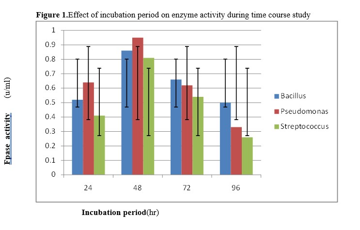
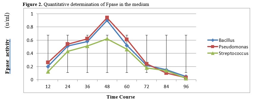
Figure 2. Quantitative determination of Fpase in the medium
DISCUSSION
The morphology of the cow dung samples observed were shown in table 1. The observation agrees with the observation obtained by [6] in their study on the current status of cow dung as a bioresources for sustainable growth. Table 1 also shows that cow dung occurs in watery, slurry and solid form which could easily have a direct or indirect contact with water causing an environmental pollution of water bodies as was described by [20]. This result also agreed with the studies carried out by [21], [22] and [23] which showed the presence of bacteria in the cow dung. . The colonial characteristics of the selected isolates are shown in Table 2. These characteristics agree with the bacterial characteristics guidelines published by [18]. The work done by [19] confirms the identification of the bacteria from the cow dung used in the present study that animal excreta are major source of intestinal tract pathogens which could be shed in feces. These pathogens may be persist for a period of days, weeks or months depending on the type of pathogen and the environmental condition in which they could have the potential to increase the risk of occurrence of infections and diseases that pose huge threat to human health.
The isolated bacteria strains were screened for cellulose enzyme production in a cellulose differentia medium in a submerged process according to the process described by [10]. Table 3 reported seven bacteria and their zone of inhibition as Bacilus subtilis(11mm),E.coli(2mm), Streptococcus mutans (10mm), Serratia spp(6mm),Salmonda typlu(2mm), Pseudornonas aeroginsa(12mm), during the screening process as incubated in carboxy-methyl cellulose agar (CMC) for 48h.The supernatant was found to bear cellulolytic enzymes, supporting other previous studies that indicated the ability by known members of the Bacillus genus to secrete proteins extracellularly [15]. Three bacterial isolates (Pseudornonas aeroginsa 12mm), Bacilus subtilis 11mm) and Streptococcus mutans (10mm), were found to have the highest potential to produce cellulase, an enzyme that have the potential of degrading cellulose in cow dung, to ascertain the degradation potentials of these isolates in a biomass material. Table 4. shows the zone of clearance, diameter and the zone of colonies of the three bacteria isolates that will be optimized after flooding the medium with congo red. Pseudornonas aeroginosais the highest bacteria with a zone of clearance of 48mm, Diameter of 13mm and zone of colony of 3.7mm, followed by Streptococcus mutans with zone of clearance of 46mm, diameter of 20mm and zone of colonies of 2.3mm while Bacilus subtilishad the least zone of clearance of 40mm, diameter of 12mm and 1.3mm of zone of colonies. Thus, the result revealed certain bacteria have high potential to degrade cellulose. This result agrees with the work done by Umaru, (2013) who identified Bacillus spp, Pseudomonas spp, Serratia spp and some alcaligenes as suitable microorganisms for cellulose degradation of animal excreta. The result is also in line with the work done by [24], who reported that out of 368 isolates obtained from cow dung which he studied the degradation abilities of some bacteria isolated from cow dung and goat droppings, Bacillus stearothermophilus was predominant with a wide range of 70mm, thus contrary to the present study that observed that Pseudomonas aeroginosa was the predominant cellulose degrader. [25] reported that P. aeruginosa, Bacillus spp and serratia spp have the ability to secrete Cellulolytic enzyme like Endo-1,4-β-glucanases, Exo-1,4-β-glucanases that cleave the internal bonds in the cellulose chain there by reducing them to simple sugars.
The increase level of cellulolytic activity during the incubation period can be attributed to the efficient utilization of the carbon and nitrogen sources in the biomass material, thus indicating a maximum production of cellulase as shown in figure 1, where at 48hr P.aeruginosa yielded a cellulase of 0.95u/ml, followed by B. subtilis which yielded 0.85u/ml and S. mutans had a relatively close yield of 0.8u/ml. This report is similar with the report by [16] that cellulose degraders in the cow dung could be mesophilic or slightly thermophilic temperatures of incubation to be able to optimize a significant cellulase production.
Fpase was determined in the medium for cellulolytic activity and monitoring through a time course study as shown in figure 2. Using a line graph, the rate of Cellulolytic activity, showed that at 48h, P. aeruginosa yielded 0.94u/ml which is the climax of enzymatic production, followed by a slightly lower production seen in B. Subtilis at 48h Streptococcus mutans showed a sharp drop in the cellulolytic activity with a range of 0.62u/ml at 48h, which can be attributed to some certain enzymatic reaction. There was significant degradation of biomass material (wood pulp) by the bacterial isolates from cow dung in the study conducted by [17] on the wood pulp degradation for paper industries. Using a cellulose medium incubated in a shaking water bath, He found out that Pseudomonas aeruginosa was observed to be the most effective bacterial isolate (highest degradation potential) followed by Lactobacillus and Serratia spp, thusconforms to the present study. [15] noted that Proper knowledge of bacterial degradation will help to eliminate or reduce environmental pollution from biomass material such as cow dung.
CONCLUSION
The result of estimation of the bacterial isolates from cow dung reveals that it can be effective when applied to any biomass material as a bioremediation technique or conversion of toxic or hazardous material to a friendlier and less life threatening raw material for industrial purposes. Application of cow dung in proper and sustainable way can enhance productivity of yield in farming system, thereby minimizing the chances of bacterial and fungal pathogenic disease. Based on this research, it can be concluded that cow dung can be used as a source for isolation of cellulase producing bacteria
REFERENCES
- Shankar, T., Mariappan, V., and Isaiarasu, L. Screening cellulolytic bacteria from the mid-gut of the popular composting earthworm, Eudriluseugeniae (Kinberg). World Journal of Zoology. 2011: 6: 142-148.ISSN 1817-3098. http://idosi.org/wjz/wjz6(2)11/6.pdf.
- Balachandrababu, A., Revathi, M. M., Yadav, A., and Sakthivel, N. Purification and characterization of a thermophilic Paenibacillus barcinonensis. Journal of Microbiological Biotechnology, 2012:185(20): 5907-5914.
- Sreeja, S.J., Jeba-Malar, P. W., Sharmila-Joseph, F. R., Steffi, T., Immanuel, G., and Palavesam, A. Optimization of cellulase production by Bacillus altitudinis APS MSU and Bacillus licheniformis APS2 MSU, gut isolates of fish Etroplus suratensis. International Journal of Environmental Biology, 2013.:2: 401-406.
- Doi, H, Cellulases of Mesophilic Microorganisms: cellulosome and non-cellulosome producers. Annals of the New York Academy of Sciences, 2008:11:267–279. https://doi.org/10.1128/JB.185.20.5907-5914.2003.
- Tong, Z., Linlin, L., Zilin, S., Guangxin, R., Yongzhong, F., Xinhui, H. and Gaihe, Y. Biogas Production by Co-Digestion of Goat Manure with Three Crop Residues. Journal of Biotechnology. 2013:8(6):845.
- Garg, Y.,and Mudgal S.O. Cellulosomes from Mesophilic Bacteria. Journal of Bacteriology.2007:185(20): 5907-5914.
- Randhawa. T, and Kullar. K. “Plastics of the future? The impact of biodegradable polymers on the environment and on society”.International Education in English. 2011:58 (1): 50–62.
- Sewant, M., Hanreich, A., Klocke, M., Schluter, A., Bauer, C., and Perez, C.M. Towards molecular biomarkers for biogas production from lignocellulose-rich substrates. Anaerobe, 2007: 29: 10-21.
- National Population Commission, Nigeria census as estimated 2006 hand book. 2006: Pg: 41.
- Andro, T., Chambost, P., Kotoujansky, A., Cattano, J. and Barras, F. Mutants of Erwiniachrysanthemi defective in secretion of pectinase and cellulase. Journal of Bacteriology. 2004: 160:1199-1203.
- Zymo research services. Zymo research corp, Primary 17062 Murphy Ave Irvine, California. 2020. 92614, USA.
- Olutiola, P.O., Famurewa, O., and Sonntag, H.G. An Introduction to General Microbiology, a practical approach. Heldelbayer verlag sansalfluid Druckard GmbH-heldeberg, Germany. 2011: Pp 157-179.
- Babu, M. K. and Satyanarayare. H. A. Cellulases and related enzymes in Biotechnology Advances. 2006: 18, 355-383.
- Lynd, L.R, VanZyl, W.H., McBride, J.E. and Laser, M. Consolidated bioprocessing of cellulosic biomass: an update. Current Opinion Biotechnology. 2005: 16(5):577-583.
- Lal, T., and Rattan. Y. “Composting”. Pollution a to Z. Retrieved from http:Pollution science.2003:V(3). Pg:34.
- Barber, K. and Ferry, K. Woody and non-woody biomass utilization for fuel and implications on plant nutrients availability in the Mukehantuta watershed in Ethiopia. African Crop Science Journal. 2013:21(3): 625-636.
- Hahnemueche, .L. Environmental Biotechnology: Principles and Applications, McGraw-Hill Book Co.: New York, NY, USA. 2015: pp. 768.
- Health Care Canada. Guidelines for Canadian Drinking Water Quality: Technical Guideline Document-Bacterial Waterborne Pathogens-Current and Emerging Organisms of Concern, Water Quality and Health Bureau, Healthy Environment and Consumer Safety Branch, Health Canada: Ottawa, ON, Canada. 2006: pp. 1–34.
- Eriksson, O., Reich, M.C., Frostell, B., Bjorklund, A., Assefa, G., Sundqvist, J.-O., Granath, J., Baky, A.,and Thyselius, L. Municipal solid waste management from a systems perspective. Journal of Cleaning Products.2005:13: 241–252.
- Kumari, P., Mathanker, G.K., Sharma, B. and Maurya, B.R. Effect of organic amendments on microbial population and enzyme activities of soil. Journal of Crop and Weed. 2014:10(1): 64-68.
- Jenkins, S.R., Armstrong, C.W., and Monti, M.M. Health Effects of Biosolids Applied to Land: Available Scientific Evidence. Virginia Department of Health. Available online. 2007.
- Carbone, R., Da Silva, F.M., Tavares, C.R.G., and Dias, .P. Bacterial population of a two-phase anaerobic digestion process treating effluent of cassava starch factory. Environmental. Technology. 2002:23: 591–597. . https://doi.org/10.1080/09593332308618386
- Seppälä, M., Pyykkönen, V., Väisänen, A.,and Rintala, J. Biomethane production from maize and liquid cow manure-effect of the share of maize, post methanation potential and digestate characteristics. Journal of Biosciences. 2013: 107: 209–216. https://doi.org/10.1016/j.fuel.2012.12.069.
- Orji, H. Celluases of bacterial origin. Enzyme and Microbial Technology. 2011: 11(10):626–644.
- Akullian, A., Karp, C., Austin, K. and Durbin, D. Plastic Bag Externalities and Policy in Rhode Island” (PDF). Brown Policy Review. An Article on polymer Chemistry.2006: PP: 34-50. Retrived: 17th Oct. 2018.