Assessment of the Activity of Creatine Kinase in Rabbits Fed with Extracts of Cocos Nucifera Milk, Cocos Nucifera Water, Vernonia Amygdalina and Prosopis Africana
- Daikwo M.A
- Omattah P.E
- Abdullahi H.S
- Abdullahi M.H
- James G.O
- 39-51
- Oct 1, 2023
- Education
Assessment of the Activity of Creatine Kinase in Rabbits Fed with Extracts of Cocos nucifera Milk, Cocos nucifera Water, Vernonia amygdalina and Prosopis africana
Daikwo M.A1., Omattah P.E2., Abdullahi H.S1., Abdullahi M.H1 and James G.O3
1Department of Biochemistry and Molecular Biology, Nasarawa State University, Keffi-Nigeria
2 Department of Biochemistry, Kogi State University, Anyigba-Nigeria
3 Department of Biochemistry, Federal University of Agriculture, Makurdi-Nigeria
DOI: https://doi.org/10.51584/IJRIAS.2023.8906
Received: 17 July 2023; Accepted: 14 September 2023; Published: 01 October 2023
ABSTRACT
Despite more specific and sensitive cardiac biomarkers, creatine kinase isoenzyme creatine kinase-muscle/brain (CK-MB) in serum or plasma is still an important parameter for diagnostics of the origin of CK elevations and for disease monitoring. The aim of this study is to investigate the effect of varying doses of coconut milk, coconut water, Vernonia amygdalina (bitter leaf) and Prosopisafricana on creatine kinase. Fifteen adult male rabbits weighing between 1.5kg-2.5kg were divided into five groups of three rabbits each. Group 1 was the reference/control group which received no extract, while groups 2,3,4 and 5 received 5ml/kg per body weight (b.wt) of coconut milk, coconut water, bitter leaf and P. africana extracts respectively. Results obtained revealed that creatine kinase concentration in group 2 was significantly (p<0.05) increasedcompared to the control for the 10, 20, and 30 days of administration respectively. Groups 3, 4and 5 showed a significant decrease for the first 10 days. Although, group 3 shows an increase within 20 and30daysadministration as compare to the control. Also creatine kinase concentration shows no significantly difference in group 4 and 5 at 20 days administration but an increase after 30 days administration. Phytochemical studies on P. africana and V. amygdalina revealed the presence of bioactive components comprising alkaloids, flavonoids, tannins, saponins, terpenoids, steroids and glycosides. The bioactive compounds may be responsible for the medicinal property of these plants that form the basis of their use in herbal medicine.
Key words: Phytochemical, Coconut milk, Coconut water, Bitter leaf, Creatine kinase
INTRODUCTION
Despite more specific and sensitive cardiac biomarkers, creatine kinase isoenzyme creatine kinase-muscle/brain (CK-MB) in serum or plasma is still an important parameter for diagnostics of the origin of CK elevations and for disease monitoring (Thygesenet al., 2012). Creatine kinase (also known as Adenosine-5-triphosphate, creatine phosphotransferase, creatine phosphokinase or phosphocreatine phosphokinase) play a central role in the energy metabolism of cells that have high and fluctuating energy requirements by catalysing the reversible transfer of the phosphoryl group from phosphocreatine to ADP to generate ATP (Kazak and Cohen, 2020). The product phosphocreatine or creatine is a readily energy source for cells. CK is present in many tissues but skeletal and heart muscles contain the highest concentrations. The normal serum value for CK is 15 – 100U/L for males and 10 – 80U/L for females (Vasudevan and Sreekumari, 2007). CK released from skeletal muscle accounts for almost all of the CK activity detected in the plasma of healthy individuals. Circulating CK is cleared by degradation in the liver and reticuloendothelial system and has a circulating half–life of 12 hours. Occasionally, measurement of creatine kinase isoenzymes may be helpful in elucidating the origin of an unexplained or persistently elevated total creatine kinase.
In human tissues, CK exists in three different isoenzymes. CK is a dimmer; each subunit has a molecular weight of 40KD. The subunits are CK-M (Muscle type) and CK-B (brain type), which are combined into three distinct isoenzymes, CK-MM, CK-MB, and CK-BB. Phosphocreatine serves as an easily diffusible energy storage metabolite to regenerate ATP on demand by cytosolic creatine kinase isoforms. It has previously been shown that the phosphagen system is exploited by breast cancer cells to meet their high energy demands (Qianet al., 2012), and that creatine can affect SMAD2/3 phosphorylation in colorectal cancer to promote metastasis (Zhanget al., 2021). Also, heterocyclic aromatic amines, derived from creatine and other dietary sources following cooking at high temperatures, have been associated with increased cancer risk for multiple cancer types including colorectal and prostate cancers (Bylsmaand Alexander, 2015).
Down-regulation of the CK system is a hallmark of heart failure regardless of aetiology (Ventura-Clapieret al.,2004). For example, total creatine is reduced by up to 56 % and CK activity by up to 48 % (Yeet al.,2001), and low PCr/ATP ratio is a predictor of mortality in patients with dilated cardiomyopathy, contributing to the hypothesis that the failing heart is energy starved (Neubauer, 2007). Also, Until the mid-1990s, determination of serum CK levels was a key tool in the diagnosis of myocardial infarction (MI) in patients presenting with chest pain in emergency departments. Subsequently, the diagnostic role has been replaced, to a certain extent, by the muscle protein troponin. However, raised levels of serum CK are still closely associated with cell damage, muscle cell disruption, or disease. These cellular disturbances can cause CK to leak from cells into blood serum (Brancaccio et al., 2007). Measurement of serum CK activity and determination of isoenzyme profiles are still an important indicator of the occurrence of muscle cell necrosis and tissue damage due to disease or trauma (Totsuka et al., 2002).
Cocos nucifera (L.) is an important member of the family Arecaceae (palm family) popularly known as coconut, coco, coco-da-bahia, or coconut-of-the-beach (Aragão, 2002). All parts of the fruit of the coconut tree can be used. Both the green coconut water and solid albumen ripe fruits are used industrially and in home cooking in many ways (Rosa et al.,2001). Additionally, several parts of the fruit and plant have been used by people in different countries for the treatment of various pathological conditions. In Brazil, extract from the husk fiber of C. nucifera is used to treat diarrhea (Esquenaziet al., 2002). In Papua New Guinea, the leaves and roots of young plants are chewed as treatment for diarrhoea and stomachaches (Holdsworth, 1992). In Fiji, coconut oil is used to prevent hair loss and coconut water is used to treat renal disease (Singh, 1986). In Ghana, people use coconut milk to treat diarrhoea (Yarteyet al.,1993). In Guatemala, the husk fiber extract is used as an antipyretic, to reduce renal inflammation, and as a topic ointment for dermatitis, abscesses, and injuries (Caceres et al.,1987). In Haiti, a decoction of the dry pericarp is used for oral treatment of amenorrhea, and the oil is applied as an ointment to burns (Wenigeret al.,1986); an aqueous extract from the husk fiber is also used for oral asthma treatment (Hope et al.,1993). In India, infusions made with the coconut inflorescence are used for the oral treatment of menstrual cycle disorders (Bhandaryet al.,1995).
Vernonia amygdalina, commonly known as bitter leaf, It is a perennial plant with height between 1 m and 6 m (Nwosu et al., 2013) in the African tropics and other parts of Africa, particularly, Nigeria, Cameroon and Zimbabwe. It is reputed to have several health benefits. The organic fraction extracts of the plant were shown to possess cytotoxic effects towards human carcinoma cells of the nasopharynx (Kupchanet al., 1969). It is effective against amoebic dysentery (Moundipaet al., 2000), gastrointestinal disorders (Akahand Ekekwe, 1995), and has antimicrobial and antiparasitic activities (Hladiket al., 2005). The biologically-active compounds of Vernonia amygdalina are saponins and alkaloids (Murainaet al., 2010), terpenes, steroids, coumarins, flavonoids, phenolic acids, lignans, xanthones and anthraquinone (Cimangaet al., 2004), edotides(Izevbigie, 2003) and sesquiterpenes (Kupchanet al., 1969).
Prosopis africana is one of the plants used to stimulate wound healing in traditional medicine of south-eastern Nigeria. It is a tree with a very hard wood (Burkart, 1976) and easily distinguishable by its dark rough bark, pale drooping foliage with small pointed leaflets and sausage-shaped fruit. The morphological features have been described in detail (Keayet al., 1964). In Nigeria, P. africana is variouslycalled Okpei (Igbo), Ayan (Yoruba), Okpeghe (Idoma and Tiv), and Kiriya or Kiriaya (Hausa). A poultice of the boiled seeds is usually applied externally to relieve sore throat, while the fermented seed is used as a seasoning agent in food (Oguntoyinboet al., 2007). In traditional medicine practice, juice expressed from the stem bark is applied on open wounds as an astringent and to cleanse the wound surface. The bark is also crushed to a pulp and placed on the wound surface as a dressing. Due to its use in wound treatment in traditional medicine, we evaluated the potentials of this plant as wound care agent by studying the effects of the stem bark extract on different parameters of wound care.
Objective of Study
This study is aimed at evaluating the phytochemical constituents of Vernonia amygdalina and Prosopis africanaas well as assessing the activity of Creatine kinase in the serum of rabbitsfed with aqueous extracts of Cocos nucifera milk and water, V. amygdalina and Prosopis africana.
MATERIALS AND METHODS
ANIMAL
The experimental animals used for this study were fifteen (15) adult male rabbits of between 6 to 8 months old with average weights of 1.5kg to 2.5kg. The rabbits were obtained from the animal house of the Veterinary Research Institutes, Jos, Plateau State, Nigeria. The animals were acclimatized for 7 days under standard environmental conditions with a 12-hour light/dark cycle maintained on a regular feed (Vital feed) and water ad libitum.
Plant Materials
Coconut (Cocos nucifera)
The coconuts were obtained from the environs of Anyigba, Kogi State, Nigeria and were identified and authenticated in the Department of Botany, Kogi State University, Anyigba, Nigeria.
Vernonia amygdalina (Bitter leaf)
Fresh leaves of V. amygdalina were collected from Idah Avenue, Kogi State University, Anyigba, Nigeria and were identified and authenticated in the Department of Botany, Kogi State University, Anyigba, Nigeria.
Prosopis africana
Fresh leaves of P. africana, the seeds and bark were obtained from the environs of the Faculty of Agriculture, Kogi State University, Anyigba, Nigeria and were identified and authenticated in the Department of Botany, Kogi State University, Anyigba, Nigeria.
Fermented Condiment of P. africana
The fermented condiments of P. africana, commonly known as okpehe by the local dialect among the Igala people of Kogi State in Nigeria, were obtained from the source of production in Anyigba, Kogi State and were properly identified and authenticated in the Department of Food Nutrition and Home Sciences, Faculty of Agriculture, Kogi State University, Anyigba.
Equipment and Apparatus
The equipment used for refrigerator, water bath, weighting balance laboratory mill, test-tubes and its racks, beakers, needle and syringes, cotton wools, measuring cylinders, laboratory spatula, gloves, sample bottles, EDTA bottles, knife, white cotton cloth, homogenizer, distilled water and separating funnel.
Reagents/Chemicals
2% HCl, Drangendorff reagent, Ethylacetate, Ammonia, Sulphuric acid, 20% Potassium hydroxide, Fehlings solution A and B, ethanol, Chloroform and Ferric chloride.
Experimental Design and Procedure
Animal protocol: A total of fifteen (15) adult male rabbits (1.5kg – 2.5kg) weight range were divided into five (5) experimental groups of three (3) rabbits each and administered (by oral intubation) varying doses of coconut milk, coconut water, extracts of V. amygdalina and Prosopis africanafor 30 days.
Group 1 (Normal/standard control): Rabbits were administered with a normal diet and water ad libitum.
Group 2: Rabbits were administered with a daily dose (5ml/kg body weight) of coconut milk via intubation.
Group 3: Rabbits were administered with a daily dose (5ml/kg body weight) of coconut water via intubation.
Group 4: Rabbits were administered with a daily dose (5ml/kg body weight) of bitter leaf extract via intubation.
Group 5: Rabbits were administered with a daily dose (5ml/kg body weight) of okpehe extract via intubation.
The duration of the administration lasted 30 days. The dose of extracts administered was 5ml/kg body weight for the first 10 days, 10ml/kg body weight and 15ml/kg body weight for 20 days and 30 days respectively.
Animal Sacrifice and Collection of Blood Sample
Administration lasted 30 days. One (1) rabbit per group was sacrificed after the first 10 days. The same process was carried out on the second and the last 10 days of administration. The blood samples were collected through the jugular vein of the rabbits and immediately transferred into sterile ethylene diamine tetra-acetic acid (EDTA) containing tubes. Blood samples were then transferred into labeled centrifuging tubes and spinned at 3500 rpm for 15 minutes. The plasma collected was assayed for creatine kinase.
METHODS
Preparation of Coconut Water and Coconut Milk
The nuts of Cocos nucifera were opened with a sharp knife and the water inside was drained out and filtered to avoid impurities from contaminating the experimental analysis. The shell was separated from the pulp and the pulp was obtained. The brown skin adhering to the pulp was removed by scraping. The pulp was washed in running water to remove any residue. The coconut pulp was finely grated with a grater and the milk was expressed by squeezing with cheese cloth. During the process, the desired coconut milk was obtained in the form of droplets. The husk was later squeezed with a clean separating white cloth to obtain more of the coconut milk. This was repeated daily to avoid contaminations and auto-oxidation of labile substances.
Preparation of Aqueous Extracts of Vernonia amygdalina
Fresh leaves of the plant were thoroughly washed with distilled water and then squeezed in distilled water. The suspension was filtered through cotton gauze and the liquid extracts was collected.
Preparation of Okpehe Extract (P. africana)
Okpehe was dissolved in distilled water and then filtered using a white cloth. The filtrate was finally made up to the required volume with distilled water.
Preparation of Samples for Phytochemical Analysis
The seeds, bark and leaves of Prosopis africanaand the leaves of V. amygdalina were washed with distilled water to remove dirt. They were air-dried in the laboratory bench for two weeks and then ground into uniform powder using a Thomas Wiley Mill Machine (Model Ed – 5 USA). The process is known as pulverization. The powdered materials for each plant parts were stored in airtight bottles and labeled appropriately for chemical analysis.
Water Extraction (Aqueous)
A small quantity of milled leaves, seeds and bark of Prosopis africana and the leaves of V. amygdalina were extracted in cold distilled water and used for the analysis.
Phytochemical Analysis (Qualitative)
The phytochemical tests below were carried out on the extract of Prosopis africana and V. amygdalina to determine the bioactive constituents according to the procedure and methods in Trease and Evans (1983) and Harbone (1973). These phytochemical tests were done to detect the presence of secondary metabolites, such as alkaloids, flavonoids, tannins, saponins, resins, steroids, glycosides, and terpenoids in the leaves, seeds, and bark of the plants under investigation.
Test for Alkaloids (Trease and Evans, 1983)
A quantity (0.2g) of the samples was boiled with 5ml of 2% HCl in a steam bath. The mixture was filtered and a 1ml portion of the filtrate was measured into four test tubes. Each of the 1ml filtrates was treated with 2 drops of Drangendorff reagent: A red precipitate indicates the presence of alkaloids.
Test for Flavonoids (Trease and Evans, 1983)
A quantity (0.2g) of each of the extracts was heated with 10ml of ethylacetate in boiling water for 3 minutes. The mixture was filtered differently and the filtrates used for the ammonium test: A quantity (4ml) of each of the filtrates was shaken with 1ml of dilute ammonia solution (1%). The layers were allowed to separate. A yellow coloration observed at the ammonia layer indicates the presence of flavonoids.
Test for Glycosides (Trease and Evans, 1983)
Dilute sulphuric acid (5ml) was added to 0.1g each, of the extracts in a test tube and boiled for 15 minutes in a water bath. It was then cooled and neutralized with a 20% potassium hydroxide solution. A mixture of 10ml of equal parts of Fehling’s solution A and B was added and boiled for 5 minutes. A more dense red precipitate indicates the presence of glycosides.
Test for Steroids and Terpenoids (Trease and Evans, 1983)
A quantity (9ml) of ethanol was added to 1g of each of the extracts refluxed for a few minutes and filtered. Each of the filtrates was concentrated to 2.5ml in a boiling water bath. Distilled water, 5ml was added to each of the concentrated solutions, each of the mixtures was allowed to stand for 1 hour and the waxy matter was filtered off. Each of the filtrates was extracted with 2.5ml of chloroform using a separating funnel. To 0.5ml each of the chloroform extracts in a test tube was carefully added 1ml of concentrated sulphuric acid to form a lower layer. A reddish-brown interface shows the presence of steroids.
To another 0.5ml each of the chloroform extract was evaporated to dryness in a water bath and heated with 3ml of concentrated sulphuric acid for 10 minutes on a water bath. A grey color indicates the presence of terpenoids.
Test for Saponins (Harbone, 1973)
A quantity (0.1g) of each of the extracts (aqueous and n-hexane) was boiled with 5ml of distilled water for 5 minutes. The mixture was filtered while still hot and the filtrates were used for frothing test. A quantity (1ml) of the different filtrates was diluted with 4ml of distilled water. The mixture was shaken vigorously and then observed on standing for a stable froth.
Test for Tannins (Harbone, 1973)
A quantity (2g) each, of the extracts (n-hexane and water) was boiled with 5ml of 45% ethanol for 5 minutes. Each of the mixtures was cooled and filtered. The different filtrates were subjected to a ferric chloride test; A quantity (1ml) of each of the filtrates was diluted with distilled water and 2 drops of ferric chloride. A transient greenish-black color indicates the presence of tannins.
Test for Resins (Harbone, 1973)
The precipitate test was carried out to test the presence of resins in the seed, leaves and bark under investigation.
A quantity (0.2g) of each of the extracts was treated with 15ml of 96% ethanol. The alcoholic extract was then poured into 20ml of distilled water in a beaker. A precipitate occurring indicates the presence of resins.
DETERMINATION OF CREATINE KINASE
The activity of serum creatine kinase levels was conducted following a standard procedure (Grant, et al., 2000).
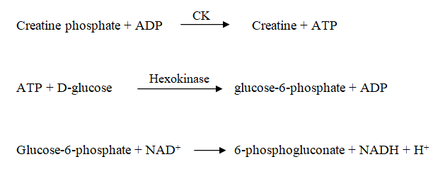
Assay Principle
Creatine kinase catalyzes the conversion of creatine phosphate and ADP to creatine and ATP. The ATP and glucose are converted to ADP and glucose-6-phosphate by hexokinase (HK). Glucose-6-phosphate dehydrogenase (G-6-PDH) oxidizes at the D glucose-6-phosphate and reduces the nicotinamide adenine dinucleotide (NAD). The rate of NADH formation, measured at 340nm is directly proportional to serum creatine kinase activity.
Reagent composition
| Creatine phosphate: | 30mmol/L |
| ADP: | 2mmol/L |
| D-glucose: | 20mmol/L |
| NAD+: | 2mmol/L |
| N- acetylcysteine: | 20mmol/L |
| Hexokinase: | 3000U/L |
| G-6-PDH: | 3000U/L |
Method
A specified volume (500µL) of the working reagent from the creatine kinase kit and 20µL of each serum sample were mixed. Each mixture was transferred into a measuring cuvette. Absorbance was read after 4 minutes in a spectrophotometer at 340nm. Further readings were taken during the next 4mins at 1min intervals. The change in the concentration of creatine kinase per minute (ΔE/min) for every reading was recorded and the mean values were determined.
Statistical Analysis
Data are reported as means ± standard deviation, where appropriate. One-way analysis of variance (ANOVA) was used to compare the measurement obtained after each treatment with control measurements. Differences were considered significant when P≤0.05.
RESULTS
Table 1: Effects of coconut (Cocos nucifera) milk and water, bitter leaf and okpehe extracts on serum concentration of creatine kinase for 30 days.
|
Groups |
Creatine kinase values (U/L) | |||
| Treatments | 10 days | 20 days | 30 days | |
| 1 | Control | 4.02±0.01* | 2.50±0.14* | 1.65±0.07* |
| 2 | Coconut milk | 5.25±0.07* | 3.70±0.14* | 2.10±0.14* |
| 3 | Coconut water | 0.85±0.07* | 4.10±0.14* | 2.82±0.02* |
| 4 | Bitter leaf | 2.45±0.07* | 2.45±0.07* | 3.74±0.01* |
| 5 | Okpehe (P.africana) | 1.44. ±0.01* | 2.10±0.14* | 4.28±0.01* |
(n = 3)* P < 0.05 Vs control
Effect of coconut water on serum creatine kinase (CK):
From Table 1, a significant decrease (P<0.05) was observed in the serum creatine kinase of the test group when compared with that of the control.
Effect of coconut milk on serum creatine kinase
Creatine kinase decreased significantly (P<0.05) in the test groups administered with high doses (10 and 15ml/kg b.wt) of coconut milk. A significant different (P<0.05) in the creatine kinase of the experimental animals when compared with the control was observed.
Effect of bitter leaf on serum creatine kinase:
Asignificant decrease (P<0.05) was observed in serum creatine kinase of the test group when compared to the control. The creatine kinase level increased significantly (P<0.05) in the test group administered high doses (10 and 15ml/kg b.wt) of bitter leaf extract.
Effect of Okpehe (P. africana) on Serum Creatine Kinase:
There is a significant decrease (P<0.05) of serum creatine kinase in the test group when compared with the control.
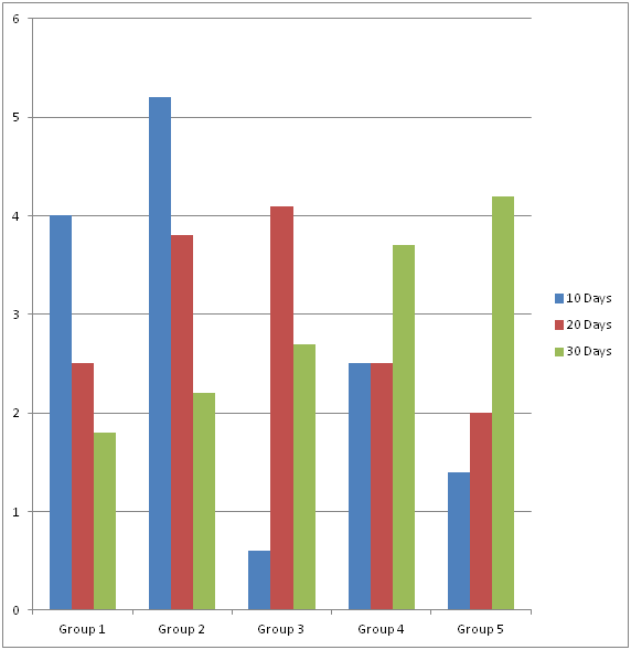
Figure 1: Effect of different plant extracts on creatine kinase activity in rabbits
The phytochemical contents of the milled seeds, leaves and bark of P. africanausing a polar solvent are presented in Table 2.
Table 2: Phytochemical constituents of P. africana
| Plant Parts | Alkaloids | Flavonoids | Glycosides | Saponins | Tannins | Steroids | Resins | Terpenoids |
| Seed | ++ | + | – | – | + | + | – | ++ |
| Leaves | +++ | + | + | – | + | + | – | ++ |
| Bark | + | ++ | ++ | + | + | + | – | + |
+++present in highconcentration
++ presentin moderate concentration
– absent
Phytochemical screening of the seeds, leaves and bark of P. africanashowed the presence of alkaloids, flavonoids, glycosides, saponins, steroids and terpenoids. From the results, Tannins and Steroids had the least concentration in the seeds, leaves, and bark of P. africana. Flavonoids and glycoside were present in a moderate concentration in the bark while alkaloids were present in high concentration in the leaves. Saponins were present in low concentration in the bark and absent in the seeds and leaves. Terpenoids were moderately present in the seeds and leaves while resin was absent in the seeds, leaves, and bark of P. africana.
Table 3: The phytochemical compositions of the leaves of Vernonia amygdalina.
| Phytochemicals | Intensity |
| Alkaloids | ++ |
| Flavonoids | + |
| Glycosides | + |
| Saponins | ++ |
| Tannins | ++ |
| Steroids | ++ |
| Resins | – |
| Terpenoids | ++ |
Key
– Absent
+ Present in low concentration
++ Present in moderate concentration
+++ Present in highconcentration
From the results, alkaloids, saponins, tannins, steroids, and terpenoids were moderately present in V. amygdalina as compared to P. africanawhich also contained moderate concentration of flavonoids and glycosides in the bark and moderate concentration of terpenoids in the seeds and leaves. Resins were absent while flavonoids and glycosides are present in low concentration in V. amygdalina.
DISCUSSION
The results in Table 1 show that coconut milk significantly increased (P<0.05) the serum levels of creatine kinase in the groups treated with the varying doses (5, 10, 15ml/kg. body weight respectively) of the coconut milk. This trend was quite interesting due to its dose-dependent effect.
The evidence that the concentrations of creatine kinase decrease as the dose of coconut milk increases is striking especially as Lawrence et al., (1979) showed that creatine kinase is found in an abnormal amount in patients with untreated prostatic carcinoma.
The mean creatine kinase value of the group fed with coconut water significantly decreased (P<0.05) when the control was compared to the test group and was significantly (P<0.05) increased as high doses were administered for 20 days. The increased levels of creatine kinase of the test group when compared to the control may be due to the presence of hydrolytic cellulose, which breaks down the wall of the kernel to liberate the oil upon which lipase acts to release free fatty acids and glycerol (Pohowich, et al., 2000).
Creatine kinase activity was essentially similar in the test group fed with a bitter leaf between 10 and 20 days of administration. Creatine kinase was significantly (P<0.05) increased after 20 days. This may be attributed to the non-pathologic experimental rabbits used in this work since creatine kinase traces could be used to monitor and assess the progression of prostate disorders in humans or animals.
The administration of Okpehe (P. africana) significantly (P<0.05) reduced the concentration of creatine kinase when the control was compared to the test group. Also, the creatine kinase concentration of the test group significantly (P<0.05) increased when compared to the control after 20 days.
This result is again striking as the presence of increased creatine kinase in most malignant fluids suggests the use of creatine kinase as an adjunct to the cytologic or histologic diagnosis of cancer (Coolen, 1976).
The phytochemical analysis of the seeds, leaves and bark of P. africana and the leaves of V. amygdalina showed the presence of alkaloids, flavonoids, tannins, saponins, glycosides, terpenoids, and steroids; and also possess medicinal properties as well as physiological activity (Sofowora, 1993). The presence of flavonoids in low concentration in the leaves and seeds and the moderate concentration of flavonoids in the bark of P. africana confers on it various biological function which include protection against allergies, platelet aggregation, microbes, ulcers, hepatoxin, viruses and tumors (James et al., 2003). Flavonoids reduce the risk of estrogen-induced cancers by interfering with the enzyme that produces estrogen (Okwu and Omodamiro, 2005). Moderate concentration of alkaloids in the leaves of V. amygdalina and the seeds and leaves of P. africana gives it the ability to function as spasmolytic, anti-cholinergic, and anesthetic agents (Amakoha, et al., 2002).
The presence of saponins in low and moderate concentration in the seeds, leaves, and bark of P. africana and V.amygdalina may be responsible for the hemostatic activity of these plants where they arrest bleeding from damaged or injured vessels by precipitating proteins to form vascular plugs (Duke, 1992). The saponins in V. amygdalina are used in veterinary vaccines as adjuvants (e.g., foot and mouth disease vaccines) helping to enhance the immune response. They are also mild detergents and are used commercially as well as in research. The presence of steroids in the plants under investigation enables the plants to be of interest in pharmacy due to their relationship with such compounds as sex hormones (Okwu, 2001).
The identification of moderate concentration of tannins in the leaves of V. amygdalina makes it useful in the tanning of leather, application on burns to heal injury and cuts to stop bleeding. Tannins are widely used as mouth washes, eyewashes, and vaginal douches and also treat rectal disorders. Tannins anti-inflammatory effect has helped in controlling gastritis, oesophagitis, and irritating bowel disorders (Chun, et al., 2003).
The moderate concentration of glycosides in the bark of P. Africana confers on its abilities that account for its use as drugs in the treatment of congestive heart failure and cardiac arrhythmia. They work by inhibiting the Na*/K+ pump. This causes an increase in the level of calcium ions. This inhibition increases the amount of Ca++ ions available for contraction of the heart muscle, improves cardiac output, and reduces distention of the heart. These glycosides (ouabain and some frog poisons) are used in Africa as arrow-poisons for hunting.
Terpenoids function in communication and defense. Terpenoids contribute to the scent of flowers and are under investigation for antineoplastic, and other pharmaceutical uses (Swiezewska and Danikiewicz, 2005).
CONCLUSION
This study shows that repeated administration of moderate doses (10 ml/kg b.wt.) of V. amygdalina and P. africana(Okpehe) extract significantly (p<0.05) reduce the serum levels of creatine kinase. This indicates the possible usage of creatine kinase in the prevention, treatment and management of kidney impairment, heart diseases, muscle diseases and prostate cancer and may also be used as a possible marker for the diagnosis of kidney diseases, muscle diseases and prostatic carcinoma.
The results obtained from the phytochemical analysis of the milled seeds, bark and leaves of P. africana(Okpehe) and the leaves of V. amygdalina showed the presence of alkaloids, flavonoids, saponins, tannins, glycosides, steroids, and terpenoids. The presence of these bioactive constituents justifies the use of these plants in the treatment of many debilitating ailments like cancer, diabetes, ulcers, obesity, heart diseases, and infections due to microorganisms. Comprehensive pharmacological and phytochemical investigation is however required to determine the exact mechanism of action of the anti-tumor activity of the extracts as well as the isolation and characterization of the bioactive constituents responsible for the effect observed.
REFERENCES
- Akah P.A., Ekekwe R.K. (1995). Ethnopharmacology of some of the asteraceae family used in the Nigerian traditional medicine. Fitoterapia. 1995;66:352–355.
- American Cancer Society (2010). What is Prostate Cancer? Information and Resources for Cancer: Breast, Colon, Prostate, Lung and other Forms.
- American Cancer Society. (2009). Cancer Facts and Figures 2009. Atlanta, G: American Cancer Society.
- Amokoha, R.A., Ubwa, S. Otokpa, T. and Shenge, G. (2002) Urology. 54(6), 1001-1201.
- Aragão W.M. (2002). Côco: pós-colheita. Série frutas do Brasil. Brasília: EmbrapaInformação Tecnológica; http:// livraria.sct.embrapa.br / iv_resumos/pdf/00070000.pdf
- Aumuller, G. (1979). Prostate Gland and Seminal Vesicles. Berlin-Haidelberg: Spring Verlag.
- Bate-smith, F. and Swam, G. (1992). Flavonoid compounds in Comparative Biochemistry. Florkin M. Maso H.S. Eds. Vol. III, 75-809. Academic Press. New York.
- Bhandary M.J., Chandrashekar K.R., Kaveriappa K.M. (1995). Medical ethnobotany of the Siddis of Uttara Kannada district, Karnataka, India. The Journal of Ethnopharmacology. 1995;47:149–158. doi: 10.1016/0378-8741(95)01274-H.
- Brancaccio P., Maffulli N., Limongelli F.M. (2007). Creatine kinase monitoring in sport medicine. British Medical Bulletin. 2007;81-82(1):209–230.
- Braun, K., Ehemann, V., Wiessler, M., Pipkorn, R., Didinger, B. Mueller, G., Waldeck, W. (2009). “High resolution flow cytometry: a suitable tool for monitoring aneuploid prostate cancer cells after TMZ and TMZ Bioshuttle treatment. Int. J. Med. Sci. 6(6): 338-47.
- Burkart A. A. (1976). monograph of the genus Prosopis (Mimosoideae). Journal of the Arnold Arboretum. 1976;57:216–525.
- BylsmaL.C., AlexanderD.D. (2015). A review and meta-analysis of prospective studies of red and processed meat, meat cooking methods, heme iron, heterocyclic amines and prostate cancer. The Nutrition Journal. 2015;14:125.
- Caceres A., Giron L.M., Alvarado S.R., Torres M.F. (1987). Screening of antimicrobial activity of plants popularly used in Guatemala for the treatment of dermato-mucosal diseases. The Journal of Ethnopharmacology. 1987;20:223–237. doi: 10.1016/0378-8741(87)90050-X.
- Calle, E.E., Rodriguez, C., Walker Thurmond, K., Thun, M.J. (2003). Overweight, Obesity and Mortality from Cancer in a Prospectively Studies Cohort of U.S.A adults. N. Engl. Med. 348(17): 1625-38.
- Chun, O.K., Kim, D.O., Lee, C.Y. (2003). Superoxide radical scavenging activity of the major polyphenols in fresh plums. J. Agric Food Chem. 51 (27): 8067-8072.
- Cimanga R.K., Tona L., Mesia K., Musuamba C.T., De Bruyne T., Apers S., Hernan N., Miert V.S., Pieters L., Totte J., Vlietink A.J. (2004). In vitro anti plasmodia acivity of extravts and fractions of seven medicinal plants used in the democratic republic of Congo. The Journal of Ethnopharmacology. 2004;93:27–32.
- Djulbegovic, M., Beyth, R.J. and Neuberger, M.M. (2010). “Screening for Prostate Cancer: Systematic Review and Meta-analysis of Randomized Controlled Trials. BMJ 31: 4543.
- Duke, J. (1992). Handbook of Biological Active Phytochemicals and their Activities BOCA Ration (FL) CRC Press, P. 99-131.
- Esquenazi M.D., Wigg M.M., Miranda, Rodrigues H.M., Tostes J.B.F., Rozental S., (2002). Antimicrobial and antiviral activities of polyphenolics from Cocos nucifera Linn. (Palmae) husk fiber extract. Research in Microbiology. 2002;153:647–652. doi: 10.1016/S0923-2508(02)01377-3.
- Essink.Bot, M.L., De Koning, H.J., Nijs, H.G., Kirkels, W.J. Van der Maas, P.J., Schroder, F.H. (1998). Short term effects of population-based screening fro prostate cancer on health – related quality of life. J. Natl. Cancer Inst. 90(12): 925-31.
- Grant, Ownby, C. and Peel, R. (2000). Comparison of the neutralizing abilities of three antivenom preparations. XIIIthWorld Congress of the International Society on Toxinology. Paris, P. 228.
- Harbone, J.B. (1973). Phytochemical Methods: A Guide to Modern Techniques in Plant Analysis. Chapman and Hall, London; 221-32.
- Hladik C., Krief S., Haxaire C. (2005). Ethnomedicinal and bioactive properties of plants ingested by wild chimpanzees in Uganda. The Journalof Ethnopharmacology. 2005;101:1–5.
- Holdsworth D. (1992). Medicinal plants of the Gazelle peninsula, New Britain Island, Papua New Guinea, Part I. International Journal Of Pharmacognosy. 1992;30:185–190. doi: 10.3109/13880209209053992.
- Hope B.E., Massey D.G., Fournier-Massey G. (1993). Hawaiian materia medica for asthma. The Hawai’i Medical Journal. 1993;52:160–166.
- Izevbigie E.B. (2003). Discovery of water-soluble anticancer Agents (Edotides) from a vegetable found in Benin City, Nigeria. Experimental Biology and Medicine. 2003;228:293–298.
- Kazak. L., Cohen, P. (2020). Creatine metabolism: energy homeostasis, immunity, and cancer biology. Nature Reviews Endocrinology. 2020;16:421–36.
- Keay R.W., Onochie C.F., Stanfold D.P. (1964). Nigerian Trees. 2 nd ed. Ibadan: Fed Dept of Forest Research; 1964. pp. 113–4.
- Kupchan S.M., Hemmnigway R.J., Karim A., Werner D. (1969). Tumor inhibitors. XLVII Vernodalin and Vernomygdin. Two new cytotoxic sesquiterpene lactones from Vernonia amygdalina Del. The Journal of Organic Chemistry. 1969;34:3908–3911.
- Moundipa F.P., Kamini G., Melanie F., Bilong F.C., Bruchhaus I. (2000). In vitro amoebic activity of some medicinal plants of the Bamun region (Cameroon) African Journal of Traditional, Complementary and Alternative Medicines. 2000;62:113–121.
- Muraina I.A., Adaudi A.O., Mamman M., Kazeem H.M., Picard J., McGaw L.J., Eloff J.N. (2010). Antimycoplasmal activity of some plant species from northern Nigeria compared to the currently used therapeutic agent. Pharmaceutical Biology. 2010;48:1103–1107.
- Neubauer, S. (2007). The Failing Heart — An Engine Out of Fuel. New England Journal of Medicine, 356(11), 1140–1151. doi:10.1056 /nejmra063052
- Nwosu, S.I., Stanley, H.O., Okerentugba, P.O. (2013). Occurrence, types and location of calcium oxalate crystals in Vernonia amygdalina Del (Asteraceae) The International Journal of Natural Sciences. 4 (3) (2013), pp. 533-537
- Oguntoyinbo F.A., Sanni A.I., Franz C.M., Wilhelm H., Holzapfel W.H. (2007). In vitro fermentation studies for selection and evaluation of Bacillus strains as starter cultures for the production of okpehe, a traditional African fermented condiment. The International Journal of Food Microbiology. 2007;113:208–18.
- QianX.L., LiY.Q., GuF., LiuF.F., LiW.D., ZhangX.M. (2012). Overexpression of ubiquitous mitochondrial creatine kinase (uMtCK) accelerates tumor growth by inhibiting apoptosis of breast cancer cells and is associated with a poor prognosis in breast cancer patients. Biochemical and Biophysical Research Communications. 2012;427:60–6.
- Rosa M de F., Santos F.J. de S., Montenegro A.A.T., Abreu F.A.P., Correia D., Araújo F.B.S., (2001). Caracterização do pó da casca do coco verdeusadocomosubstratoagrícola. Fortaleza: Embrapa Agroindústria Tropical, Comunicado Técnico, 54; 2001. http://www.ceinfo .cnpat. embrapa. br/arquivos/artigo_2459.pdf
- Singh Y.N. (1986). Traditional medicine in Fiji: some herbal folk cures used by Fiji Indians. The Journal of Ethnopharmacology. 1986;15:57–88. doi: 10.1016/0378-8741(86)90104-2.
- Thygesen, K., Alpert, J.S., Jaffe, A.S., Simoons, M.L., Chaitman, B.R., White, H.D. (2012). Task force for the universal definition of myocardial infarction: third universal definition of myocardial infarction. Nature Reviews Cardiology.2012;9:620–33.
- Totsuka M., Nakaji S., Suzuki K., Sugawara K., Sato K.. (2002). Break point of serum creatine kinase release after endurance exercise. Journal of Applied Physiology. 2002;93(4):1280–1286.
- Ventura-Clapier, R., Garnier, A., and Veksler, V. (2004). Energy metabolism in heart failure. The Journal of Physiology, 555(1), 1–13. doi:10.1113/jphysiol.2003.055095.
- Weniger B., Rouzier M., Daguilh R., Henrys D., Henrys J.H., Anton R. (1986). [Traditional medicine in the Central Plateau of Haiti. 2. Ethnopharmacologic inventory]. The Journal of Ethnopharmacology. 1986;17:13–30. doi: 10.1016/0378-8741(86)90070-X.
- Yartey J., Harisson E.K., Brakohiapa L.A., Nkrumah F.K. (1993). Carbohydrate and electrolyte content of some home-available fluids used for oral rehydration in Ghana. The Journal of Tropical Paediatrics. 1993;39:234–237. doi: 10.1093/tropej/39.4.234.
- Ye, Y., Gong, G., Ochiai, K., Liu, J., and Zhang, J. (2001). High-Energy Phosphate Metabolism and Creatine Kinase in Failing Hearts: A New Porcine Model. Circulation, 103(11), 1570–1576. doi:10.1161/01.cir.103.11.1570
- ZhangL., ZhuZ., YanH., WangW., WuZ., ZhangF. (2021). Creatine promotes cancer metastasis through activation of Smad2/3. Cell Metabolism.2021;33:1111–23.