
Analysis of ZnO and Tio2 as An Effective Nanomaterials for the Development of DSSCs: A Review
- Azeez J.
- 208-213
- Feb 17, 2024
- Physical Education
Analysis of ZnO and Tio2 as An Effective Nanomaterials for the Development of DSSCs: A Review
Azeez J.*1, 2
1Africa Centre of Excellence in Future Energy and Electrochemical Systems (ACEFUELS), Federal University of Technology, Owerri, Nigeria.
2Department of Science Laboratory Technology, The Polytechnic, Ibadan.
*Corresponding author
DOI: https://doi.org/10.51584/IJRIAS.2024.90118
Received: 10 January 2024; Revised: 18 January 2024; Accepted: 23 January 2024; Published: 17 February 2024
ABSTRACT
Energy crisis and global warming are the two major problems facing us today. The search for the best effective photo-anode film for the development of Dye-sensitized solar cells (DSSCs) – a third generation organic cell, is very essential. ZnO and TiO2 had been reportedly used as nanomaterial for the development of DSSCs. This paper presents reviewed works on the analysis of major properties of these nanomaterials. X-ray diffraction (XRD) analysis, Microstructural and compositional analysis, and photo-electrochemical analysis were considered for these materials. Our analysis agreed well with many researchers who had claimed that TiO is the best photo-anode material for DSSCs.
Keywords: DSSCs, ZnO, TiO, Nanomaterial, photo-anode, XRD.
INTRODUCTION
Research and development of Dye-sensitized solar cells (DSSCs) have arisen as a technically and economically credible alternative to the p-n junction photovoltaic devices. Recently, great amount of research efforts has been directed toward the fabrication of DSSCs. This is because their effectiveness relies on production cost and environmental friendliness, DSSCs have attracted exceptional consideration, since their first development in 1991 [1–3]. DSSCs are termed as encouraging cost-effective solar cells that have a comparable conversion efficiency from sunlight into electricity. Numerous photovoltaic cells that guarantee solar energy conversion have been discovered in the past years. Although, significant problems are confronting their universal adoption which is limited to two important difficulties namely; cost and efficiency [1,3,4]. Four main parts of DSSCs are porous crystalline wide semiconductor band gap electrode, sensitizer, electrolyte, and counter electrode [1,4]. In DSSCs, the dye sensitization of photo-anode would account for efficient electrical energy conversion from sunlight in the cells, therefore, there is need to know the best photo-anode material.
X-ray diffraction (XRD) characterization is a powerful nondestructive technique for characterizing crystalline materials. It provides information on crystal structure, phase, preferred crystal orientation (texture), and other structural parameters, such as average grain size, crystallinity, strain, and crystal defects [19]. XRD peak intensity tells about the position of atoms within a lattice structure and peak width tells about crystallite size and lattice strain. XRD is used to study the crystalline structure of materials since the X-ray wavelengths (between 0.2 and 10 nm) are comparable to the interatomic spacing of crystalline solids. The technique measures the average spacing between layers or rows of atoms. XRD is useful for evaluating minerals, polymers, corrosion products, and unknown materials. In most cases, the samples analyzed at Element are analyzed by powder diffraction using samples prepared as finely ground powders [5].
Microstructure are material structures seen at the micro level. Specifically, they are structures of an object, organism, or material as revealed by a microscope at magnifications greater than 25 times. Simply put, anything that is not regular from a given crystalline structure is a microstructure. A microstructural analysis is performed in order to evaluate the microstructure in a metal alloy. The observation is made at different magnifications depending on the observation to be made. Microstructural analysis is used widely in industry to evaluate products and materials. Performance, response to environment and failure mechanisms are just some of the areas in which microstructural analysis can be utilized to assess and develop products [6].
Photoluminescence (PL) is a process in which a molecule absorbs a photon in the visible region, exciting one of its electrons to a higher electronic excited state, and then radiates a photon as the electron returns to a lower energy state. PL is the spontaneous emission of light from a material following optical excitation. It is a powerful technique to probe discrete energy levels and to extract valuable information about semiconductor sample composition, quantum well thickness or quantum dot sample monodispersity. Photoluminescence spectroscopy is a widely used technique for characterization of the optical and electronic properties of semiconductors and molecules. Photoluminescence spectra are recorded by measuring the intensity of emitted radiation as a function of either the excitation wavelength or the emission wavelength. An excitation spectrum is obtained by monitoring emission at a fixed wavelength while varying the excitation wavelength [7].
ANALYSIS OF ZNO AND TIO NANOPARTICLES
- X-ray diffraction (XRD) analysis of ZnO powder
In 2014 and 2021, Adedokun et. al., and Bindu & Thomas were respectively reported on the structural properties of ZnO nanoparticles. They were able to established their reports using XRD analysis. They reported, the structural properties of the ZnO nanoparticles were established using XRD analysis as shown in Fig. 1 [8]. The peaks in the XRD plot corresponded with the typical wurtzite ZnO structure (hexagonal phase, JCPDS file 75–0576). The average crystallite size, D, of 34.2 nm was calculated from the highest diffraction peak along the〈101〉plane for the ZnO nanoparticles, which further established that there is no impurity or remnant bulk materials in the ZnO powder. The XRD pattern’s peak broadening indicates the presence of small nanocrystals in the samples [9].
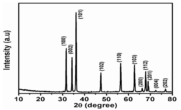
Fig. 1: X-ray diffraction (XRD) analysis of ZnO powder [8]
- Microstructural and compositional analysis of ZnO
The structural analysis of ZnO was strengthened by the transmission electron microscopy (TEM) analysis of the ZnO nanoparticles [8]. Fig. 3 shows the TEM analysis of the ZnO nanoparticles. Nanoparticles with hexagonal-typical ZnO shape were produced as found in Fig. 3(a) and the nanoparticle’s cross-sectional width were ranged from 20 nm to 50 nm. The crystalline nature of individual nanoparticles was confirmed by the HRTEM result, while from Fig. 3(b) the corresponding spacing of the (101) plane was shown. The Selected Area Electron Diffraction (SAED) pattern of the sample as shown in Fig. 3(c) describes planes of (101), (102), and (200) for ZnO nanoparticles [8].
- Microstructural and compositional analysis of TiO2
TEM was used to further examine the particle size, crystallinity and morphology of samples. TEM bright field image of TiO2 micropowder is shown in Fig 4(a) [10]. It is clearly seen that the TiO2 powders in anatase phase are mostly spherical morphology. Furthermore, it can be estimated that the particle size of samples in Fig 4(a) are microscale with the grain size about 0.3-0.7μm.
TEM bright field images of TiO2 nanopowder is shown in Fig 4(b). It can be estimated that the particle size of powders in Fig 4(b) is nanoscale with the grain size less than 10nm. The corresponding selected area electron diffraction (SAED) patterns of nano-TiO2 powders is shown in Fig 4(c). This is in agreement with XRD results in Fig (2). In Fig 4(c), the SAED patterns of nano-TiO2 powders in anatase phase shows that the brightness and intensity of polymorphic ring is weak, so they are poorly crystallized and partly amorphous. The crystallinity of nano-TiO2 powders can also be observed by phase-contrast images or Moire patterns. Fig 4(d) show crystal lattice planes of nano-TiO2 in anatase phase. It is seen that, there are many crystal lattice planes with d-spacing of 0.313nm for the plane (101).
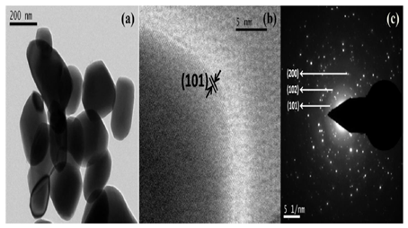
Fig. 3. (a) TEM image showing ZnO nanoparticles (b) a single nanoparticle HRTEM image, and (c) SAED pattern [8].
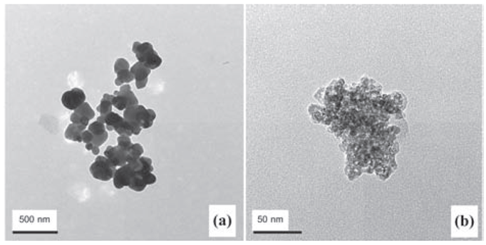
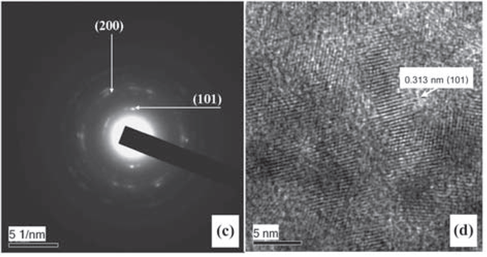
Fig. 4: Images of anatase phase. (a) TEM image of micro-TiO2 powder; (b) TEM image of nano-TiO2 powder; (c) SAED pattern of nano-TiO2 powder and (d) HRTEM image of nano-TiO2 powder [10].
- Photoluminescence (PL)/Photo-electrochemical comparison between ZnO and TiO2
Table 1.0 [8] shows photo-electrochemical comparison of natural pigments used in ZnO/TiO2-based DSSCs. The values obtained for the open circuit voltage (Voc), current density (Jsc), Fill factor (FF %) and Energy conversion efficiency ( (%)) for the TiO2-based DSSCs shows that they are more effective than ZnO-based DSSCs.
Table 1.0: Photo-electrochemical comparison of Natural Pigments used in DSSCs [8]
| Natural
Pigment |
Photoanode
Materials |
Voc
(V) |
Jsc
(mA/cm2) |
FF
(%) |
η(%) | Ref |
| Pheophytin a | TiO2 | 0.593 | 2.199 | 0.355 | 0.46 | [11] |
| 0.718 | 0.698 | 0.480 | 0.24 | |||
| Carotenoids | TiO2 | 0.43 | 2.84 | 0.46 | 0.56 | [12] |
| 0.58 | 0.45 | 0.60 | 0.16 | |||
| Bixin | ZnO | 0.32 | 0.087 | 0.37 | 0.010 | [13] |
| Anthocyanins | TiO2 | 0.644 | 0.24 | 0.49 | 0.076 | [14] |
| 0.495 | 0.94 | 0.65 | 0.301 | |||
| Chlorophyl | TiO2 | 0.36 | 0.8 | 0.69 | 0.178 | [15] |
| Carica papaya | 0.373 | 0.149 | 30.37 | 0.017 | ||
| Citrus lanatus | ZnO | 0.333 | 0.140 | 26.78 | 0.013 | [16] |
| Persea americana | 0.303 | 0.111 | 29.51 | 0.010 | ||
| Solanum melongena | 0.321 | 0.126 | 28.24 | 0.011 | ||
| Citrus fruit peel | ZnO | 0.323 | 0.299 | 29.18 | 0.028 | [17] |
| 0.205 | 0.033 | 30,40 | 0.013 | |||
| 0.291 | 0.042 | 28.12 | 0.004 | |||
| 0.207 | 0.265 | 26.40 | 0.022 | |||
| Musa paradisiaca | ZnO | 0.282 | 0.122 | 25.49 | 0.009 | [8] |
| Mangifera indica | 0.288 | 0.265 | 30.79 | 0.024 | ||
| Punica granatum | 0.283 | 0.133 | 27.51 | 0.010 | ||
| Ananas comosus | 0.214 | 0.033 | 29.45 | 0.002 |
CONCLUSION
We therefore conclude that TiO2 nanoparticle material is better than ZnO materials for the development of DSSCs. Also, due to being non-toxic and less expensive and its easy availability, TiO2 is mostly used as a semiconducting layer in DSSCs because photosensitizers are easily attached on the nanocrystalline particle of TiO2 compare to ZnO [18]. Thus, we recommend the use of TiO2 as an effective nanoparticle material for the development of DSSCs as reported in Table 1 above.
REFERENCES
- O. Adedokun, K. Titilope, A.O. Awodugba, Review on natural dye-sensitized solar cells (DSSCs), Int. J. Eng. Technol. IJET. 2 (2016) 34, https://doi.org/10.19072/ijet.96456 .
- B.Q. Liu, X.P. Zhao, W. Luo, The synergistic effect of two photosynthetic pigments in dye-sensitized mesoporous TiO2 solar cells, Dye. Pigment. 76 (2008) 327–331, https://doi.org/10.1016/j.dyepig.2006.09.004.
- B. O’Regan, M. Gratzel, A low-cost, high-efficiency solar cell based on dye-sensitized colloidal Ti02 films, Nature 353 (1991) 737–740, https://doi.org/10.1016/0146-5724(84)90144-4 .
- M. Gr¨atzel, Dye-sensitized solar cells, J. Photochem. Photobiol. C Photochem. Rev. 4 (2003) 145–153, https://doi.org/10.1016/S1389-5567(03)00026-1 .
- https://en.wikipedia.org/wiki/x-raydiffraction
- https://en.wikipedia.org/wiki/microstructuralandcompositionalanalysis
- https://en.wikipedia.org/wiki/Photoluminescence
- O. Adedokun, O.L. Adedeji, I.T. Bello, M.K. Awodele, A.O. Awodugba, Fruit peels pigment extracts as a photosensitizer in ZnO-based Dye-Sensitized Solar Cells, Chemical Physics Impact 3 (2021) 100039, www.sciencedirect.com/journal/chemical-physics-impact, https://doi.org/10.1016/j.chphi.2021.100039
- P. Bindu, S. Thomas, Estimation of lattice strain in ZnO nanoparticles: x-ray peak profile analysis, J. Theor. Appl. Phys. 8 (2014) 123–134, https://doi.org/10.1007/s40094-014-0141-9
- K. Thamaphat, P. Limsuwan, and B. Ngotawornchai, Phase Characterization of TiO2 Powder by XRD and TEM, Kasetsart J. (Nat. Sci.) 42 : 357 – 361 (2008)
- V. Shanmugam, S. Manoharan, A. Sharafali, S. Anandan, R. Murugan, Green grasses as light harvesters in dye sensitized solar cells, Spectrochim. Acta – Part A Mol. Biomol. Spectrosc. 135 (2015) 947–952, https://doi.org/10.1016/j.saa.2014.07.096.
- E. Yamazaki, M. Murayama, N. Nishikawa, N. Hashimoto, M. Shoyama, O. Kurita, Utilization of natural carotenoids as photosensitizers for dye-sensitized solar cells, Sol. Energy. 81 (2007) 512–516, https://doi.org/10.1016/j.solener.2006.08.003.
- N.M. G´omez-Ortíz, I.A. V´azquez-Maldonado, A.R. P´erez-Espadas, G.J. Mena-Rej´on, J.A. Azamar-Barrios, G. Oskam, Dye-sensitized solar cells with natural dyes extracted from achiote seeds, Sol. Energy Mater. Sol. Cells. 94 (2010) 40–44, https://doi.org/10.1016/j.solmat.2009.05.013.
- N.J. Cherepy, G.P. Smestad, M. Gr¨atzel, J.Z. Zhang, Ultrafast electron injection: implications for a photoelectrochemical cell utilizing an anthocyanin dye-sensitized TiO2 nanocrystalline electrode, J. Phys. Chem. B. 101 (1997) 9342–9351, https://doi.org/10.1021/jp972197w.
- G. Calogero, I. Citro, G. Di Marco, S. Armeli Minicante, M. Morabito, G. Genovese, Brown seaweed pigment as a dye source for photoelectrochemical solar cells, Spectrochim. Acta – Part A Mol. Biomol. Spectrosc. 117 (2014) 702–706, https://doi.org/10.1016/j.saa.2013.09.019.
- O. Adedokun, M.K. Awodele, Y.K. Sanusi, A.O. Awodugba, Natural dye extracts from fruit peels as sensitizer in ZnO-based dye-sensitized solar cells, IOP Conf. Ser. Earth Environ. Sci. (2018) 173, https://doi.org/10.1088/1755-1315/173/1/012040.
- O. Adedokun, O.L. Adedeji, M.K. Awodele, I.T. Bello, A.O. Awodugba, Citrus fruit peels extracts as light harvesters for efficient ZnO-based dye-sensitized solar cells, J. Phys. Conf. Ser. 1299 (2019), 012010, https://doi.org/10.1088/1742-6596/1299/1/012010.
- K. Sharma, V. Sharma and S. S. Sharma, Dye-Sensitized Solar Cells: Fundamentals and Current Status, Nanoscale Research Letters (2018) 13:381, https://doi.org/10.1186/s11671-018-2760-6.
- Bunaciu AA, Udriştioiu EG, Aboul-Enein HY. X-ray diffraction: instrumentation and applications. Crit Rev Anal Chem. 2015;45(4):289-99. doi:10.1080/10408347.2014.949616. PMID: 25831472.