
Effect of Petrol on the Electrocardiogram, Cardiac Troponin-I and Body Weight of Wistar Rats and Mitigation by Moringa Oleifera Leaf Extract
- Ashiru Dahiru
- Bashir Sa’idu
- Zaid Shehu
- Ibrahim Hassan Maina
- Shuaib Zakariya Adam
- Ja’afaru Ishaq Abdullahi
- Nafisat Abdulazeez
- Nicholas Nathaniel Pilau
- 455-464
- Jun 21, 2024
- Economics
Effect of Petrol on the Electrocardiogram, Cardiac Troponin-I and Body Weight of Wistar Rats and Mitigation by Moringa Oleifera Leaf Extract
Ashiru Dahiru1*, Bashir Sa’idu1, Zaid Shehu2, Ibrahim Hassan Maina1, Shuaib Zakariya Adam1, Ja’afaru Ishaq Abdullahi1, Nafisat Abdulazeez1, and Nicholas Nathaniel Pilau2
1Department of Veterinary Physiology and Biochemistry, Usmanu Danfodiyo University, Sokoto. Nigeria.
2Department of Veterinary Medicine, Usmanu Danfodiyo University, Sokoto. Nigeria.
*Correspondence Author
DOI : https://doi.org/10.51584/IJRIAS.2024.905040
Received: 11 November 2023; Accepted: 22 November 2023; Published: 21 June 2024
ABSTRACT
Petrol is commonly used as a fuel for internal combustion engines. Humans and animals continued to be exposed to petrol vapor in the environment, occupationally by petroleum attendants and refinery workers, at home and on the roads. Fumes from petrol products cause environmental pollution that has resulted in various health challenges, including increased blood pressure and heart rate, which may lead to hypertension. This study is aimed at evaluating the effects of petrol on the electrocardiograms (ECG) and Cardiac troponin of Wistar rats and possible mitigation by Moringa oleifera extract. A total of 15 adult Wistar rats were randomly divided into three groups of five each: the control group (not given anything), the group exposed to petrol only and the group treated with Moringa oleifera extract after exposure to petrol. Rats were exposed to petrol vapor daily for 5 minutes over four weeks in a fume chamber with monitored room conditions, resulting in an average dosage exposure of 0.008 cm3/min/rat. Ketamine was used as an anesthetic agent, and the ECGs of the rats were measured with EDAN 10 veterinary electrocardiography. Serum cTn-I assay was analyzed at the end of the four weeks. The results showed an increase in heart rate, reduced QRS complex, increased in P wave and PR interval. Even though the cTn-I has considered revising but not statistically significant. It is concluded that petrol has effects on the electrocardiogram and cardiac troponin I of Wistar rats, and treatment with Moringa oleifera showed no remediating effect.
Key word: ECG, Petrol, Cardiac Troponin-I, Body weight, and Moringa oleifera
INTRODUCTION
Petrol
Petrol is a complicated mixture of light hydrocarbons with a boiling range of (15 OC to 190 OC) and a carbon atom count of 5 to 11. It’s one of the byproducts of petroleum distillation and refinement. In the past, organic lead compounds were frequently added to gasoline to prevent engine knock, but this practice has decreased as a result of environmental concerns. To further stabilize petrol and enhance its appearance, fragrance, and color, additional compounds are also added [1]. Initially, crude oil was simply distilled to create petrol without the aid of any chemical conversion procedures. The percentage of atmospheric distillation known as light, medium, and heavy naphtha is a possible source of gasoline [1]. Petroleum products have been demonstrated to result in tachycardia, dysrhythmia, dizziness, pulmonary hypertension, pulmonary aspiration, severe respiratory failure, cardiac failure, poor regulation of vascular tone, and endogenous fibrinolysis [2].
Electrocardiography
Today, a key component of the initial assessment for patients presenting with cardiac symptoms is electrocardiography. In particular, it is crucial as a non-invasive, economical method to assess arrhythmias and ischemic heart disease [3]. In the 1950s and 1960s, electrocardiography was used to diagnose cardiac illness in numerous species, and it is still a crucial diagnostic procedure today [4]. An ECG recording shows the electrical activity of the heart and can give valuable information on the myocardium’s anatomical and functional properties [5]. In fundamental cardiovascular research, electrocardiography in rats is a commonly used experimental technique. ECG recording methods are straightforward, however, it can be difficult to understand the electrocardiographic characteristics. This is due to the possibility that experimental circumstances like the type of anesthetic, the strain, or the age of the animal may skew the analysis [5]. Compared to bigger animals, rat and mouse models provide a number of benefits, including reduced costs, less variability, the availability of transgenic models, and a wide range of research instruments [6]. Modern day advances in small rodent electrocardiography have enabled assessments in conscious unrestrained animals and improved ECG interpretation [7]. Rat ECGs have been used in fundamental cardiovascular research examining the function of the heart under physiological circumstances and in animal models of cardiovascular disorders [5].
Cardiac Troponin-I
The sarcomere, the basic contractile unit of the heart, consists of thick and thin filaments composed of myosin and actin, respectively. The thin filament contains tropomyosin and troponin, which regulate the interaction between actin and myosin in a calcium-dependent manner [8]. This regulatory site is the troponin complex, a tadpole-shaped heterotrimer immobilized on the thin filament, which acts in an allosteric manner to regulate the Ca2+-dependent interaction of actin and myosin filaments [9]. Acute coronary syndrome (ACS), including ST-segment elevation MI (STEMI), non-ST-segment elevation MI (NSTEMI), and unstable angina, is characterized by myocardial ischemia resulting from atherosclerotic plaque rupture or erosion [8]. This leads to partial or complete vascular occlusion, microembolization, or clotting cascade activation, causing localized ischemia and infarction. Myocardial ischemia is the first step in developing an MI and occurs because of an oxygen supply-demand mismatch [10]. or reduced coronary flow [11]. Cardiac troponin (cTnI/T) is commonly used as a specific biomarker for myocardial injury in diagnosing AMI, but it can also be released due to non-ischemic and extra-cardiac conditions [12]. The sensitivity of cTnI/T for myocardial injury is well established, but claiming specificity for any disease is not feasible [10].
Wistar Rats
An outbred albino rat is the Wistar rat. At a time when laboratories mainly used the house mouse, this breed was created at the Wistar Institute in 1906 for use in biological and medical research. It is noteworthy because this breed was the first rat to be created to serve as a model organism (Mus musculus). The original colony of laboratory rats was started by physiologist Henry Herbert Donaldson, scientific manager Milton J. Greenman, and geneticist/embryologist Helen Dean King. More than half of all laboratory rat strains are descended from this colony [13]. The Wistar rat has been utilized extensively in biomedical research during the 20th century, and numerous strains have been developed to meet the needs of researchers in various fields [13]. ECG analysis in tiny rodents was used in a wide range of works as part of many research themes. Compared to larger animals, the rat and mouse models for research include benefits such as cheaper costs, less physiological variability, and the flexibility to use transgenic models [14].
MATERIALS AND METHODS
Study Area
The study was conducted in the laboratory of the Department of Veterinary Physiology and Biochemistry, Faculty of Veterinary Medicine, Usmanu Danfodiyo University, Sokoto, Sokoto State. It lies between latitude 13° 3’ 490N and longitude 5° 14’ 890E at an altitude of 272 m above sea level [15]. The state is located in the extreme northwest of Nigeria. It shares common borders with the Niger Republic to the north, Kebbi State to the south-west, and Zamfara State to the east [16].
Study Design
An experimental study was conducted, and the random sampling technique was employed. A total of 15 male and female Wistar rats weighing between 130 and 250g were randomly divided into three groups of five rats each. The first group served as the control. The second group was exposed to petrol vapor. The third group exposed to petrol then later treated with Moringa oleifera leaf extract.
Experimental Animal/Acclimatization
A total of 15 males and females are adults. Wistar rats weighing between 130 and 250g were acquired from the animal house of the Department of Pharmacology and Toxicology, Faculty of Pharmaceutical Sciences, Usmanu Danfodiyo University, Sokoto. They were housed in a well-ventilated cage (figure 1). The rats were on standard rat chow and tap water ad libitum. They were acclimatized for two weeks before the experimental period. Procedures involving animals and their care were performed in accordance with the National Institute of Health (NIH) guidelines for the care and use of animals by National Research Council [17]. Ethical approval was sought from the Faculty Animal Research and Ethics Committee (FAREC) of the Faculty of Veterinary Medicine, Usmanu Danfodiyo University, Sokoto.
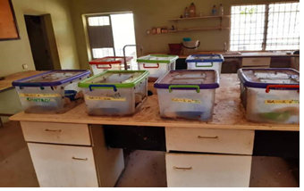
Fig 1: showing rats in their cages
Plant Materials/Preparation of Plant Extract
Moringa oleifera leaves were obtained from Danchadi village, Bodinga LGA, Sokoto State (figure 2). The plant was identified at the herbarium unit of the Department of Biological Sciences, Usmanu Danfodiyo University, Sokoto (PCG/UDU/SOR1/0001). The 80% methanol cold extraction method was used [18]. 40mg/kg/rat was used as the dosage throughout the study period; this was obtained after a toxicity study [19].

Fig 2: Moringa oleifera leaves and stems
Exposure to Petrol
The petrol used was purchased from the NNPC Mega Station, close to FGC, Sokoto, and Sokoto State. Rats in group A (control) were kept in a petrol vapor-free section of the experimentation room. Rats in group B were placed in the fume chamber (figure 4). The fume chamber is a 20-liter bucket with a very tight lid [20]. During the exposure period, the rats were placed in the chamber. The nebulizer cup was filled with petrol and the liquid petrol turned to vapor as soon as the nebulizer (figure 3) was switched on. They were allowed to stay in the fume chamber for 5 minutes and then were removed back to their cages in the vapor-free section of the experimentation room. This was done every day for four weeks. The room condition was monitored and maintained at a temperature of 28 ± 2 ºC. The average dosage exposure was 0.008cm3/min/rat.
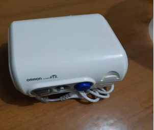
Fig 3: showing human nebulizer
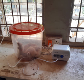
Fig 4: rats undergoing exposure to petrol vapor
Measurement of Electrocardiography
The ECGs of the rats were measured with EDAN 10 Veterinary Electrocardiography (ECG), made by Ronseda Electronics CO., LTD., China (figure 5). At the end of the four weeks, each rat was anesthetized with ketamine intramuscularly. Once a rat was anesthetized, it was placed on a white board. The EDAN electrode clips for the right forelimb, left forelimb, right hind limb, left hind limb, and the heart were put in place after rubbing the site with an adequate quantity of gel [21]. The EDAN was connected to the laptop, and information about each rat was recorded and saved. This was followed by ECG recording for one minute and saving it until it was done for rats in the two groups.
Lead II
Heart signals are picked up by an ECG machine. Electrodes were placed externally at specific body locations, i.e., the fore and hind limbs, and then heart signals of very small amplitude (few millivolts) were recorded [22]. Lead II records the electrical activity as seen from the inferior (diaphragmatic) surface of the heart. This lead is often used for rhythm monitoring [23]. Lead II, also known as a rhythm strip, is useful in evaluating heart rate and rhythm because it gives you a longer recording in which to evaluate the pattern of ECG waveforms [24]. Lead II is used here because it is diagnostic.
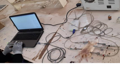
Fig 5: showing rat undergoing ECG
Determination of Cardiac Troponin I
The differences in amino acid sequences consider the development of quantitative assays for cTnI/T (figure 6). Most cTn assays are non-competitive enzyme-linked immunosorbent assays (ELISA) based on the sandwich principle, utilizing the high specificity and affinity of antibodies [25]. The assay is based on a capture antibody which binds to a specific epitope of cTn, and a detection antibody which binds to a separate epitope. The epitopes are often closely spaced to prevent a proteolytic cleavage event diminishing sensitivity. Furthermore, the most stable regions of cTn are selected as epitopes; regions that are not susceptible to cleavage or post-translational modifications, e.g., phosphorylation [26]. The detection antibody is linked to a signal-generating system to enable quantification. Signal amplification is achieved by using an enzyme which can cleave multiple molecules of a substrate over a given time-interval, or by using other detection methodologies such as gold micro particles.
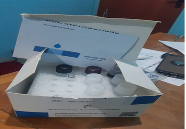
Fig 6: Rat Tn-1 ELISA Kit
Determination of Body Weight
The body weight was determined daily throughout the study period using a weighing scale.
Statistical Analysis
Data are expressed as means ± standard error of means (SEM); statistical analysis was done using a one-way ANOVA (Tukey-Kramer Multiple Comparisons Test), and body weight data were analyzed using a 2-way repeated measures mixed approach. P <0.05 was considered significant. All analysis was done using InVivoStat software (Version 4.2.0).
RESULTS
ECG Result of Group A (control)
The result of the ECG of rat in group A (control) showing normal waves (P, QRS and T) as indicated in lead II presented in figure 7.
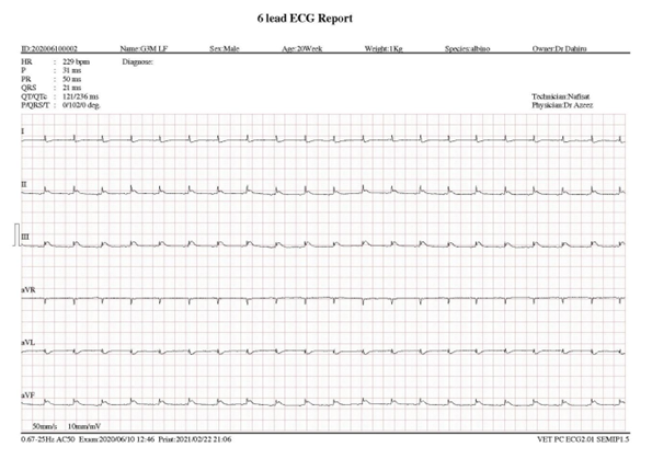
Figure 7: shows group A ECG result with regular P, QRS and normal PR interval.
Key: HR (Heart rate), P (Atrial depolarization), PR (Period), QRS (Ventricular depolarization)
ECG Result of Group B (exposed group)
The result of the effect of petrol on ECG of rats in group B (exposed group) showing abnormal P, QRS and T waves as indicated in figure 8. The result indicated that ECG recording showed a significant increase in the heart rate of the rat exposed to petrol (322 beats/min) compared to the heart rate of the rat in the control group, as seen in Figure (7) (229 beats/min). The QRS complex was significantly narrower in the petrol-exposed group (15m) compared to the control group (17m). There was significantly (p < 0.05) increase in the duration of atrial depolarization (P) in the exposed group (51ms) compared to the control group (24ms). There was an increase in the PR interval of the exposed group (62ms) compared to the control group (48ms).
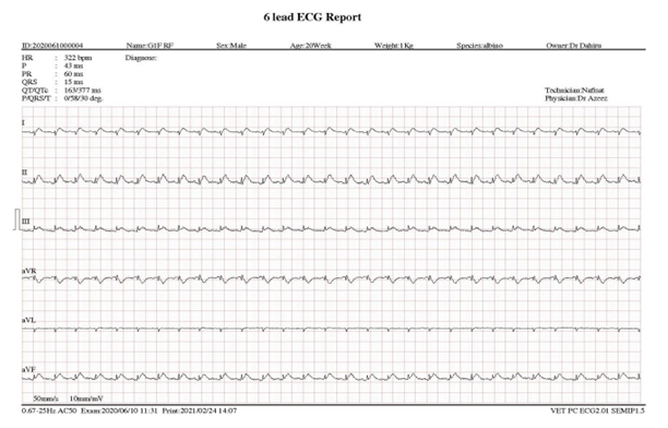
Fig 8: showing result of ECG of group B (exposed group)
Key: HR (Heart rate), P (Atrial depolarization), PR (Period), QRS (Ventricular depolarization)
Result of Cardiac Troponin Concentration in Wistar Albino Rat exposed to petrol and its Mitigation by Moringa oleifera leaf extract is shown in Table 1a and 1b. One-way Analysis of Variance (ANOVA). The P value is 0.0824, considered not quite significant. Variation among column means is not significantly greater than expected by chance. Tukey-Kramer Multiple Comparisons Test. If the value of q is greater than 4.232 then the P value is less than 0.05.
Table 1a: Effect of Petrol Fumes on Cardiac Troponin Concentration in Wistar Albino Rat and its Mitigation by Moringa oleifera leaf extract. (N = 15)
| Comparison | Mean Difference | Q | P value. |
| A vs B | 0.07160 | 0.5041 ns | P>0.05 |
| A vs C | 0.3978 | 2.801 ns | P>0.05 |
| B vs C | -0.3262 | 2.297 ns | P>0.05 |
KEY: A: General Control; B: Petrol; C: Petrol then Moringa. While ns: not significant and *: significant. Analysis was done using one-way ANOVA and Tukey-Kramer Multiple Comparisons Test.
Table 1b. Means and standard error of means of cardiac troponin-I of rats exposed to petrol and administered Moringa oleifera leaf extract. (N = 15)
| Parameter | A | B | C |
| cTn-I (Hg/l) | 0.67 ± 0.20b | 1.07 ± 0.08a | 1.07 ± 0.12a |
KEY: A: General Control; B: Petrol; C: Petrol then Moringa. abMeans with different superscript is considered statistically significant (P <0.05). Using one-way ANOVA and Tukey-Kramer Multiple Comparisons Test
The result of the study on the effect of petrol on body weight of rats is presented in Table 2. There were no significant (P > 0.05) difference between control and other groups.
Table 2: Means and standard error of means of body weight of rats exposed to petrol and administered Moringa oleifera leaf extract. (N = 15)
| Weeks | A | B | C |
| W1 (g) | 194.40 ± 4.07 | 234.60 ± 20.75 | 236.20 ± 24.30 |
| W2(g) | 200.20 ± 4.78 | 232.80 ± 1.31 | 239.60 ± 23.70 |
| W3(g) | 206.20 ± 5.20 | 234.60 ± 20.16 | 236.40 ± 23.71 |
| W4(g) | 203 ± 6.47 | 242.00 ± 24.46 | 242.20 ± 27.00 |
| W5(g) | 203.80 ± 6.58 | 238.20 ± 21.58 | 237.80 ± 23.59 |
KEY: A: General Control; B: Petrol only; C: Petrol then Moringa. W1: Week 1; W2: Week 2; W3: Week 3; W4: Week 4 and W5: Week 5. abcMeans in a row with a different superscript differ significantly (P < 0.05). The data were analyzed using a 2-way repeated measures mixed approach.
DISCUSSION
This study showed that inhalation of petrol vapor for 10 minutes every day for four weeks caused no mortalities in the experimental rats. This is consistent with previous findings by [20]. A research study found that rats survived between 6 and 13 weeks of exposure to petrol vapor [27]. However, the study showed that petrol had an effect on the ECG of exposed Wistar rats.
The significant increase in the heart rate observed in this study is consistent with the work of [20] who reported that inhalation of hydrocarbons elicited vasoconstriction and impaired vascular tone, which might occur as a result of elevation of arterial blood pressure, respiratory depression, hypoxia, and hypercapnia induced by petrol fumes. This is a serious medical condition because an increase in heart rate is associated with an increase in blood pressure, which is a major predisposing factor to hypertension. This study showed an increase in the duration of atrial depolarization (P wave) and is consistent with the work of [28], who reported that prolongation of the P wave may be associated with increased susceptibility to supraventricular arrhythmias in Wistar rats after myocardial infarction. This study has revealed a narrowed QRS complex, which is consistent with the study of [5], who reported QRS narrowing can be seen in supraventricular arrhythmias, heart failure, and myocardial ischemia. This might be due to the activity of the hydrocarbon component of petrol which inactivated the sodium current and thus interfered with the proper firing of the pacemaker cells and the myocytes (hence myocardial ischemia).
The study also showed that there was an increase in the PR interval, which is consistent with the report of [5] that the PR interval always increases with a reduced QRS complex and it’s similar to the findings of [28] who reported that prolongation of the P wave may be associated with increased susceptibility to supraventricular arrhythmias in Wistar rats after myocardial infarction.
Originally the rationale behind the cTn-I assay was relatively simple: myocardial necrosis leads to membrane disruption causing troponin release which is detected in serum. The elevation of serum cTn-I level was used to confirm the histopathological changes on the myocardium. Interestingly, this study revealed an increase in cardiac troponin I level after exposure to petrol which might be due to damage of cardiac muscle as shown in other parameters. However, from this study Moringa oleifera has no effect on the cTn-I level and it is similar to the findings of [29] and [30] that uses it to assess cardiac injury in rats.
This study showed that inhalation of petrol fumes for 5 minutes daily for four weeks caused no significant changes in body weight when compared between control and exposed groups. This is contrary to the findings of [31], [27] and [20] who reported exposure to petrol vapor 10 minutes every day for four weeks causing a very significant reduction in body weight . This may be due to differences in the duration of the study.
CONCLUSION
The ECG recording in this study showed that exposure to petrol vapor had negatively affected atria contraction (P), reduced the time of ventricular contraction (QRS), and increased the heart rate (HR). However, cTn-I level has not significantly affected and treatment with Moringa oleifera shows no results.
ACKNOWLEDGMENT
We acknowledged the support of all staff of the Department of Physiology and Biochemistry, Faculty of Veterinary Medicine, Usmanu Danfodiyo University, Sokoto.
REFERENCES
- Al-Amshawee, S. (2014). Improvement of gasoline production. Technical Report, 5(3), 20–25.
- Azeez, O. M., Akhigbe, R. E., Anigbogu, C. N., Ige, S. F., and Saka, W. A. (2012). Variability in cardiovascular function and baroreflex sensitivity following inhalation of petroleum hydrocarbons. Journal of Cardiovascular Disease Research, 3(2), 99–103.
- Alghatrif, M., and Lindsay, J. (2012). A brief review: history to understand fundamentals of electrocardiography. Journal of Community Hospital Internal Medicine Perspectives, 9666(2), 1.
- Buchanan, J. W., and Sci, M. M. (2018). The history of veterinary cardiology. Journal of Veterinary Cardiology, 15(1), 65–85.
- Konopelski, P., and Ufnal, M. (2016). Electrocardiography in Rats: A Comparison to Human. Physiol, 8408(9), 717–725.
- Ubah, F. E., Akpanabiatu, M. I., and Atangwho, I. J. (2008). Effect of Vitamin A on Weight Loss and Haematotoxicity Associated with Gasoline Vapor Exposure in Wistar Rats. International Journal of Pharmacology, 4(1), 40–45.
- Farraj, A. K., Hazari, M. S., and Cascio, W. E. (2011). The Utility of the Small Rodent Electrocardiogram in Toxicology. Toxicological Sciences, 121(1), 11–30.
- Roffi M., Patrono C., Collet J-P., Mueller C., Valgimigli M., Andreotti F., (2016). 2015 ESC Guidelines for the management of acute coronary syndromes in patients presenting without persistent ST-segment elevation: Task Force for the Management of Acute Coronary Syndromes in Patients Presenting without Persistent ST-Segment Elevation of the European Society of Cardiology (ESC). Eur Heart J; 37:267–315.
- Baker J. O., Reinhold J., Redwood S. and Marber M. S. (2011). Troponins: redefining their limits. Heart; 97:447–452.
- Thygesen K., Alpert J. S., Jaffe A.S., Simoons M. L., Chaitman B. R., White H. D., et al (2012). Joint ESC/ACCF/AHA/WHF Task Force for the Universal Definition of Myocardial Infarction. Third universal definition of myocardial infarction. Eur Heart J; 33:2551–2567.
- Heusch G. (2016). Myocardial ischemia: lack of coronary blood flow or myocardial oxygen supply/demand imbalance? Circ Res; 119:194–196.
- Agewall S., Giannitsis E., Jernberg T., Katus H. (2011). Troponin elevation in coronary vs. non-coronary disease. Eur Heart J. 32:404–411b.
- Clause, B. T. (2016). The Wistar rat as a right choice: Establishing mammalian standards and the ideal of a standardized mammal. Journal of the History of Biology, 10(2), 15–24.
- Arini, P. D., Liberczuk, S., Mendieta, J. G., María, M. S., and Bertrán, G. C. (2018). Electrocardiogram Delineation in a Wistar Rat Experimental Model. Research Article, 1–10.
- Nata’ala, S. U., Abdullahi, K., Abubakar, U., and Daneji, A. I. (2009). Malaria in Sokoto, Northwestern Nigeria. 8(24), 7101–7105.
- ICT Directorate, Sokoto State Government (2023). https://sokotostate.gov.ng/history-of-sokoto/the-land/.
- National Institute of Health (1996). GUIDE FOR THE CARE AND USE OF LABORATORY ANIMALS. Eighth Edition; page 11-236
- Dahiru, A., and Ishaq, A. (2021). Ameliorative Effect of Moringa oleifera Leave Extract on Kerosene Induced Hematological, Serum Biochemical and Histological Changes in Wistar Rats. American Journal of Applied Chemistry, 9(6), 202–206.
- Ibrahim Maina Hassan, Bashir Saidu, Jaafaru Abdullahi Ishaq, Ashiru Dahiru, Nafisat Abdulazeez, Halima Ibrahim Yusuf, Umar Aliyu Dikko, Bulama Ibrahim, Adama Musa Abdullahi (2021). Toxicity Study to Determine Changes in Physiological Vital Parameters and Lethal Dose of Moringa Oleifera Found in Sokoto Metropholis Using Albino Rats Models. Biochemistry and Molecular Biology. (ISSN: 2575-5048). 6(1): 7-10. Index in Cross Ref. doi:10.11648/j.bmb.202110601.12
- Azeez, O. M., Adah, S. A., Olaifa, F. H., Basiru, A., and Abdulbaki, R. (2017). The ameliorative effects of Moringa oleifera leaf extract on cardiovascular functions and osmotic fragility of wistar rats exposed to petrol vapour. Sokoto Journal of Veterinary Sciences, 15(2), 36–42.
- Tontodonati, M., Fasdelli, N., and Dorigatti, R. (2011). An improved method of electrode placement in configuration Lead II for the reliable ECG recording by telemetry in the conscious rat. Journal of Pharmacological and Toxicological Methods, 63(1), 1–6.
- Dhiman, A., Singh, A., Dubey, S., Jain, S., and August, J. (2016). Design of Lead II ECG Waveform and Classification Performance for Morphological features using Different Classifiers on Lead II. Research Journal of Pharmaceutical, Biological and Chemical Sciences Design, 7(1226), 1226–1231.
- Sampson, M., and McGrath, A. (2015). Understanding the ECG. Part 2: ECG basics. British Journal of Cardiac Nursing, 10(12), 588–594.
- Mewton, N., Strauss, D. G., Winslow, R., Soliman, E. Z., and Mcclelland, R. (2012). Prognostic value of electrocardiogram QRS score in asymptomatic individuals without clinical cardiovascular disease: insights from the multi-ethnic study of atherosclerosis. European Heart Journal, 34(8), 910–911.
- Melanson S. E., Tanasijevic M. J. and Jarolim P. (2007). Cardiac troponin assays: a view from the clinical chemistry laboratory. Circulation. 116: e501–e504.
- Thygesen K., Mair J., Katus H., Plebani M., Venge P., Collinson P. et al. (2010). Recommendations for the use of cardiac troponin measurement in acute cardiac care. Eur. Heart J. 31:2197–2204.
- Abubakar, M. B., Abdullahi, W. Z., and Amrah, S. (2015). The Effects of Exposure to Petrol Vapours on Growth, Haematological Parameters and Oxidative Markers in Sprague-Dawley Male Rats. Malays J Med Sci, 22(1), 23–31.
- Milliez P, Deangelis N, Rucker-Martin C, Leenhardt A, Vicaut E, Robidel E, Beaufils P, Delcayre C. & Hatem S.N. (2005). Spironolactone reduces fibrosis of dilated atria during heart failure in rats with myocardial infarction. Eur Heart J 26: 2193-2199.
- Pokrzywinski, K. L., Rosen E. T., Bonanno L., Biel, T., Moshkelani, D., Aryal, B., Mog, S., and Rao, V. A. (2016). Reproductive hormone levels modulate doxorubicin induced cardiomyopathy in female tumor-bearing spontaneously hypertensive rats. Cancer research, 76(14) Abstract nr 4639.
- Reagan, W. J., Barnes, R., Harris, P., Summers, S., Lopes, S., Stubbs, M., Blackwell, D. and Steidl-Nochos, J. (2017). assessment of cardiac troponin I responses in nonhuman primates during restraint, blood collection and dosing in preclinical safety studies. Toxicologic pathology, 45 (2); 335-343.
- Ubah F. E., Akpanabitu M.I., Ndem J.I., Alozie Y. and Ebong P.E. (2009). Comparative nephrotoxic effect associated with exposure to diesel and gasoline vapours in rats. J. Toxicol Environ Health Sci., 1:68-74.