Synergistic Efficacy, Antimicrobial and Wound Healing Potentials of Milled Persea Americana Seed / Syzygium Aromaticum Oil Against Albino Rats and Wound Isolates
- Yusuf-Babatunde, M. A.
- Osuntokun, O. T.
- Olubena, O. O.
- 554-576
- Jun 26, 2024
- Microbiology +1 more
Synergistic Efficacy, Antimicrobial and Wound Healing Potentials of Milled Persea Americana Seed / Syzygium Aromaticum Oil Against Albino Rats and Wound Isolates
1Osuntokun, O. T., 2*Yusuf-Babatunde, M. A. and 3Olubena, O. O.
1Department of Microbiology, Adekunle Ajasin University, Akungba-Akoko, Ondo State, Nigeria.
2Department of Pharmacy Technician, Ogun State Polytechnic of Health and Allied Sciences, Ilese-Ijebu, Nigeria
3Department of Public Health Technician, Ogun State Polytechnic of Health and Allied Sciences, Ilese-Ijebu, Nigeria.
*Corresponding Author
DOI: https://doi.org/10.51584/IJRIAS.2024.905049
Received: 05 May 2024; Revised: 20 May 2024; Accepted: 24 May 2024; Published: 26 June 2024
ABSTRACT
This study aimed to investigate the effects of oil avocado extracts, vegetable oil, Goya oil, and clove oil and its healing potential on male and female albino rats and their antibacterial activity against surgical wound isolates. The antimicrobial efficacy of avocado seed oil and cloves extract against surgical wound isolates was investigated using the agar well diffusion method and a microplate reader to detect the killing time rate of each isolate against oil extracts applied topically to the wounds of albino rats. The following bacteria were isolated, Escherichia coli, Pseudomonas aeruginosa, Klebsiella oxytoca, Chrysobacterium meningosepticum, Klebsiella oxytoca, Escherichia coli, Acinetobacter baumannii and Bergeyella zoochelum. The wound healing process was evaluated by measuring the serum and tissue bioassays and tissue repair enzymes. It was observed that Avocado seed oil infused in vegetables has the highest micro-titre reading against Bergeyella zoochelum (0.199) and Avocado oil infused in vegetable oil with cloves (0.100) against Chrysobacterium meningo septicum at 24 and 48 hours, respectively, this denotes bacteriostatics and Avocado oil infused in Goya with cloves against Bergeyella zoochelum (0.046) has the lowest value and Avocado oil infused in n-hexane against Escherichia coli has the highest value (0.080); this denotes bacteriocide activity of extracted oil. It was observed that the surgical wound on the albino rats closed within seven days and the measured tissue and serum bioassay were relatively under standard. The results of this study suggest that avocado seed oil and clove oil have potential natural alternatives to synthetic antimicrobial agents and treatment of wounds and wound infection in albino rats, as the avocado seed oil and clove used in this study inhibited all the surgical wound isolates.
Keywords: Synergistic Efficacy, Antimicrobial, Wound Healing Potentials
INTRODUCTION
Wound healing is a complex process involving different cell types, cytokines, growth factors, and the extracellular matrix with the purpose of swiftly reestablishing skin integrity [1]. This wound healing process occurs in three overlapping phases: inflammation, proliferation, and remodeling [2]. Homeostasis is followed after several hours by an inflammatory stage, during which cytokines and growth factors are secreted, and leucocytes and to a lesser extent other cell types are recruited to clean the wound. In the proliferative phase Previous studies have shown that the healing process may be modulated by fatty acids [3]. Linolenic, linoleic and oleic acids are precursors of eicosapentaenoic (EPA), arachidonic (AA), and eicosatrienoic acids (ETA) which are part of the structure of cell membrane phospholipids and serve as substrates for the synthesis of eicosanoids (inflammatory mediators), such as prostaglandins, thromboxanes, prostacyclins (via cyclooxygenase), and leukotrienes (via lipooxygenase) [4]. Eicosanoids formed from arachidonic acid, prostaglandin E2, thromboxane B2, and leukotriene B4 are proinflammatory inducers, more potent than those formed from EPA, prostaglandin E3, thromboxane B3, and leukotriene B5, which have anti-inflammatory effects. Considering that these families of fatty acids compete for the same enzyme, the proper balance is of great importance. Depending on the ratio of the diet more proinflammatory or anti-inflammatory eicosanoids can be synthesized. Besides modulating the inflammatory response, eicosanoids also act in immunological responses, platelet aggregation, and cell growth and differentiation. Avocado (P. americana) extract and clove oil has been used in wound healing, the treatment of psoriasis, wrinkles, and stretch marks, as well as for their hepatoprotective actions. The unsaponifiable fraction of this oil has regenerative properties of the epidermis, besides improving scleroderma (Werner et al., 2003)
MATERIALS AND METHODS
Location / Sample Collection
The fruits of Persea Americana Mill (Avocado) were purchased from Ibaka market in Akungba-Akoko South-West L.G.A Of Ondo State, Nigeria in OCTOBER 2023. It was then conveyed to Microbiology Department, Adekunle Ajasin University Ondo State where it was identified authenticated. It was afterwards taken to the Microbiology Laboratory where it was allowed to ripen before it was cut open and its butter removed while the peel was washed with distilled water and air dried.
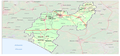
Figure 1: Map of Ondo state (OCHA 2018).
Collection of Surgical Wound Isolates
During the study, five (5) different surgical wound samples were collected aseptically in sterile universal bottles from the Microbiology section of the University’s Health Centre at Adekunle Ajasin University, Akungba-Akoko in Ondo. The samples were then transported to the laboratory within 15 minutes for microbial analysis.
Extraction Process of Sample
Solvent Extraction (N-Hexane)
After collection of the sample, the avocado seed was washed and air-dried for about 3 days, it was cut and diced into smaller pieces which turned bright orange due to an unusual form of oxidation. 500g of the diced avocado seeds were soaked in 800 ml of n- hexane for about 10 days. After ten days, the samples were filtered using a sterile muslin cloth, the residue was discarded and the filtrate which contains the n-hexane and the essential oil was exposed to air for evaporation of the solvent leaving the essential oil [5].
Cold Press Method of Extraction
Goya Oil
This was done using the cold press method of extraction which is one of the best methods. The avocado seed is washed and crush into smaller pieces and 70g of the crushed avocado seed is added into a clean container and 200 ml of goya oil was added into the readily prepared sample and it was covered with an aluminum foil for about 2 weeks. After 2 weeks, the avocado seed nutrients have been infused into the goya oil and shows a green color appearance which indicates the presence of the avocado seed nutrients which was then sieved into a clean McCartney bottles which makes it a combination of Goya oil and Avocado seed oil [6].
Double Boiler Method of Extraction (Vegetable Oil)
The avocado seed oil was extracted from the seed with the use of vegetable oil using a double boiler method of extraction which makes it a combination of vegetable oil and Avocado seed oil. The avocado seed is perfectly washed and grated into a powdery form, 100g of the powdered avocado seed is then measured into a clean steel container and 200 ml of vegetable oil was added and gently mixed together making sure that the vegetable oil is above the powdered avocado seed. Then boil water for about 15 minutes and put the steel container containing the mixed avocado seed and vegetable oil into the readily boiled water and apply heat. Stir the mixture very gently for every 5-10 minutes for about 1 hour and closely observe the changes in the oil as the avocado seed oil and nutrients are directly infused into the vegetable oil. Allow the mixture to cool and sieve into a clean container, then filtered directly into a McCartney bottle [5].
Combination of Goya Oil, Avocado Seed Oil and Cloves
The avocado seed oil which was extracted with the use of Goya oil, using the Cold Pressed method is used as a solvent to soak cloves for about 1 week. The cloves are grated and powdered into a powdery form and pour into the readily prepared mixture of avocado seed oil and Goya oil and soak for about 1 week. The cloves’ nutrients and potency infused into the mixture of the avocado seed oil and Goya oil. Then sieve into a clean container and filter into a McCartney bottle. The nutrients and potency of the mixture increases for essential use.
NOTE: Cloves are rich ineugenol, a compound that has anti-inflammatory and antioxidant properties. This makes cloves a great ingredient for treating wounds. They can also help to stimulate blood flowto the scalp, promoting hair growth.
Combination of Vegetable Oil, Avocado Oil and Cloves
The avocado seed oil which was extracted with the use of Vegetable oil using the Double boiler method is used as a solvent to soak cloves for about 1 week. The cloves are grated and powdered into a powdery form and pour into the readily prepared mixture of avocado seed oil and Vegetable oil and soak for about 1 week. The cloves’ nutrients and potency infused into the mixture of the avocado seed oil and vegetable oil. Then sieve into a clean container and filter into a McCartney bottle. The nutrients and potency of the mixture increases for essential use.
NOTE: Avocado fat content, antioxidant and anti-inflammatory properties, and minerals also make a great ingredient for your hair and skin and especially for wound healing. Avocado oil may also help to stimulate blood flow and unclog blocked follicles. These effects may help fight hair loss.
Isolation, Identification and Characterization of Surgical Wound Organisms
Preparation of Wound Samples for Culture
9ml of sterile physiological saline was aseptically dispensed into five sterile test tubes and the mouth was corked with cotton wool wrapped with aluminum foil, each test tube was then labeled as 10-1 -10-5 respectively. Wound samples were dispensed into 9 ml of sterile physiological saline. 1ml of the stock culture was serially transferred to other 9 ml of sterile physiological saline inother test tubes in an aliquot manner up to the fifth dilution. Pour plate method of inoculation was used for the enumeration of bacteria; 0.5ml of the five- fold dilution of 10-3 and 10-5 wound samples (inoculum) was put into sterile petri dishes.
Identification of Isolates
Microscopic Examination of Isolates from Wound Sample
Cultural and microscopic examinations were performed to determine the identity of t isolated wound microorganisms. The cultural characteristics of the isolates were examined; the creamy pigmentation, round and slightly elevated shape, irregular and thread-like some of which are swarmy, with no distinct colony and cellular morphology characteristics. Furthermore, conventional identification of the isolates was carried out using various biochemical tests such as, indole, motility, gram staining, fermentation of sugars (Sucrose, Lactose, Dextrose), Urease, Hydrogen sulfide, gelatin liquefaction and nitrate reduction test.
Gram Staining Technique
Sterilized inoculating loop was used to make a smear of the culture onto a clean, grease-free slide. The slide was then labeled with each isolate code and heat-fixed. The smear was flooded with crystal violet (primary stain) for 60 seconds and rinsed with water. Lugol’s Iodine was applied to the slide as a mordant and left to stay for 1 minute. After that, it was rinsed with water and left for 30 seconds. 70% ethanol was used to decolorize the smear for 15 seconds, and it was immediately rinsed off in gently running tap water to remove the ethanol effect. The slide was then counterstained with safranin for 60 seconds, rinsed with water and blot dried. It was reported that the slides were viewed under the microscope using oil immersion (X100). It was observed that Gram-positive cells appeared purple since they retained the purple color of the primary stain (crystal violet) as they were not decolorized by alcohol. In contrast, Gram-negative cells appeared pink as the alcohol removed the crystal violet-iodine complex [7].
Antimicrobial Assay of the Oil Extract Preparation and standardization of Inoculum Suspension
Direct colony suspension technique was used for the standardization and preparation of inoculum. Pure cultures of the test organisms were transferred into sterile screw-capped McCartney bottles containing normal saline (0.90% w/w) using a flamed inoculating loop. A suspension with a turbidity equivalent to 0.5 McFarland standard was also prepared at the same time to serve as a reference for turbidity. To achieve equal turbidity, both the reference and inoculum suspensions were placed against a white card with black stripes. Turbidity was observed with the unaided eye. Standardizes inoculum were refrigerated [8].
Inoculation and Incorporation of Test Organisms and Oil Extracts
Antimicrobial susceptibility test was carried out using Mueller-Hinton agar and the organisms were seeded on the Mueller-Hinton agar using sterile swab-sticks. Sterile swab-sticks were used to pick inoculum from standardized inoculum suspension to sterile petri-dishes containing molten Mueller-Hinton agar. Agar well diffusion method was used to dispense antimicrobials (oil extracted). A sterile cork borer of 1mm diameter was used to bore wells on the agar medium that has been seeded with test organisms. Using syringe, the graded concentrations (100, 50,25,12.5,6.25mg/ml and 3.125mg/ml) of the extracted oil were gently dispensed into each designated wells. Positive controls (Amoxicillin) were also incorporated into one of the designated wells. After 24 hours the results were taken by observing, measuring and recording the zone of inhibitions formed around the agar wells [9].
Measurement of Death Rate of the Isolates Using a Microplate Reader
This procedure used by Osuntokun (2019) was used during this test. Growth dynamic test was done to determine the rate of growth of the isolates as well as their killing time in due time. A loopful colony was picked from the stocked culture slant and inoculated into 9ml nutrient broth which was incubated for 24 hours at 370C. A loopful of the each organism was picked from the broth culture into another 9ml nutrient broth in three sets which are labelled as set A, B, C and D respectively. The growth rate was measured using ultraviolet (UV-vis) spectrophotometer; the spectrophotometer was set at 620λ wavelength, warmed up for15 minutes and calibrated; during the determination of the growth, sterilized nutrient broth was used to calibrate the spectrophotometer for set A. during the determination of the killing rate sterilized nutrient broth, in which the oil to be assay has been incorporated into was used. The first reading was taken at zero hour, twenty-four hour and it continues after every twenty-four hours for five times.
Animal Care
Experimental Design
Thirty two (32) healthy male and female albino rats were used for the experiment. The rats were divided into eight groups of four (4) rats per group and housed in aluminum cages throughout the research. All the animals were kept in a holding room illuminated with 12 h light/dark cycles at room temperature of 23 ± 2 °C with relative humidity of 45% to 55%.
Biochemical Assays
The following biochemical assay were carried out to evaluate the safety or toxicity risk of Persea americana Mill and Syzygium aromaticum oil extracts.
Toxicological Assay of Persea Americana Mill and Syzygium Aromaticum Oil on Experimental Animal
The hair on the limb region was shaved 24 h before the test. The rats were generally anesthetized with intraperitoneal injection of ketamine: xylazine (0.4 mL: 0.1 mL, 40 mg/kg: 10 mg/kg). Ethanol (70%) was used as an antiseptic for the shaved region before introducing the wound. A circular excision wound was made using a biopsy punch (4 mm in diameter) as mentioned by Shailajan et al. (2011) [10]. The wounding day was counted as day zero. The wounds were treated topically twice daily with the oil samples respectively until the 7th day of the experiment. Wound size was observed, measured and photographed. On the final day the rats were sacrificed, blood was taken and small portion of the wound area was extracted, rinsed in saline buffer which was kept in freezer for further analysis.
Assay of Serum Enzymes and Tissue Functions
The serum enzymes, alanine amino transferase (ALT), aspartate amino tranferase (AST), alkaline phosphatase (ALP), total protein, albumin and urea assays were carried out using enzymes kit by Randox Laboratories Ltd, United Kingdom. The blood samples in the EDTA tubes were centrifuge for 15 minutes at 40oC and analyzed according to the manufacturer description
RESULTS
Table1: Shows the number of bacterial colonies found on nutrient agar from surgical wound isolates collected from four different locations (Hospitals) in four towns namely: Owo, Akure, Ondo and Okitipupa, Southwest, Nigeria. The results showed that the colony counts varied widely across the sample codes, ranging from 56 to 140. The highest colony counts were observed in AK24/ONDOT30, which has a dilution factor of 10-3, while the lowest colony count was observed in OK11, which has dilution factor of 10-5. AK24/ONDOT30 has the highest colony count (140) but also the lowest dilution factor 10-3, which means that the sample was not diluted much before being cultured. This suggests that AK24/ONDOT30 has a very high bacterial load in the wound sample, which could indicate a serious infection. On the other hand, OK11 has the lowest colony count (56) but also the highest dilution factor 10-5, which means that the sample was diluted a lot before being cultured. This suggests that OK11 has a very low bacterial load in the wound sample, which could indicate a mild infection. The sample codes with higher colony counts and lower dilution factors (OWO17 and AK24/ONDOT30) has higher bacterial loads and potentially higher infection risks than the sample codes with lower colony counts and higher dilution factors (such as, OWO19, OWO10, OK11, and ONDOT4/AKR23).
Table 1: Dilution Factors and Number of Colony Found on Nutrient Agar
| Sample code | Dilution factor | Number of colony |
| AK24/ONT30 | 103 | 140 |
| ONT4/AKR23 | 105 | 83 |
| OWO17 | 103 | 103 |
| OK11 | 103 | 56 |
| OWO10 | 105 | 90 |
| OWO19 | 103 | 76 |
Sample code Dilution factor Number of colony
Keys: AK: State Hospital Akure. ONT: Ondo State Teaching Hospital.
OW: Federal Medical Centre, Owo. OK: Okitipupa General Hospital.
Table 2: Morphology and Colonial Characteristics of Surgical Wound Isolate on Agar
| Isolate code | Colony color | Elevation | Opacity | Margin | Colony shape | Grams reaction | Shape |
| AK24/ONT30 | Whitish | Raised | Opaque | Lobate | Irregular | Positive | Cocci |
| ONT4/AKR23 | Creamy | Raised | Opaque | Entire | Circular | Positive | Cocci |
| OWO17 | Whitish | Convex | Opaque | Lobate | Circular | Negative | Rod |
| OK11 | Creamy | Flat | Opaque | Entire | Circular | Positive | Rod |
| OWO10 | Pink | Flat | Translucent | Entire | Circular | Negative | Cocci |
| OWO19. | Whitish | Convex | Opaque | Lobate | Irregular | Negative | Rod |
Keys: AK: State Hospital Akure.
ONT: Ondo State Teaching Hospital.
OW: Federal Medical Centre, Owo.
OK: Okitipupa General Hospital.
Table 2: Shows the morphology and colonial characteristics of test isolates under microscope. The morphology which includes; the color, elevation, opacity, margin, colony shape for proper identification. From the table it was observed that AK24/ONDOT30 and OWO17 has whitish colonies, ONDOT4/AKR23 and OK11 has creamy colonies while OWO10 has apink colony. All the isolated colonies were opaque except OWO10 which appeared translucent. The table indicates that AK24/ONDOT30 and ONDOT4/AKR23 has raised elevation, OK11 and OWO10 has flat elevation while OWO17 and OWO19 has convex elevation. All the isolates are circular in shape except AK24/ONDOT30 that is irregular in shape while OK11, OWO10 and ONDOT4/AKR23, AK24/ONDOT30 and OWO17 have lobate margin. Also, it was observed in this table that AK24/ONDOT30, ONDOT4/AKR23, OK11, OWO19, OWO17 and OWO10 are Gram negative with a rod shape.
Table 3: Preliminary identification using Biochemical, Motility Test, Gas Production and Sugar fermentation Result of the Surgical Wound Isolate
| Motility | Biochemical tests | G. P | Sugar Fermentation | |||||||||||||
| Isolate code | Motility | Indole | Methyl red | Catalase | Citrate | Oxidase | Urease | Voges proskaeur | Coagulase
|
H2S | Mannitol | Glucose | Lactose | Dextrose | Fructose | Sucrose |
| AK24
ONT4 OW17 OK11 OW10 AK23 ONT30 OW19 |
+
_ + + + + + +
|
+
_ + _ _ + + + |
_
_ _ _ _ _ _ _ |
+
+ + + + + + + |
+
+ + + + + + + |
+
+ _ + + + _ _ |
+ + + + + + + +
|
_
_ + _ + _ _ + |
_
+ _ _ + _ _ + |
_
+ + + + + + _ |
+
+ + + + + _ + |
+
+ + + + + + + |
_
_ _ _ _ __
|
+
+ + + + + + + |
+
+ + + ++ + +
|
+
+ + + + + + +
|
Key: (+) = Positive, (-) = Negative, the isolates from Ondo State Teaching Hospital (ONT), Federal Medical Centre, Owo (OW), General Hospital, Okitipupa (OK), and State Hospital, Akure (AK).
Table 3: Shows the preliminary biochemical test of the isolates. In this table, ONDOT4/AKR23, OK11 and OWO17 were negative for indole while AK24/ONDOT30 and OWO10 were positive. All isolates were positive for catalase, citrate and urease test and negative to methyl red test respectively. It was observed in this table that AK24/ONDOT30, ONDOT4/AKR23, OK11 and OWO17 were Voges-Proskauer negative while OWO10 were positive. OWO17 and ONDOT4/AKR 23 were negative for motility test while OK11, OWO10 and AK24/ONDOT30 were positive. It was observed that OK11, OWO10 and ONDOT4/AKR23 were oxidase positive while OWO17 and AK24/ONDOT30 were negative. Also, it was observed that all isolates were positive for mannitol, glucose, fructose, sucrose and dextrose fermentation test and negative for lactose fermentation respectively. It was observed that ONDOT4:AKR23, OWO10, OK11, OWO17 and OWO19 were positive for hydrogen sulfide test while AK24/ONDOT30 was negative. The table shows that OWO10, OK11, OWO17, OWO19 and AK24 were negative for coagulase test while ONT24/AKR23 was positive and OK11 and AK24/ONDOT30 were positive for gas production while OWO10, ONDOT4/AKR23, OWO19 and OWO17 were negative for gas production.
Table 4: List of Surgical Wound Isolate Characterize Using Bergey’s Manual of Determinative Bacteriology
| Isolate code | Probable isolates |
| AK 24 | Klebsiella oxytoca |
| ONDOT4 | Escherichia coli |
| OWO17 | Pseudomonas aeruginosa |
| OK 11 | Acinetobacter baumannii |
| OWO10 | Bergeyella zoochelum |
| OWO19 | Chrysobacterium meningosepticum |
| AKR23 | Escherichia coli |
| ONDOT30 | Klebsiella oxytoca |
Keys: AK: State Hospital Akure, ONT: Ondo State Teaching Hospital, OW: Federal Medical Centre, Owo., OK: Okitipu pa General Hospital
Table 4: Using Bergey’s Manual, Escherichia coli, Acinetobacter baumanii, Klebsiella oxytoca, Bergeyella zoochelum, Pseudomonas aeruginosa and Chrysobacterium Meningo septicum were identified.
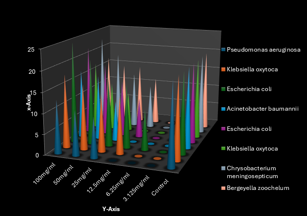
Fig 2; Zone of Inhibition of N-Hexane Avocado Oil seed Extracts (L/E) at 100- 3.125mg/ml concentrations against surgical Wound Isolates
Fig 2; Antimicrobial screening of avocado seed oil extracted with solvent indicated as (L/E) at a concentration of 100mg/ml – 3.125mg/ml tested against each isolates. The zone of inhibition at concentration of 100mg/ml as the highest Antimicrobial zone which has a range of (35mm) against E. coli and at a lower concentration of 3,125mg/ml, the zone of inhibition range is (0.00mm) tested against each isolates.
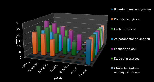
Fig 3; Zone of Inhibition of Avocado Oil seed (Persea Americana mill) infused in vegetable oil (A/V) at 100-3.125mg/ml 3.125mg/ml concentrations against surgical Wound Isolates
Fig 3; Antimicrobial screening of avocado seed oil infused with vegetable oil indicated as (A/V) at a concentration of 100mg/ml – 3.125mg/ml tested against each isolates. The zone of inhibition at concentration of 50mg/ml as the highest Antimicrobial zone which has a range of (24mm) against E.coli and at a lower concentration of 3,125mg/ml, the zone of inhibition range is (0.00mm) tested against each isolates
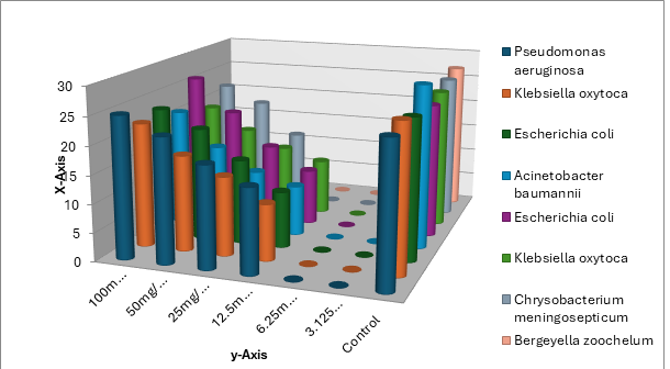
Fig 4; Zone of Inhibition of Avocado seed Oil (Persea Americana mill) infused in Goya oil (A/G) at 100 – 3.125mg/ml 3.125mg/ml concentrations against surgical Wound Isolates
Fig 4: Antimicrobial screening of avocado seed oil infused with Goya oil indicated as (A/G) at a concentration of 100mg/ml – 3.125mg/ml tested against each isolates. The zone of inhibition at concentration of 100mg/ml as the highest Antimicrobial zone which has a range of (26mm) tested against E. coli and at a lower concentration of 3,125mg/ml, the zone of inhibition range is (0.000mm) tested against each isolates.
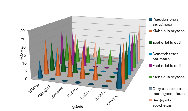
Fig 5; Zone of Inhibition of Avocado seed Oil (Persea Americana mill) infused in vegetable oil with cloves (Syzygium aromaticum) indicated as (C/A/G) at 100- 3.125mg/ml concentrations against surgical Wound Isolates
Fig 5; Antimicrobial screening of avocado seed oil infused with Goya oil and cloves indicated as C/A(G) at a concentration of 100mg/ml – 3.125 mg/ml tested against each isolates. The zone of inhibition at concentration of 50mg/ml as the highest Antimicrobial zone which has a range of (25mm) tested against E. coli and at a lower concentration of 3,125mg/ml, the zone of inhibition range is (0.0mm) tested against each isolates.
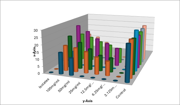
Fig 6; Zone of Inhibition of Avocado seed Oil (Persea americana mill) infused in vegetable oil with cloves (Syzygium aromaticum) indicated as (C/A/V) at 100– 3.125mg/ml concentrations against surgical Wound Isolates
Fig 6; Antimicrobial screening of avocado seed oil infused with vegetable oil and cloves indicated as C/A(V) at a concentration of 100mg/ml – 3.125mg/ml tested against each isolates. The zone of inhibition at concentration of 50mg/ml as the highest Antimicrobial zone which has a range of (28mm) tested against E. coli and at a lower concentration of 3,125mg/ml, the zone of inhibition range is (0.0mm) tested against each isolates.
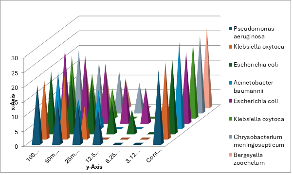
Fig 7; Zone of Inhibition of Amoxicilin as positive control indicated as (A/A) at 100 – 3.125mg/ml concentrations against surgical Wound Isolates
Fig 7: Antimicrobial screening of amoxicillin used as positive control at a concentration of 100mg/ml – 3.125mg/ml tested against each isolates. The zone of inhibition at concentration of 100mg/ml as the highest Antimicrobial zone which has a range of (43.6mm) against E.coli and at a lower concentration of 3,125mg/ml, the zone of inhibition range is (0.0mm) tested against each isolates except E.coli which as (5.0mm) at a lower concentration of 3.125mg/ml
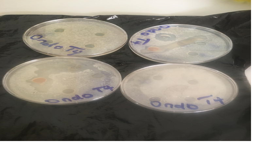
Plate 1: Zone of inhibition of oil extract and clove against tested isolates between 100 and 3.125 mg/ml concentrations against surgical Wound Isolates
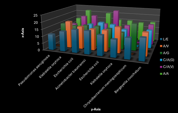
Fig 8a; Minimum Inhibitory Concentration of Avocado (Persea Americana Mill) seed extracts against surgical wound isolates at a concentration of 6.25mg/ml concentrations
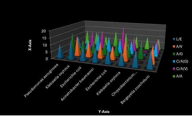
Fig 8b; Minimum Bactericidal Concentration of Avocado (Persea Americana Mill) seed extracts against surgical wound isolates at a concentration of 3.125mg/ml
Key; A/v: avocado seed oil infused in vegetable A/G: Avocado seed oil infused in Goya oil L/E: Avocado oil infused in n-hexane C/A(G): Avocado oil infused in Goya with cloves C/A(V): Avocado oil infused in vegetable oil with cloves
Fig 8 a & b: show the MIC and MBC of all the oil extracts against the surgical wound isolates examined. The MIC of the extracts against the isolate was observed in the table ranges between 3.125mg/ml to 100mg/ml at exactly 6.25mg/ml, while it has MBC of 3.125mg/ml concentration on Escherichia coli.
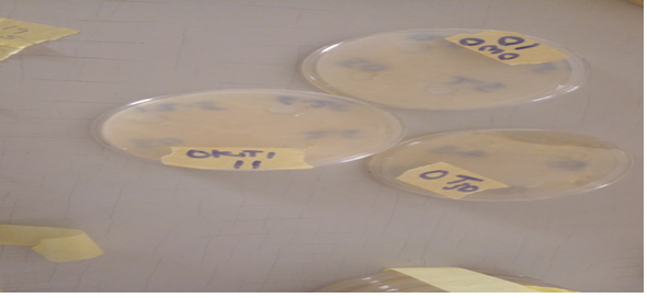
Plate 2: Serial dilution of concentration at 3.125mg/ml-100mg/ml of avocado seed extracts and clove for minimum and maximum inhibitory concentration
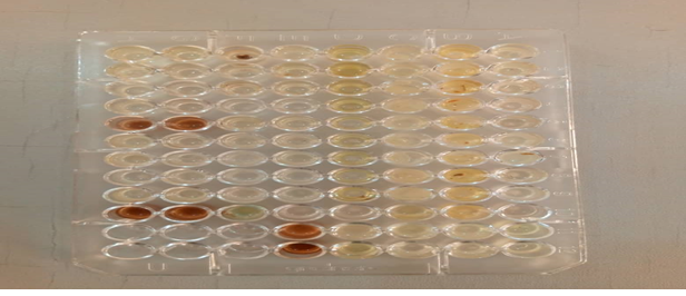
Plate 3: A micro-titer well containing a nutrient broth and oil extracts at different concentration to measure killing kinectics of isolate
Table 5: killing time of surgical wound isolates against avocado seed oil / cloves at (0hrs) using Microtitre-plate reader
| Isolates | A/V | A/G | L/E | C/A(G) | C/A(V) |
| Bergeyella zoochelum | 0.243 | 0.243 | 0.260 | 0.248 | 0.251 |
| Escherichia coli | 0.247 | 0.256 | 0.249 | 0.252 | 0.244 |
| Pseudomonas aeruginosa | 0.259 | 0.260 | 0.246 | 0.255 | 0.258 |
| Klebsiella oxytoca | 0.248 | 0.243 | 0.259 | 0.256 | 0.251 |
| Acinetobacter baumannii | 0.244 | 0.247 | 0.247 | 0.252 | 0.245 |
| C.meningo septicum | 0.253 | 0.257 | 0.258 | 0.249 | 0.243 |
| Escherichia coli | 0.245 | 0.253 | 0.256 | 0.259 | 0.248 |
| Klebsiella oxytoca | 0.243 | 0.249 | 0.259 | 0.251 | 0.255 |
Table 5: shows the growth dynamics and killing time of surgical wound isolates using a microplate reader with a 96 well which contains a nutrient broth at a concentration of 25mg/ml and addition of Avocado seed extracts In this table, it was observed that at 0 hours, Bergeyella zoochelum has the lowest growth rate of 0.243λ while Pseudomonas aeruginosa has the highest growth rate of 0.260 λ which decreases with time.
Table 6: killing time of surgical wound isolates against avocado seed oil at (24hrs) using Microtitre-plate reader
| Isolates | A/V | A/G | L/E | C/A(G) | C/A(V) |
| Bergeyella zoochelum | 0.199 | 0.188 | 0.189 | 0.172 | 0.174 |
| Escherichia coli | 0.184 | 0.175 | 0.179 | 0.180 | 0.190 |
| Pseudomonas aeruginosa | 0.192 | 0.197 | 0.187 | 0.183 | 0.181 |
| Klebsiella oxytoca | 0.178 | 0.176 | 0.174 | 0.194 | 0.198 |
| Acinetobacter baumannii | 0.199 | 0.175 | 0.186 | 0.172 | 0.177 |
| C.meningo septicum | 0.195 | 0.179 | 0.189 | 0.182 | 0.192 |
| Escherichia coli | 0.188 | 0.176 | 0.172 | 0.191 | 0.196 |
| Klebsiella oxytoca | 0.174 | 0.178 | 0.184 | 0.188 | 0.194 |
Table 6; shows the growth dynamics and killing time of surgical wound isolates using a microplate reader with contain a nutrient broth at a concentration of12.5mg/ml and addition avocado seed extracts with cloves In this table, it was observed that at 24 hours, Bergeyella zoochelum and Acinetobacter buamannii had the highest growth rate of 0.199λ while Escherichia coli had the lowest growth rate of 0.172 λ which decreases more with time.
Table 7: killing time of bacteria isolates against avocado seed oil at (48hrs) using microtitre plate reader
| Isolates | A/V | A/G | L/E | C/A(G) | C/A(V) |
| Bergeyella zoochelum | 0.100 | 0.130 | 0.108 | 0.127 | 0.104 |
| Escherichia coli | 0.125 | 0.121 | 0.108 | 0.105 | 0.129 |
| Pseudomonas aeruginosa | 0.130 | 0.126 | 0.123 | 0.106 | 0.112 |
| Klebsiella oxytoca | 0.119 | 0.115 | 0.111 | 0.124 | 0.107 |
| Acinetobacter baumannii | 0.116 | 0.125 | 0.130 | 0.129 | 0.109 |
| C.meningo septicum | 0.100 | 0.104 | 0.126 | 0.129 | 0.120 |
| Escherichia coli | 0.102 | 0.108 | 0.125 | 0.121 | 0.104 |
| Klebsiella oxytoca | 0.127 | 0.113 | 0.107 | 0.116 | 0.128 |
Keys; A/v: avocado seed oil infused in vegetable A/G : Avocado seed oil infused in Goya oil L/E: Avocado oil infused in n-hexane C/A(G) : Avocado oil infused in Goya with cloves C/A(V): Avocado oil infused in vegetable oil with cloves
Table 7: shows the growth dynamics and killing time of surgical wound isolates using a microplate reader with contain a nutrient broth at a concentration of12.5mg/ml and addition avocado seed extracts with cloves In this table, it was observed that at 48 hours, Bergeyella zoochelum had the lowest growth rate of 0.100λ while Pseudomonas aeruginosa had the highest growth rate of 0.130 λ which decreases more with time.
Table 8: killing time of bacteria isolates against avocado seed oil at (72hrs) using microtitre plate reader
| Isolates | A/V | A/G | L/E | C/A(G) | C/A(V) |
| Bergeyella zoochelum | 0.046 | 0.080 | 0.071 | 0.078 | 0.061 |
| Escherichia coli | 0.048 | 0.056 | 0.059 | 0.078 | 0.072 |
| Pseudomonas aeruginosa | 0.051 | 0.055 | 0.062 | 0.080 | 0.065 |
| Klebsiella oxytoca | 0.061 | 0.074 | 0.066 | 0.053 | 0.047 |
| Acinetobacter baumannii | 0.049 | 0.057 | 0.051 | 0.046 | 0.066 |
| C.meningo septicum | 0.048 | 0.056 | 0.077 | 0.072 | 0.074 |
| Escherichia coli | 0.068 | 0.079 | 0.080 | 0.046 | 0.061 |
| Klebsiella oxytoca | 0.058 | 0.064 | 0.071 | 0.057 | 0.049 |
A/v: avocado seed oil infused in vegetable A/G : Avocado seed oil infused in Goya oil L/E: Avocado oil infused in n-hexane C/A(G) : Avocado oil infused in Goya with cloves C/A(V): Avocado oil infused in vegetable oil with cloves
Table 8: shows the serum biochemical parameters of albino rats treated with Avocado seed (Persea Americana Mill) extracts and Syzygium aromaticum. The serum biochemical parameters investigated in this table are Total protein TP (mg/dl), Aspartate amino AST (U/L), Alkaline Phosphatase ALP (U/L), Alanine Aminotransferase ALT (U/L), Alkaline Albumin ALB (mg/dl), Glutathione GSH (μg/dl), Globulin GLB (mg/dl). The table compares the values of these parameters among eight groups of rats, each treated with different doses (50mg/kg and 100mg/kg) of Avocado seed extracts (oil) and clove oil extracts with positive and negative controls. The positive control groups (G and H) have no wound, while the negative control groups (E and F) have wound with no treatment. The other groups (A, B, C, and D) have wound and are treated with 50mg/kg and 100mg/kg doses avocado seed oil infused with Goya and cloves and also with avocado seed oil infused with vegetable oil with cloves. Based on the table, The groups of albino rat treated with Avocado seed oil infused with Goya oil and clove oil extracts at 50mg and 100mg (A and B) and Avocado seed oil infused with vegetable oil and clove oil extracts (C and D) have higher ALT, ALP, AST, TP, GSH, ALB, and GLB levels than the negative control groups (E and F), which may suggest that the extracts has ability to heal wound and improve the immune function of the rats.
Table 9: killing time of bacteria isolates against avocado seed oil at (96hrs) using microtitre reader
| Isolates | A/V | A/G | L/E | C/A(G) | C/A(V) |
| Bergeyella zoochelum | 0.010 | 0.040 | 0.018 | 0.015 | 0.028 |
| Escherichia coli | 0.038 | 0.031 | 0.019 | 0.012 | 0.017 |
| Pseudomonas aeruginosa | 0.022 | 0.035 | 0.027 | 0.011 | 0.026 |
| Klebsiella oxytoca | 0.032 | 0.016 | 0.019 | 0.026 | 0.025 |
| Acinetobacter baumannii | 0.036 | 0.039 | 0.024 | 0.020 | 0.013 |
| C.meningosepticum | 0.026 | 0.018 | 0.021 | 0.019 | 0.013 |
| Escherichia coli | 0.039 | 0.029 | 0.019 | 0.024 | 0.036 |
| Klebsiella oxytoca | 0.021 | 0.017 | 0.038 | 0.040 | 0.031 |
A/v: avocado seed oil infused in vegetable A/G: Avocado seed oil infused in Goya oil L/E: Avocado oil infused in n-hexane C/A(G): Avocado oil infused in Goya with cloves C/A(V): Avocado oil infused in vegetable oil with cloves
Table 9: shows the growth dynamics and killing time of surgical wound isolates using a microplate reader with contain a nutrient broth at a concentration of12.5mg/ml and addition avocado seed extracts with cloves in this table, it was observed that at 96 hours, Bergeyella zoochelum had the lowest growth rate of 0.010λ while Klebsiella oxytoca had the highest growth rate of 0.040 λ which decreases more with time.
Table 10i: Serum Bioassay of Albino Rats Treated with Persea Americana Mill / Syzygium aromaticum Oil Extracts
| Parameters | A | B | C | D | E | F | G | H |
| ALT µ/l | 24 | 47 | 21.5 | 43 | 55.5 | 48.5 | 42,5 | 40,9 |
| ALP µ/l | 57.96 | 179.4 | 132.48 | 259.44 | 157.32 | 226.32 | 134,6 | 223.0 |
| AST µ/l | 26.6 | 53.8 | 32.4 | 37.2 | 25.4 | 38 | 34.3 | 32.4 |
| TP mg/dl | 13.53695 | 12.82759 | 12.6502 | 12.53202 | 13.44828 | 12.97537 | 12,98 | 11.34 |
| GSH µg/dl | 17.44681 | 77.65957 | 56.38298 | 80.85106 | 59.57447 | 75.95745 | 67,23 | 71.9 |
| ALB mg/dl | 17.66852 | 18.06019 | 13.57778 | 16.79815 | 11.8805 | 20.14907 | 18,34 | 17.34 |
| GLB mg/dl | 4.13157 | 5.2326 | 0.92753 | 4.26613 | 1.56772 | 7.1737 | 3,2 | 2.34 |
Keys:
A: 50mg/kg of Avocado oil infused with Goya oil and clove oil extracts
B: 100mg/kg of Avocado oil infused with Goya oil and clove oil extracts
C: 50mg/kg of Avocado oil infused with vegetable oil and clove oil extracts
D: 100mg/kg of Avocado oil infused with vegetable oil and clove oil extracts
E: 50mg/kg of negative control (wound with no treatment)
F: 100mg/kg of negative control (wound with no treatment)
G: 50mg/kg of positive control (no wound)
H: 100mg/kg of positive control (no wound)
ALT: Alanine amino transferase
GLB: Globulin
TP: Total protein
ALB: Alkaline Albumin
ALP: Alkaline phosphatase
GSH: Glutathione
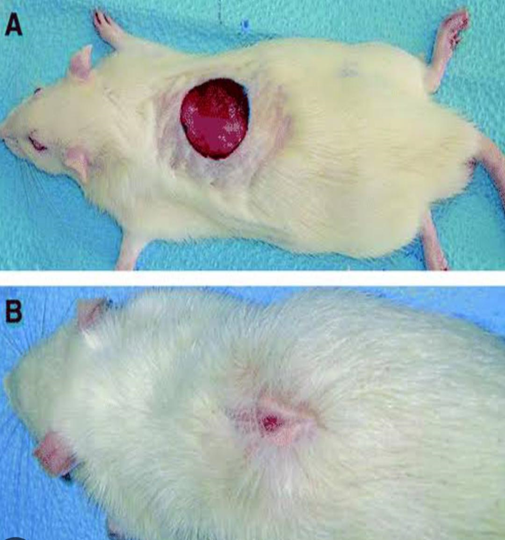
Plate 4: Serum bioassay of albino rats treated with Persea Americana and Syzygium aromaticum oil extracts
TABLE 10i: Tissue Bioassay Oo Albino Rats Treated with Persea americana Mill and Syzygium aromaticum oil Extracts
| Parameters | A | B | C | D | E | F | G | H |
| ALT µ/l | 24 | 47 | 21.5 | 43 | 55.5 | 48.5 | 22,5 | 25,0 |
| ALP µ/l | 57.96 | 179.4 | 132.48 | 259.44 | 157.32 | 226.32 | 140,6 | 145.7 |
| AST µ/l | 26.6 | 53.8 | 32.4 | 37.2 | 25.4 | 38 | 46.88 | 36 |
| TP mg/dl | 13.53695 | 12.82759 | 12.6502 | 12.53202 | 13.44828 | 12.97537 | 134.6 | 134.9 |
| GSH µg/dl | 17.44681 | 77.65957 | 56.38298 | 80.85106 | 59.57447 | 75.95745 | 45.23 | 40.9 |
| ALB mg/dl | 17.66852 | 18.06019 | 13.57778 | 16.79815 | 11.8805 | 20.14907 | 15.90 | 13.0 |
| GLB mg/dl | 4.13157 | 5.2326 | 0.92753 | 4.26613 | 1.56772 | 7.1737 | 4.90 | 3.667 |
Keys:
A: 50mg/kg of Avocado oil infused with Goya oil and clove oil extracts
B: 100mg/kg of Avocado oil infused with Goya oil and clove oil extracts
C: 50mg/kg of Avocado oil infused with vegetable oil and clove oil extracts
D: 100mg/kg of Avocado oil infused with vegetable oil and clove oil extracts
E: 50mg/kg of negative control (wound with no treatment)
F: 100mg/kg of negative control (wound with no treatment)
G: 50mg/kg of positive control (no wound)
H: 100mg/kg of positive control (no wound)
ALT: Alanine amino transferase
GLB: Globulin
TP: Total protein
ALB: Alkaline Albumin
ALP: Alkaline phosphatase
GSH: Glutathione
Table 10i: shows the tissue bioassay of albino rats treated with extracts of Persea Americana Mill and Syzygiumaromaticum. The serum biochemical parameters investigated in this table are Total protein TP (mg/dl), Aspartate amino AST (U/L), Alkaline Phosphatase ALP (U/L), Alanine Amino transferase ALT (U/L), Alkaline Albumin ALB (mg/dl), Glutathione GSH (μg/dl), Globulin GLB (mg/dl). The table compares the values of these parameters among six groups of rats, each treated with different doses (50mg/kg and 100mg/kg) of avocado seed oil and clove oil extracts with positive and negative controls. The positive control groups (E and F) have no wound, while the negative control groups (C and D) have wound with no treatment. The other groups (A, and B) have wound and are treated with 50mg/kg and 100mg/kg doses of avocado seed oil infused with Goya oil and clove oil extracts and avocado seed oil infused with vegetable oil and clove oil extracts.
Table 10ii: The table shows that the oil extracts of avocado and clove have different effects on the tissue bioassay parameters, depending on the dose and the form of the oil extracts. The negative control groups (C and D) have higher levels of CREATININE and UREA than the positive control groups (E and F), indicating that the oil extracts are not toxic. The avocado oil infused with Goya oil and clove oil extracts (A) have lower levels of CREATININE, UREA, ALT, and AST than the avocado seed oil infused with vegetable oil and cove oil extracts B), suggesting that the extracts infused with vegetable oil have fewer toxic effects on the rats. Also, the oil of both extracts and clove have lower levels of GSH and PROT than the positive control groups (E and F ) at 50mg/kg, indicating that the extracts may reduce the antioxidant and protein levels in the tissues while the oil extracts of avocado seed oil infused with Goya oil and clove have higher levels of ALP than the positive control groups (E and F ), suggesting that the extracts may stimulate wound healing.
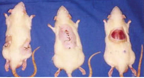
Figure 8: Macroscopic observation of excision wound inflicted on albino rats
Plate 5: Macroscopic observation of excision wound inflicted on albino rats.
Plate 5: Macroscopic observation of excision wound inflicted on albino rats.
DISCUSSION
This study aimed to investigate the effects of oil avocado (Persea Americana) extracts, vegetable oil, Goya oil, and clove oil (Syzygium aromaticum) and its healing potential on male and female albino rats and their antibacterial activity against surgical wound isolates. Infection in wound delays healing and may cause wound breakdown and complete wound dehiscence. Despite progress in development of antibacterial agents, there are still special needs to find new antibacterial agents due to development of multidrug resistant bacteria [11]. Essential oils are potential source of antibacterial compounds especially against pathogenic bacteria.
The antimicrobial activity of Persea Americana Mill and Syzygium aromaticum oils extracted with different solvents and concentrations was evaluated against eight surgical wound isolates from four selected general hospitals in Ondo State, Nigeria. In table 4, the results showed that the oils exhibited varying degrees of inhibition against the tested bacteria, depending on the type of solvent, concentration, and bacterial strain. The diced avocado seed (L/E) soaked in N-hexane showed the highest antimicrobial activity among the oils, followed by the avocado seed oil infused with Goya oil with clove while all the other oil extracts also showed antimicrobial activity against each isolates except the negative controls (vegetable oil and Goya oil) which shows no zone of inhibition . The avocado seed oil extracted with solvent inhibited all the bacterial strains, with zones of inhibition ranging from 10.0 mm to 35.0 mm at 100 mg/ml concentration. The avocado oill infused with Goya with cloves oil inhibited all bacterial strains, with zones of inhibition ranging from 10.0 mm to 28.0 mm at the same concentration. The solvent extracted oil and avocado oil infused with goya with clove oils also showed some activity at lower concentrations at (50 mg/ml, 25 mg/ml, and 12.5mg/ml) some organisms were not susceptible to the oils as some of them have no zone of inhibition at (6.25mg/ml and 3.125mg/ml). At 6.25mg/ml, 40% of the organisms did not have zone of inhibition. At 3.125 mg/ml, 90% of the organisms showed no zone of inhibition to the oil extracts. The minimum inhibitory concentration (MIC) of avocado seed oil were 100 mg/ml for Klebsiella oxytoca and Escherichia coli, 25mg/ml for Bergeyella zoochelum and Acinetobacter baumanii and 12.5 mg/ml for Chrysobacterium meningosepticum while the minimum bactericidal concentration (MBC) of the avocado seed oil with cloves were 50mg/ml and 25 mg/ml, respectively, for most of the bacterial strains, The MIC of the avocado seed oil were 25mg/ml, 50 mg/ml and 100 mg/ml, respectively, for all the bacterial strains and MBC of the avocado seed oil were 50 mg/ml and 100 mg/ml, respectively.
The avocado seed oil extracted with solvent, avocado oil infused with Goya oil, avocado oil infused with vegetable oil, avocado oil infused with Goya with cloves, and avocado oil infused with vegetable oil and cloves showed antimicrobial activity against any of the bacterial strains at any concentration. This suggests that the olive oil may have interfered with the extraction or diffusion of the active compounds from the plant materials. Alternatively, the olive oil may have acted as a nutrient source for the bacteria, enhancing their growth and reducing the inhibitory effect of the oils. The N-hexane extract of Persea americana Mill was found to be much more effective indicating that solvent extraction method is more better than olive and vegetable oil (lipids)as an extraction solvent of the antibacterial chemical component. In fact, most of the antimicrobial chemicals that were identified were water insoluble, which makes organic solvents more suitable for their extraction [12].
The positive control (Amoxicilin) showed strong antimicrobial activity against all the bacterial strains, with zones of inhibition of 25mm at 100 mg/ml concentration. The zone of inhibition refers to the clear zone around the agar well or antimicrobial disk. Also, the results obtained from the growth dynamics and killing time of the bacterial isolates shows that the effects of Persea Americana Mill oil extracts on bacterial growth varied depending on the isolate. Some isolates, such as Escherichia coli and klebsiella oxytoca showed a significant inhibition of growth in the presence of the oil extracts, while others were less affected by the oils. The antimicrobial activity of Persea americana Mill used in this study might act as gram-negative bacteria by generating through the cell walls of the bacteria. The antimicrobial properties of avocado oil maybe due to bioactive compounds like tocopherols, carotenoids, beta-sitosterol, and terpenoids [13].
The antibacterial properties of avocado seed oil consist of tannins, catechin flavones, and polyphenolic compounds which are often found in the tissues and seed of the avocado fruit. The pharmacodynamic and pharmacokinetic mechanisms of the oil extracts of avocado and clove on wound healing activity was explored in this study. The oil extracts modulated the biochemical markers of inflammation and oxidative stress, such as ALT, AST, ALP, GSH, TP, ALB, and GLB, which are involved in the protection and repair of the wounded tissues. These effects are consistent with previous studies that reported the anti-inflammatory and antioxidant activities of avocado and clove extracts in various animal models of wound healing [14]
The phytochemical constituents of the oil extracts, such as,tannins, catechin , flavones, and polyphenolic compounds could be responsible for these effects, as they have been shown to affect the expression and activity of various enzymes, cytokines, and transcription factors involved in the inflammatory and oxidative pathways. The oil extracts also improved the immune functions by reducing the levels of creatinine, urea, ALT, AST, and ALP. These effects indicate that the oil extracts enhanced the clearance and detoxification of the oil extracts and their metabolites, and prevented the accumulation of toxic substances in the blood and tissues. These effects are in agreement with previous studies that demonstrated the nephroprotective and hepatoprotective effects of avocado and clove extracts in various animal models injury [15].
The oil extracts could modulate the activity and expression of various enzymes and transporters involved in the renal and hepatic excretion and biotransformation of the oil extracts and their metabolites, such as cytochrome P450, uridine diphosphate glucuronosyltransferase, multidrug resistance protein, and organic anion transporter. The toxicological analysis show that the serum level of ALT and AST were not altered by Syzygium aromaticum and Persea Americana Mill oil extracts. Level of other biochemical parameters was not significantly altered suggesting that the oil extracts were not hepato-toxic, which may be due to the presence of antioxidant in Syzygium aromaticum and Persea Americana Mill [16]. Avocado seed consists of various extracts which can be used for different applications. Starch, oil, lipid, protein, crude fibre, antioxidants, vitamins and minerals are among the most know extracts which can be obtained from avocado seed [17]. Avocado seeds are high in phytochemicals and are utilized for medicinal purposes. Avocado-seed extracts also have many health-related bioactive properties, such as anti-hyperglycaemic, anticancer, anti-hypercholesterolemia, antiioxidant, anti-inflammatory, and anti-neurogenerative effects are clearly demonstrated how these properties can be used to formulate or fortify food [18].
In conclusion, based on the results obtained from this study, it can be concluded that P.americana mill and S.aromaticum has a potential antimicrobial effect against surgical wound organisms and low toxicity as well as serve as an important source of antibacterial compounds that may provide renewable sources of useful antibacterial drugs against bacterial infections in humans and animals [19]. The oils from P.Americana mill and Syzygium aromaticum showed varying degrees of antibacterial activity against clinical isolates [20]. From the study it can be inferred that the oil extracts shows significant growth inhibiting effect on Gram-negative bacteria (Escherichia coli). Also the oil extracts of avocado and clove have pharmacodynamic and pharmacokinetic effects that improve the wound healing process in rats by modulating the serum and tissue bioassay parameters related to inflammation, oxidative stress, renal function, liver function, and protein synthesis [21]. Furthermore, the toxicity of the oil extracts is dose dependent and the efficacy of this Persea Americana Mill and Syzygium aromaticum against these microorganisms provide a scientific ground for the application of medicinal plants in the prevention and treatment of bacterial infections caused by pathogenic bacteria such as Escherichia coli, Acinetobacter baumanii and Klebsiella oxytocawhich have the ability to develop resistance to antibiotics [22].
The recommendation from the results, it is evident that avocado seed oil and clove have significant antimicrobial properties as a result of their polyphenolic components. Avocado is also rich in unsaponifiable compounds such as sterols, mainly β-sitosterol, vitamins, carotenoids, tocopherols, and the phenolic compounds are of significant interest, for their antioxidant and anti-inflammatory properties). In addition to its high content of good fats, avocado pulp also contains several hundred phytochemical molecules that may play a role in cancer prevention.
In addition to certain molecules that are widespread in the plant world such as flavonoids (quercetin, luteolin, apigenin, etc.) or coumarins (scopoletine), avocados have the particularity of containing alkanols, a class of fat-soluble molecules that show a great inhibitory activity on cancer cells These antimicrobial and wound healing properties hold immense promise for developing novel therapeutic strategies against a variety of microbial infections. Its multifaceted mode of action, targeting multiple aspects of microbial survival and virulence, presents significant advantages over conventional antibiotics. Therefore, I would recommend that extensive research be carried out on synergistic effect of avocado seed oil and cloves.
Funding
This work was self supported
ACKNOWLEDGEMENTS
The authors wish to express appreciation to all the technical staff of the laboratory unit of the Department of Microbiology, Faculty of Science, Adekunle Ajasin University, Akungba-Akoko, Ondo State, Nigeria.
Competing Interests
Authors have declared that no competing interests exist
Dr Oludare temitope osuntokun
https://orcid.org/0000-0002-3954-6778, Web of science Your ResearcherID -L-4314-2016
REFERENCES
- Heilborn, J. D. Weber, G., A. Gronberg, C. Dieterich, and M. ¨ Stahle, (2010) Topical treatment with the vitamin D analogue calcipo- ˚ triol enhances the upregulation of the antimicrobial protein during wounding in human skin in vivo,” Experimental Dermatology, (19): 4, 332–338.
- Templin, C. K. Grote, K. Schledzewski. (2009) Ex vivo expanded haematopoietic progenitor cells improve dermal wound healing by paracrine mechanisms, Experimental Dermatology, 18: 5, 445–453.
- McDaniel, J. C. Belury, K. Ahijevych, and W. Blakely, (2008) Omega-3 fatty acids effect on wound healing, Wound Repair and Regeneration, 16: 3, 337–345.
- Y and K. Yoshikawa, (2001) Cutaneous wound healing: an update,Journal of Dermatology,.28: 10. 521–534.
- Yang H, Wang W, Romano KA, Gu M, Sanidad KZ, Kim D, Yang J, Schmidt B, Panigrahy D, Pei R, Martin DA, Ozay E, Wang Y, Song M, Bolling BW, Xiao H, Minter LM, Yang GY, Liu Z, Rey FE, Zhang G (2018). A common antimicrobial additive increases colonic inflammation and colitis-associated colon tumorigenesis in mice, Sci Transl Med.10 (443). pii: eaan4116. doi: 10.1126/scitranslmed. aan4116.
- Çakaloğlu, B., Vasfiye H. Ö. and Semih Ö. (2018). Cold press in oil extraction. A review, Ukainian Food journal, 7(4): 640-654.
- Fawole, M. O., and Oso, B. A. (2007). Laboratory manual for microbiology (7th ed.). Spectrum Books Ltd.
- Osuntokun, O.T., Azuh, V.O., Adejoro, B.F., Akele, E.O. (2021). Antimicrobial Spectrum, Growth/Killing Kinetics, Conventional/Molecular Assay of Characterizing Non-Leguminous Endophytic Bacteria and Fungi from Helianthus annuus, Carica papaya and Lycoperesicum solanum. J Biomed Res Environ Sci., 2(10) :1018-1034. doi: 10.37871/jbres1345, Article ID: JBRES1345.
- Humphries, R., Bobenchik, A. M., Hindler, J. A. and Schuetz, A. N. (2021). Overview of changes to the clinical and laboratory standards institute performance standards for antimicrobial susceptibility testing, M100, 31st edition. Journal of Clinical Microbiology. 59(12): 00213-21.
- Shailajan, S., Menon, S., Pednekar, S. and Singh, A. (2011). Wound healing efficacy of JatyadiTaila: In vivo evaluation in rat using excision wound model. International Journal of Pharmacy and Pharmaceutical Sciences. 3(2): 112-115.
- Weidner, M. S. and Sigwart, K. (2001). Investigation of the teratogenic potential of a Zingiber officinale extract in the rat. Reproductive Toxicology. 15(1): 75-80.
- Santos, R. G., Lopes, G. S. and Bianchini, A. (2015). Toxicity of clove essential oil and its ester eugenyl acetate to aquatic organisms. Environmental Toxicology and Pharmacology. 40(3): 914-922.
- Myung, N.; Kim, S. (2005) The Beneficial Effect of Avocado on Skin Inflammation in a Mouse Model of AD-like Skin Lesions. Korean J. Plant Reources 2019, 32, 705–713
- Verderio, E.A.M.; Johnson, T.S.; Griffin, M. Transglutaminases in Wound Healing and Inflammation. Transglutaminases, 38, 89–114.
- Salinas-Salazar, C.; Hernández-Brenes, C.; Rodríguez-Sánchez, D.G.; Castillo, E.C.; Navarro-Silva, J.M.; Pacheco, A. (2017) Inhibitory Activity of Avocado Seed Fatty Acid Derivatives (Acetogenins) Against Listeria Monocytogenes. J. Food Sci., 82, 134–144.
- Showande S.J., Fakeye T.O., Kajula M., Hokkanen J., Tolonen A. (2019). Potential inhibition of major human cytochrome P450 isoenzymes by selected tropical medicinal herbs-Implication for herb–drug interactions Food Sci Nutr, 7 (2019), pp. 44-55.
- Benavente-García, O.; Castillo, J. (2008) Update on Uses and Properties of Citrus Flavonoids: New Findings in Anticancer, Cardiovascular, and Anti-inflammatory Activity. J. Agric. Food Chem., 56, 6185–6205.
- Zhang, Q.; Yang, J.; Wang, J. (2016). Modulatory effect of luteolin on redox homeostasis and inflammatory cytokines in a mouse model of liver cancer. Oncol. Lett., 12, 4767–4772.
- Brizi C, Santulli C, Micucci M, Budriesi R, Chiarini A, Aldinucci C, Frosini M (2016) Neuroprotective effects of Castanea sativa mill. Bark extract in human neuroblastoma cells subjected to oxidative stress. J Cell Biochem 117:510–520.
- Lu, J., Getz, G., Miska, E. A., Alvarez-Saavedra, E., Lamb, J., Peck, D., Sweet- Cordero, A., Ebert, B.L., Mak, R.H., Ferrando, A. A., Downing, J. R., Jacks, T., HorvitzH. R. and Golub, T. R. (2005). MicroRNA expression profiles classify human cancers. Nature 435, 834–838. https://doi.org/10.1038/nature03702.
- Ogunmoyole, T., Dada, I. & Adebamigbe, O.A. (2021). Ameliorative potentials of Persea americana leaf extract on toxicants – induced oxidative assault in multiple organs of wistar albino rat. Clin Phytosci 7, 27.
- Mahadeva U.R. and Bizuneh A. (2011). Remnant B-Cell Stimulant and Anti-oxidative effects of Persea Americana Fruit extract studied in Rats introduced into Streptozotoc induced Hyperglycaemic State. African Journal of Traditional, Complementary and Alternative Medicines (AJTCAM). 8(3):210-217.