Phytochemical and Anti-Diarrhoeal Potential of Ipomea batatas Leaf Solvent Extract on Castor Oil Induced Diarrhoeal Rat Model
- Muazu Alkali ADAMU
- Suleiman Ali
- Musa Magaji Jawa
- 271-286
- Jul 6, 2024
- Chemistry
Phytochemical and Anti-Diarrhoeal Potential of Ipomea batatas Leaf Solvent Extract on Castor Oil Induced Diarrhoeal Rat Model
Muazu Alkali ADAMU*, Suleiman Ali, Musa Magaji Jawa
Department of Science Laboratory Technology, School of Sciences, Yobe State College of Agriculture Science and Technology, Gujba, Yobe, Nigeria.
*Corresponding author
DOI: https://doi.org/10.51584/IJRIAS.2024.906025
Received: 28 May 2024; Accepted: 04 June 2024 Published: 06 July 2024
ABSTRACT
The folkloric claim of Ipomoea batatas extracts in the management of diarrhoea is yet to be substantiated or refuted with scientific data. Therefore, the present study was aimed to screen the extracts of I. batatas for both its secondary metabolites, acute toxicity, and antidiarrhoeal activity at 125, 250, and 500 mg/kg body weight in rats and elemental analysis of the most active extract. The most active extract was screened for elemental analysis. Secondary metabolites were screened using standard methods while the antidiarrhoeal activity was done by adopting the castor oil-induced diarrhoeal and castor oil-induced enteropooling, models. The extracts contained flavonoids, phenolics, saponins, alkaloids, tannins, terpenes, and steroids were detected. The LD50 of extracts were found to be greater than 5000mg per kg body weight of the rats. The elemental analysis revealed the presence of potassium, zinc, sodium, and chloride. In the castor oil-induced diarrhoeal model, the extracts significantly (𝑃 < 0.05) prolonged the onset time of diarrhoea, decreased in a number of faeces, fresh weight of faeces, and water content of faeces, and increased the inhibition of defecations. Na+/K+-ATPase, and alkaline phosphatase activities in the small intestine increased significantly (𝑃 < 0.05) whereas the extract produced dose-specific changes on intestinal nitric oxide content decreased. The decreases in the masses and volumes of intestinal fluid by the crude extracts was accompanied by increase in inhibition of intestinal fluid content in the enteropooling model.
Keywords: Phytochemical compounds, diarrhoea, antidiarrhoea, castor oil, Ipomoea.
INTRODUCTION
Diarrhoea is a global health issues affecting all regions and populations, particularly in low and middle income countries of sub-Saharan Africa and Asia from which very young and old aged are more vulnerable (Turyare et al., 2021). Diarrhoea is an alteration in bowel movement, characterized by increased water content, volume and frequency of stooling, usually occurring three or more times a day, whilst the frequent passing of loose ‘pasty’ stools or formed stools by breastfed babies is not regarded as diarrhoea (Omole et al., 2019). It is a major cause of morbidity and mortality affecting more children than adults (UNICEF, 2019).
Globally, an annual incidence of about 1.7 billion childhood diarrhoeal episodes has been reported in humans, with mortality of 760, 000 children under the age of five (Abuzerr et al., 2019). Most of the deaths from diarrhoea occur amongst children aged less than 2 years living in South Asia and sub-Saharan Africa (UNICEF, 2019). Of this, Nigeria accounts for an estimate of 150 000 diarrhoeal deaths (Charyeva et al.,2015; Peter and Umar 2018).
Management strategies of diarrhoea have been to prevent dehydration via the use of oral rehydration solutions, intravenous fluids, conventional drugs such as loperamide or its combination with simethicone and antibiotics such as azithromycin and ciprofloxacin. Several options employed in the management of the diseases include oral agents such as metronidazole, antibiotics, and oral rehydration therapy (ORT) (Charyeva et al., 2015). With over a decade of the practice and promotion of ORT, diarrhoea is still the second among the causes of child death (WHO, 2017). In addition, these management options which often fail during high stool output state are also associated with undesirable side effects such as headache, convulsion, stomach cramp, vomiting, constipation, and hallucination (WHO, 2017).
However, these options are now known to be accompanied by severe constipation, vomiting, uncomfortable fullness and pain of the stomach, fast or irregular heartbeat and occasionally syncope. In addition, because of the continuous escalation in the healthcare cost, perceived efficacy, the emergence of resistant pathogenic strains, potential to exhibit bioactivity through multiple mechanisms and financial austerity in major developing countries, the interest in natural product-based drug development needs to be revitalized. Therefore, exploring alternative and complementary options in medicinal plants for the management of diarrhoea will not be out of place (WHO, 2017).
The plant kingdom have been a treasure house of potential drugs. In the recent years there has been an increasing awareness about the importance of medicinal plants. Drugs from the plants are easily available, less expensive, safe, and efficient and rarely have side effects. Plants are the richest resource of drugs of traditional systems of medicine, modern medicines, nutraceuticals, food supplements, folk medicines, pharmaceutical intermediates and chemical entities for synthetic drug (Pritish et al., 2015).
Ipomoea batatas Linn. (family, Convovulaceae), also commonly called sweet potatoes (English), “dun,adun odunkun” (Yoruba), “dankalin Hausa” (Hausa), and “nduku uto” (Igbo), is native to South East Asia and India but is now extensively cultivated in the tropical and subtropical regions (Morales et al., 2017). I. batatas is a herbaceous perennial vine, bearing alternate heart-shaped or palmately lobed leaves and medium-sized sympetalous flowers. The stems are usually crawling on the ground and form adventitious roots at the nodes. The leaves are screwed along the stems. The leaf stalk is 5 to 20 inches long (Morales et al., 2017).
Various parts of the plant have been claimed to be ethno-pharmacologically relevant in the management of several ailments. For example, a cold infusion of the root is used to treat venereal diseases, anaemia, scabies, leprosy, and skin diseases (Vishnu et al., 2019). The leaves have also been as used for managing cold, bronchitis, and eye infections and as an abort if ancient. Furthermore, I. batatas leaf extract has also been claimed to be used as a remedy for diarrhoea, and dysentery, diabetes, and cancer (Vishnu et al., 2019). I. batatas aqueous leaf extract were reported to be alterative, aphrodisiac, astringent, bactericide, demulcent, fungicide, and tonic, sweetpotato leave is a folk remedy for asthma, bugbites, burns, catarrh, ciguatera, convalescence, diarrhea, dyslactea, fever, nausea, renosis, splenosis, stomach distress, tumors, and whitlows (Duke and Wain, 1981).
Extracts of I. batatas leaf have been reported to exhibit antibacterial activity against Staphylococcus aureus, Bacillus cereus, and E.coli O157:H7 (Islam et al., 2015). Methanol extract of sweet potato roots showed gastroprotective activity against aspirin-induced ulcer in Wistar rats in a dose dependent manner (Panda and Sonkamble, 2012). Similarly, an earlier study reported a significant increase in packed cell volume, white blood cells and platelets of rabbit fed with sweet potato extract (Osime et al., 2008). Moreover, the I. batatas leaf extracts depress neuroinflammatory responses in lipopolysaccharide-activated BV-2 microglia cells by inhibiting the production of pro-inflammatory mediators, such as inducible nitric oxide synthase (iNOS), cyclooxygenase 2 (COX-2), nitric oxide (NO), and TNF-α. The anti-euro-inflammatory potential of I. batatas leaf extract was considered to be related to its strong antioxidant properties (Kang et al., 2014).
Despite several scientific studies that have validated the folkloric uses of different parts of I. batatas, there is no study in the open scientific literature that has provided scientific evidence to the acclaimed use of the leaf extract of I. batatas in the management of diarrhoea. Therefore, this study was aimed at validating or not, the acclaimed use of I. batatas leaf extract in the management of diarrhoea using chemically induced diarrhoeal models in rats.
MATERIALS AND METHODS
Collection and Identification of Plant Sample
The leaves of Ipomoea batatas used in this research were collected at 6.45 am around Model primary school located in Damaturu Local Government Area of Yobe State, Nigeria 11.7470° “N, 11.9662° “E and were identified at the Herbarium Unit of the Department of Plant Biology, Bayero University Kano Accession Number BUKHAN 409 was assigned to it.
Extraction of Ipomoea batatas (Sweet potato) Leaves
Soxhlet extraction method
Exhaustive Soxhlet extraction was performed using classical apparatus with 50 g of each of the powdered leaf samples of I. batatas. Extraction was performed with n-Hexane (1:10 w/v) (Peter and Umar, 2018). After extraction, the n-Hexane solvent was evaporated completely to concentrate it in a vacuum by using rotary evaporator at 400C under reduced pressure. The solvent-free n-Hexane extract was thereafter evaluated for the study.
Two hundred grams (200g) of dry powder of the plant material was subjected to successive soxhlet extraction with solvents of increasing polarity (n-Hexane and methanol). First 50g of the powdered plant material was placed in the extraction chamber of the soxhlet apparatus. The extracting solvent (n-Hexane) in the flask was heated until clear liquid contents of the chamber siphoned into the flask. Each time 50g of the powdered plant material was extracted with 200 ml of solvent in the soxhlet extraction process (Rahman et al., 2015). The n-Hexane extract was evaporated completely to concentrate it in a vacuum by using a rotary evaporator (Buchi labortechnik AG, Switzerland) under reduced pressure set at 400C followed by oven at room temperature for 12 h (Zavala et al., 2013). The residue was collected and dried at room temperature to remove n-Hexane.
The plant material was then dried and extracted using methanol following the same procedure as described before to get the methanol extract. Finally, the residue of methanol extract was collected and dried at room temperature. After drying, the percentage yield of all extract were determined and the yield were calculated for n-Hexane and methanol extracts respectively.
Cold maceration method
Maceration was carried out in a closed conical flask for 7 days at room temperature with frequent agitation (Handa et al., 2008) using 1000 g powdered leaf samples of I batatas and methanol (1:10 w/v). The extraction was done in an aliquot. The marc were filtered with Whatman No.1 filter paper and the filtrate was concentrated by open air evaporation. The concentrate (extract) was weighed, the percentage yield calculated, labeled, and stored at 40C in a refrigerator until required. The solvent-free methanol extracts obtained were similarly evaluated.
Phytochemical screening
Qualitative Screening of Secondary Plant Metabolites. A known volume 1.0 mL of the extract of I. batatas was screened for the presence of some secondary metabolites as described for alkaloids (Hanieh et al., 2010) steroids, anthraquinones, (Oladiji et al., 2010) saponins (Wall et al., 1954), phenolics and flavonoids (Awe and Sodipo, 2001) cardiac glycosides (Awe and Sodipo, 2001), tannins and triterpenes (Odebiyi and Sofowora, 1978) as follows.
Experimental Animals
Experiments involving animal ethics care and oral administration of various extracts were reviewed and approved by the Animal Care and Use Research Ethics Committee (ACUREC), Bayero University Kano. The protocol of animal study on Wister rats was based on the guidelines given by the Law of Animal Protection Act of the institution with approval number BUK/ DRIP/AUREC/00507. Healthy, Wistar rats of both sexes (Rattus norvegicus) weighing 140.51 ± 6.53 g were obtained from AKTH Kano, Kano State, Nigeria. The animal were then transported to Damaturu, Yobe State, Nigeria. The animals were housed in clean wooden cages placed in well-ventilated housing conditions (temp: 25oC – 27oC; photoperiod: about a 12 h light and dark cycle; relative humidity: 45% – 50%). The animals were allowed unrestricted access to clean rat pellets (Top Feeds Nigeria Limited, Ibadan, Nigeria) and tap water. The cages were cleaned on a daily basis.
Antidiarrheal Activity (Castor Oil-Induced Diarrhoeal Model)
Castor Oil-Induced Diarrhoea in Rats. Thirty-six, healthy Wistar rats were fasted for 8 hours prior to the experiment but allowed free access to water. The experimental rats were completely randomized into nine groups of four animals each. The procedure described by Bajad et al. (2001) was adopted with slight modification. Animals in group I (administered 1.0 mL of distilled water) served as normal control, animals in group II (administered 1.0 mL of distilled water), as negative control while those in groups III (positive control), IV, V, and VI (test groups) received 1.0 mL each corresponding to 2.5 mg/kg body weight of loperamide (a reference drug), 125, 250, and 500 mg/kg of the extracts respectively. Thirty minutes after administration, all the animals were orally administered 1 mL of castor oil and thereafter placed in cages lined with a pre-weighed transparent paper. Except the normal control group 1. During the 6-hour observation period, the time of onset of diarrhoea, the total number of faeces, diarrhoeal faeces, total weight of faeces, and percentage inhibition of diarrhoeal defecation in each group were computed. The weight of the faeces was obtained from the difference in the preweighed transparent paper and fresh weight of the stool. The dry weight of the faeces was obtained by drying the fresh faeces in the laboratory oven (Uniscope Laboratory oven, SM9053, Surgifriend Medicals, England) at 100 oC until a constant weight was obtained. The difference in the fresh weight of the faeces and dry weight of the faeces gives the water content of the faeces. At the end of the 6-hour exposure period, the animals were sacrificed and small intestine supernatants prepared.
% inhibition=((Mean defecation of control group – Mean defecation of treated group) )/(Mean defecation of control group)×100 (1)
Preparation of small intestine supernatants.
The procedure described by Akanji and Yakubu (2000) was adopted for the preparation of small intestine supernatants. Briefly, under ether anaesthesia, the animals were dissected and small intestine was removed. Thereafter, the contents of the small intestines were squeezed out, blotted in blotting paper, and homogenized in 0.25 M sucrose solution (1:4 w/v) using Teflon homogenizer. The homogenate was centrifuged at 894 ×g for 15 minutes to obtain the supernatant which was used for the determination of concentrations of protein, nitric oxide, activity of sodium potassium ATPase and intestinal alkaline phosphatase activity.
Determination of protein content of the supernatant.
The protein concentration in the small intestine supernatant of the animals was assayed, using Bradford’s reagent as described by Gornall et al., (1949) A known volume (4.0 mL) of Bradford reagent was added to 1.0 mL of the supernatant (appropriately diluted). This was mixed thoroughly by shaking and left undisturbed for 30 minutes at room temperature for colour development. The blank was constituted by replacing the supernatants with 1.0 mL of distilled water. The absorbance was read against blank at 540 nm. The concentration of protein in the supernatants was obtained from the calibration curve for bovine serum albumin using the expression bellow
Protein concentration (mg/ml)=Cs × F (2)
Where Cs corresponding protein concentration from the calibration curve; F = dilution factor.
Determination of Na+-K+ ATPase activity.
The procedure described by Bewaji, (1985) was adopted for the determination of the activity of Na+-K+ ATPase in the small intestine supernatant. Briefly, 400 𝜇L of 200 mM of NaCl, 40 mM of KCl, and 60 mM of Tris (pH 7.4) were pipetted into test tube. Thereafter, 20 𝜇L of MgCl2⋅6H2O (80 mM), 20 𝜇L of 20 mM EGTA, 240 𝜇L of distilled water, and 20 𝜇L of appro- priately diluted supernatant of the small intestine were added. The mixture was incubated at 37∘ C for 5 minutes. A known volume (100 𝜇L) of 8 mM ATP was added, mixed thoroughly, and incubated at 37∘ C for 30 minutes. Furthermore, 200 𝜇L of sodium dodecyl sulphate (5%) and 2,000 𝜇L of reagent C (mixture of ammonium molybdate/sulphuric acid solution [reagent A] and 9% ascorbic acid [reagent B] in ratio 4:1 v/v) were added. The mixture was left undisturbed for 30 minutes at room temperature for colour development. The blank was constituted in the same manner except that the small intestine supernatant was replaced with 20 𝜇L of distilled water. The absorbance of the test solution was read against that of the blank at 820 nm and then extrapolated from the absorbance obtained was then extrapolated from the calibration curve for phosphate to give the concentration of the inorganic phosphate. Thereafter, the specific activity of Na+-K+-ATPase was computed using the following expression:
Specific activity (µmole Pi/mg protein/hr){([Pi] × 2 × dilution factor)/(1000 × protein concentration (mg/mL))} (3)
Where [P𝑖] = concentration of inorganic phosphate in n moles (obtained from the calibration curve);
2 = factor introduced to obtain the amount of P𝑖 released per hour;
1000 = factor introduced to convert the P𝑖 released to 𝜇moles.
Determination of nitric oxide concentration.
The procedure described by Wo et al. (2013) was used to determine the concentration of nitric oxide in the small intestine supernatants of the animals. A known volume (0.5 mL) of the supernatant was added to 2 mL of 75 mmol/L ZnSO4 solution and 2.5 mL of 55 mmol/L NaOH. The solution was mixed thoroughly, adjusted to a pH of 7.3, incubated for 10 minutes, and centrifuged at 504 ×g for 10 minutes. The blank was constituted in a similar manner to the test except that 0.5 mL of supernatant was replaced by 0.5 mL of distilled water. Furthermore, 1 mL of glycine-NaOH buffer was added to the test sample and blank. A known volume (2 mL) of the deproteinized solution was added to the test and blank and the volume was adjusted to 4.0 mL with deionized distilled water. The reaction was initiated by the addition of freshly activated cadmium granules and, after 60 minutes, 2.0 mL each of test and blank was added to tubes containing 2.5 mL of ethylene diamine tetraacetic acid solution, 3.0 mL of 1.0 mol/L HCl, and 0.3 mL of 1.0 g/L fuchsin acid solution, mixed thoroughly and incubated for 2 minutes. Next, 0.2 mL of 0.05 mol/L resorcinol and 3.0 mL of 1.0 mol/L NH4OH were added. The absorbance of the test solution was read against the blank at 436 nm. The concentration of serum nitrite was extrapolated from the calibration curve of nitrite.
Determination of alkaline phosphatase activity
Assay of Alkaline Phosphatase (ALP): The method described by Bassey, (1946) as modified by (Wright et al., 1972) using Randox kits. In a cuvette, 10 µl of sample was mixed with 500 µl of the reagent. The initial absorbance was read at 405 nm, and subsequently over 3 minutes. The mean absorbance per minute was added in the calculation:
ALP activity (IU/l) = 2746× ΔA405 nm/min (4)
Where: 2742 = Extinction coefficient;
ΔA 405 nm/ min = change in absorbance per minute for the homogenate sample.
Castor oil-induced enteropooling.
The procedure described by Chitme et al. (2004) was adopted for the castor oil-induced enteropooling study. The animals were fasted without food for 6 h prior to the experiment but were allowed free access to water. Four animals were randomly selected for each group and placed in their respective cages. Animals in the normal control group (group 1) received 1.0 mL of distilled water, animals in the negative control (group 2) received 1.4 ml while those in the Castor oil + Loperamide group. (Group 3) received 1.0 mL of atropine sulphate (2.5 mg/kg body weight). Rats in groups 4, 5, and 6 (test groups) were orally administered 125, 250, and 500 mg/kg of the extracts, respectively. Immediately afterward, 1.0 mL of castor oil was administered orally to each of the rats in all the groups except Group 1 Normal control. After 30 minutes, each rat was sacrificed according to the method described by (Akanji and Yakubu, 2000) and the ends of the pylorus and caecum of the small intestine were tied. The small intestine was dissected, and its intestinal content squeezed into a measuring cylinder. The volumes and the masses of the intestinal contents were noted and used to compute the percentage of inhibition of intestinal content.
Inhibition of intestinal content %=Mass of intestinal fluid (g) {(Control – Treated)/Control} × 100 (5)
Elemental Analysis
The five (5 g) for each of the each of the cold macerated Ipomoea batatas methanol leaf extract, were ashed in an oven at 540oc for three hours, before subjecting it to various organic solvents, 0.5g each of the cooled ashed extracts were digested by heating for two hours with a mixture of 10 mls Hydrochloric acid (HCl) Nitric acid (HNO3) and perchloric acid (HClO4). The digested mixtures were evaporated down to 5mls using rotary evaporator; they were then made up to 10 mls with 2M HNO3, and to which were added 30 mls of distilled water and kept in a 100mls beaker. The resulting solutions were used for the elemental analysis using atomic absorption spectrophotometer (AAS) (A. Analyst 400 Model) at an appropriate wavelength, temperature and lamp-current for elements. Reagent blank samples were also prepared, these sample were analyzed for: zinc (Zn) sodium (Na) chloride (Cl) and potassium (K) were determined for each of the Ipomoea batatas methanol extracts (leaf), (AOAC, 1998).
Statistical Analysis
The result of the study were managed and analyzed by using Statistical Package for Social Sciences (SPSS) software version 23. The outcome from the SPSS analysis were presented as mean ± standard deviation (S.D). The statistically significant difference between the group and within the group was carried out using One-Way Analysis of Variance (ANOVA). Followed by Tukey’s post hoc multiple comparison test. The result was considered statistically significant when the p value was less than 0.05 at 95% confidence interval.
RESULTS AND DISCUSSIONS
In the present study, the traditionally acclaimed use of I. batatas extract in the management of diarrhea was substantiated with scientific evidence using chemically induced diarrheal models using castor oil-induced diarrhoea and enteropooling.
Phytochemical Screening
Results from the phytochemical study revealed the presence of alkaloids, flavonoids, tannins, phenols, saponins, cardiac glycosides, terpenoids, steroids, and polyphenols (methanolic extract). Anthraquinones, anthocyanins, cardiotonic heterosides, and coumarins were not detected in our experimental conditions (methanolic extract) (Table 1) while Terpenoids were detected in n-Hexane extract, remaining were found to be negative. Our results are in good agreement with Luo et al., (2005) for the presence of flavonoids whose some were isolated such as tiliroside, astragalin, rhamnocitrin, rhamnetin, and kaempferol, Ling-Yuz et al., (2009) for the presence of steroids, terpenes and flavonoids among which some were isolated like tetracosane, myristic acid, beta-sitosterol, beta-carotene, daucosterol and quercetin, Yin et al., (2008) for the presence of polyphenols among which citrusin, caffeicacid, 3,4-di-O-caffeoylquinic acid, 1,2,3,4-tetrahydro-beta-carboline-3-carboylic acid were isolated and Panda and Sonkamble, (2012) and Hossain, (2019).
Table 1: Qualitative phytochemical constituents of methanol and n-hexane leaf extracts of Ipomoea batatas L
| Constituents | Inference | |
| IBMLE | IBnHLE | |
| Alkaloids | + | – |
| Cardiac glycosides | + | – |
| Coumarins | + | – |
| Flavonoids | + | + |
| Phenolics | + | – |
| Saponins | + | – |
| Tannins | + | – |
| Terpenoids | + | + |
Key: IBMLE = Ipomoea batatas Methanol Leaf Extract, IBHLE =Ipomoea batatas n-Hexane Leaf Extract,
(+) = Detected (-) = Not detected
Effects of Extracts from I. Batatas Leaves on Castor Oil-Induced Diarhoea
Castor oil (CO) had been widely used for the induction of diarrhea in antidiarrhoeal activity studies because it released ricinoleic acid, a metabolite that caused diarrhoea, upon metabolism in the gut. Ricinoleic acid initiated diarrhea via mechanisms such as irritation of gastrointestinal (GI) mucosa, leading to the release of prostaglandin which stimulated gastrointestinal motility (GIM) and electrolytes secretion, reducing electrolytes absorption from the intestine and colon, which were like the pathophysiologic processes resulting in diarrhoea (Mekonnen et al., 2018). The use of CO as diarrhoea inducer had been well-documented (Shiferie and Shibeshi, 2013, Sisay et al, 2017). When administered orally, it produced an irritant laxative effect mediated by its active metabolite ricinoleic acid released by the action of intestinal lipases and induced thus diarrhoea. Ricinoleic acid produced local irritation and inflammation of the intestinal mucosa, causing the release of prostaglandins that eventually increased gastrointestinal motility, net secretion of water, and electrolytes (Rajat et al., 2013; Sisay et al., 2017). CO-induced diarrhoeal model was designed to assess the potential of a test substance in its overall antidiarrhoeal activities. The onset time for diarrhoeal defecation, the frequency and weight of fecal outputs as well as the secreted volume of intestinal fluid were determined as the main diarrheal parameters (Sisay et al., 2017). Diarrhea induced by castor oil resulted from the action of ricinoleic acid which caused the irritation and inflammation of the intestinal mucosa leading to prostaglandins (PGE2α) release. The released PGE2 stimulated GI and secretion of water and electrolytes (Rajat et al, 2013; Wansi et al., 2014), thus inducing an increase in the peristalsis and an intestinal hyper-secretion of fluid. The inhibition of prostaglandins biosynthesis prolonged the time of induction of diarrhea by castor oil (Wansi et al., 2014).
In this experiment, it was observed that the oral administration of castor oil at a dose of 5 mg/kg body weight (bw) provoked copious and abundant diarrhoea in treated normal rats characterized by an appearance time (onset time) of diarrhoeal defecation at 59.2±0.3 minutes and an enhancement of diarrhoeic parameter levels total frequency of faeces 6.22+0.34 and water content of faeces of 2.24+0.1 respectively. This state well showed that these treated animals were in diarrhoeic state and need to be treated. Firstly, the administration of Loperamide as a reference antidiarrhoeal drug at oral dose of 2.5 mg/kg body weight (bw) induced a significant increase of the appearance time (onset time) of diarrhoeal defecation at 147.3 minutes accompanied with a marked decrease of diarrehoeic parameter levels total frequency of faeces 1.87+0.05 and water content of faeces to 0.23+0.0 when compared to negative control presenting a low onset time 59.2±0.3 minutes and significant increase of diarrhoeic parameter levels total frequency of faeces 6.22+0.34 water content of faeces in mL 2.24+0.01.
Secondly, the oral administration of methanol and n-Hexane leaf extracts at oral doses of 125 mg/kg bw caused a prominent enhancement of onset time for diarrhoea and defecation production as well as all diarrhoeic parameter levels in a dose-dependent manner (Table 2). When administered at the highest oral dose of 500 mg/kg bw, both extracts caused a significant increase of onset time for diarrhoea and defecation production to 106.50+2.62 and 79.41+2.61 minutes respectively. They at the same time carried significant reduction of all diarrhoeic parameter levels with total frequency to 3.81+0.34 and 2.28+0.10, and 5.17+0.32 and 4.11+0.05 and water content of faeces in mL of 0.64+0.02 and 0.25+0.0, 1.74+0.05 and 1.25+0.06 respectively compared to negative control with low onset time and high levels of diarrhoeic parameters (Table 2). Table 3 showed the percentage reduction of defecation by Loperamide, methanol, and n-Hexane leaf extracts administered at the oral dose of 125, 250, and 500 mg/kg bw. Loperamide administered to diarrhoeal rats at an oral dose of 2.5 mg/kg bw carried percentage reductions of 70.00% of diarrhoeal defecation. At the highest oral dose of 250 and 500 mg/kg bw to treated diarrhoeal rats, methanol and n-Hexane leaf extracts caused percentage reductions of 54.50 and 66.55%, and 24.43 and 33.92% respectively. The inhibition capacity of diarrheal defecation by methanol and n-Hexane leaf extracts seems to be a dose-dependent manner while methanol leaf extract at 500 mg/kg is more inhibited defecation. For n-Hexane, its activity was low compared to its methanol extract (Table 2). In accordance with their activity level. In addition, their activity was also related to the nature of the components that they contained i.e methanol leaf extract was rich in flavonoids, tannins, phenolic compounds, and saponins, and n-Hexane was rich in terpenoids.
The reductions exhibited by the 500mg/kg body weight of methanol leaf extract were more than the other dose levels and also compared favorably (𝑃 > 0.05) with those administered with the reference drug, loperamide. In addition, the computed inhibition of defecation also increased in a dose-dependent manner for each extract, with the most remarkable inhibition occurring in the group administered with 500mg/kg body weight of methanol leaf extract at 66.55%; this also compared well with the castor oil-induced diarrhoeal rats treated with loperamide (positive control group) 70%.
The activity of sodium potassium ATPase in the small intestine increased significantly (𝑃 < 0.05) in a dose-dependent manner following the administration of the extracts 143.72+5.93,161.86+8.21 and 173.67+5.13 for methanol leaf extracts respectively (Table 2) whereas the extract significantly (𝑃 < 0.05) and dose-dependently decreased the concentration of nitric oxide. It is equally important to note that the reduction in nitric oxide by the 500mg/kg of methanol extract of Ipomoea batatas compared well with that of loperamide-treated animals 26.07+3.39 and 26.30+2.97. This is in agreement with the research conducted by Yakubu and Salimon (2015).
Table 2: Effects of Ipomoea batatas methanol and n-Hexane leaf extracts on castor oil-induced diarrhea in albino rats.
| Groups | Extract/Drug | Doses (mg/kg body weight) | Onset time of Fa | Total Faecal frequency | Water Content of Faeces (ml) | Reduction of Defecation (%) | Intestinal nitric oxide (μmol/L) | Na+/K+ ATpase (μmol Pi/mg protein/hr) |
| I | (Distilled water) | 0 | 0 | 1.21 ± 0.03 | 0.11 ± 0.01 | 100 | 8.11±1.20 | 145.03 ± 7.48 |
| II | Distilled water + castor oil) | 0 | 59.10+0.00a | 6.22+0.34g | 2.24+0.01f | 0.00 | 127.29+8.95f | 89.18+7.36a |
| III | Loperamide + castor oil | 2.5 | 147.30+3.74g | 1.87+0.05a | 0.23+0.00a | 70.00 f | 26.07+3.39a | 170.22+8.89f |
| IV | Methanol Leaf Extract | 125 | 72.30+3.00d | 3.81+0.34d | 0.64+0.02d | 38.74d | 50.10+6.26c | 143.72+5.93d |
| V | 250 | 97.40+2.61e | 2.83+0.30c | 0.37+0.00b | 54.50e | 40.10+4.52b | 161.86+8.21e,* | |
| VI | 500 | 106.50+2.62f | 2.28+0.10b,* | 0.25+0.00a,* | 66.55 f, * | 26.30+2.97a,* | 173.67+5.13f,* | |
| VII | n-Hexane Leaf Extract | 125 | 70.10+3.67a | 5.17+0.32f | 1.74+0.05e | 17.00a | 73.67+2.28e | 115.45+6.01b |
| VIII | 250 | 72.37+2.69b | 4.70+0.21e | 1.66+0.03d | 24.43b | 58.40+3.24c | 129.54+7.30c | |
| IX | 500 | 79.41+2.61c | 4.11+0.05d | 1.25+0.06c | 33.92c | 38.52+1.32b | 141.37+7.49d |
1n=3 Values are the mean of three replicates ± S.D;
Mean values with different superscripts down the column are significantly different (p < 0.05).
* Superscripts show no significant difference as compared with the Castor oil + Loperamide group.
The intestinal alkaline phosphatase, a component of the gut mucosal defence system, has the ability to detoxify lipopolysaccharide and act as a barrier for the invasion of microbes and toxins across the gut mucosal without compromising its absorptive function (Lalles, 2014; Jan et al., 2017). During diarrhoea, there is impairment of the gut barrier function leading to reduced activity of the intestinal alkaline phosphatase. The 250 (group V) and 500 (group VI) mg/kg body weight of the crude extract (Fig.1) significantly (p < 0.05) increased the intestinal alkaline phosphatase activity whereas the enzyme activity was not significantly increased by the 125 mg/kg body weight of the crude extract. The restoration of the activity of intestinal alkaline phosphatase, in crude extract-treated diarrhoeal rats Figure 1, might be an indication that the crude extract conferred a protective effect on the intestinal mucosa, thereby alleviating the episode of diarrhoea (Yakubu and Salimon, 2020).
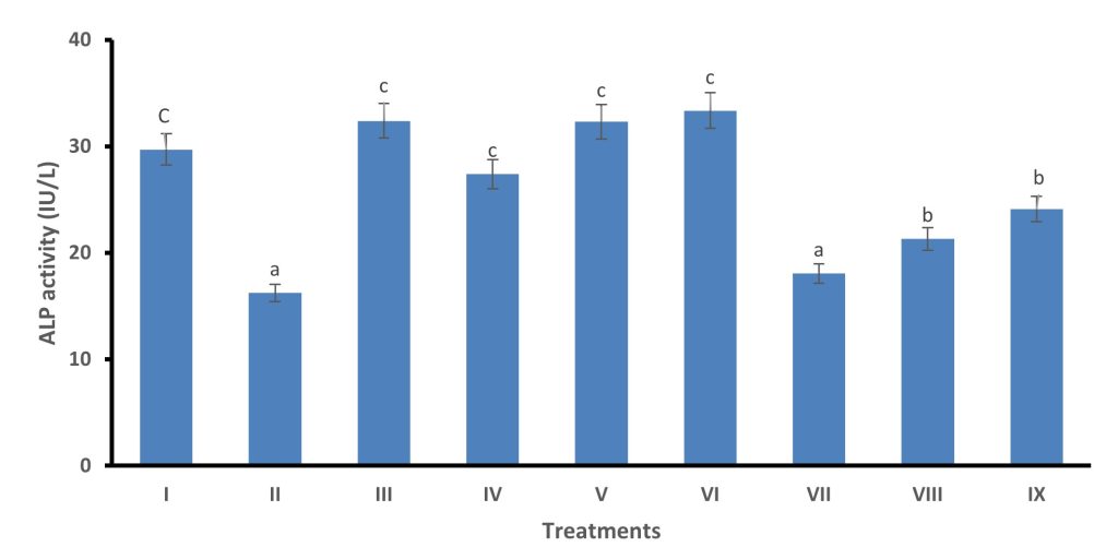
Fig.1. Effects of Ipomoea batatas methanol and n-Hexane leaf extracts on intestinal alkaline phosphatase (ALP) activity of castor oil induced diarrhoea in albino rats.
Values are mean of three replicates ± S.D;
Bars with different superscript are significantly different (p < 0.05).
Key:
Group I: Distilled water Group II: Castor oil + distilled
Group III: Castor oil + Loperamide Group IV: 125mg/kg (body weight) MLE
Group V: 250mg/kg (body weight) MLE Group VI: 500mg/kg (body weight) MLE
Group VII: 125mg/kg (body weight) n-HLE Group VIII: 250mg/kg (body weight) n-HLE
Group IX500mg/kg (body weight) n-HLE
Effects of the Methanol and n-Hexane Extracts of Ipomoea Batatas Leaf on Castor Oil-Induced Entero-Pooling in Albino Rats.
Loperamide at the oral dose of 2.5 mg/kg bw in methanol and n-Hexane leaf extracts at the highest oral dose of 250 and 500 mg/kg bw, showed significant percentage reductions in both the average volume of small intestine content (AVSIC) and an average weight of small intestine content AWSIC compared to control.
The percentage inhibitions of AVCSI for methanol and n-Hexane were 46.52 ± 1.85 and 57.18 ± 2.35%, 31.80 ± 1.98 and 33.33 ± 1.62 for Loperamide respectively, 60.16 ± 2.12% significant difference (p < 0.05. Soluble fractions also caused significant inhibitions of both parameters with percentage inhibitions from 46.52 to 57.18% for methanol leaf extract and from 31.80 to 33.33 for n-Hexane See Table 3, AWSIC; average weight of small intestine content, AVSIC: average volume of small intestine content. Methanol leaf extracts as the most active extract (35.5, 46.52 and 57.18% respectively) for AVSIC respectively), followed by n-Hexane extract (21.14, 31.80 and 33.33% respectively), for AVSIC respectively having a capacity of reducing these parameters at interesting levels. In all evaluated biological activities, methanol leaf extract exhibited high activities compared to n-Hexane extract. This organic solvent can be considered the best which can be used in further for the isolation of active constituents in high amounts. This finding clearly demonstrated the important role played by tannins in the manifestation of all evaluated activities and tannins can be considered as one of the responsible active principles for the antidiarrhoeal activity of the studied plant part. This observation was in good agreement with Labu et al., (2015); Barbara de Servi, (2017), and Cimanga et al., (2019). All evaluated biological activities in the present study generally indicated that the activities showed by all samples from I. batatas leaves were low compared to Loperamide. Again, tannins and phenolics present in the plant extracts were reported to inhibit the release of autacoids and prostaglandins, thereby inhibiting motility and secretion induced by castor oil-induced diarrhoea and exerting thus their antidiarrhoeal activity.
This activity is also promoted by antispasmodic activity recognized to tannins and phenols (Cimanga et al., 2019). The anti-diarrheal activity of flavonoids was attributed to their ability to inhibit hydroelectrolytic secretions and intestinal motility (Holowacz et al., 2016) while saponins inhibit the release of histamines, known to induce diarrhoea, whereas tannins form protein tannates that coat the surface of the intestinal mucosa, thereby reducing secretion (Hamalainen et al., 2011). The percentage of faecal output of I. batatas samples was also reduced at all different tested oral doses producing a better effect compared to the negative group (Tadesse et al., 2017). The antidiarrheal activity showed by samples from I. batatas leaves can be due to the presence of some phytochemical groups like alkaloids, flavonoids, steroids, terpenes, tannins, and saponins as evidenced by phytochemical screening since they were previously reported to exhibit this activity at different extents (Mandal et al., 2010; Kabir at al., 2015; Holowac et al., 2016; Tadesse et al., 2017; Derebe et al., 2018). The anti-enteropooling activity exhibited by the methanol leaf extract at 500 mg/kg bw in the present study compared well with the previous finding of Yakubu and Salimon (2015) in terms of volume and masses of the fluid in the small intestine, but the reduction of intestinal fluid accumulation (57.18 %) was not as high (66.45%) as that reported by Yakubu and Salimon (2020). The factors responsible for this large difference in the computed inhibition of intestinal fluid are not immediately known, but may be connected with the extraction process adopted in the present study.
Table 3. Effects of the Methanol and n-Hexane Extracts of Ipomoea batatas Leaf on Castor oil-induced entero-pooling in albino rats
| Treatments | Extract/Drug | Doses (mg/kg body weight) | Mass of Intestinal Fluids (g) | Volume of Intestinal Fluids (mL) | Reduction of intestinal Content (%) |
| Group I | Normal Control (Distilled water) | 0 | 1.03 ±0.02 | 0.23 ± 0.05 | 100 |
| Group II | Negative Control (Castor oil + distilled water) | 0.00 | 3.69 ±0.03f | 3.23 ± 0.05e | 0.00 |
| Group III | Positive Control (Atropine sulphate) | 2.5 | 1.47 ±0.03 a | 1.27 ± 0.05a | 60.16 ± 2.12 d |
| Group IV | Methanol Leaf Extract | 125 | 2.38 ±0.05d | 2.03 ± 0.05b | 35.5 ± 1.36 b |
| Group V | 250 | 1.97 ±0.03c | 1.77 ± 0.05b | 46.52 ± 1.85 c | |
| Group VI | 500 | 1.58± 0.05 a * | 1.53 ± 0.05a, * | 57.18 ± 2.35 d, * | |
| Group VII | n-Hexane Leaf Extract | 125 | 2.91 ±0.04e | 2.68 ± 0.04d | 21.14 ± 2.20 a |
| Group VIII | 250 | 2.52 ±0.03d | 2.33 ± 0.05c | 31.80 ± 1.98 b | |
| Group IX | 500 | 2.46 ±0.04 d, e | 2.27 ±0.09c | 33.33 ± 1.62 b |
1n=3 Values are mean ± S.D; Mean n=3
p < 0.05 different superscripts down the column are significantly different.
3* Superscripts show no significant different as compared with Positive control group.
The leaf of Ipomoea batatas indicates the presence of zinc (2.25mg/100g), sodium (66.75mg/100g), potassium (120.36mg/100g) and chloride (0.79mg/100g). However, all the elements are within the permissible limit of world health organization (WHO) standard. Research in children suggest that zinc supplementation (20 mg per day for 10 days in children older than two months) may play a crucial role in treating and preventing acute diarrhea, particularly in developing countries. Studies demonstrate a decrease in the risk of dehydration, duration and severity of the diarrheal episode by estimated 20% to 40% Wendy and Andrew, 2014).
Epidemiological studies and studies in animals subject to diarrhea indicated that, diets high in potassium can reduce the risk of diarrhea and hypertension. The present study reveals the potassium content of Ipomoea batatas leaves (120.36 mg/100g). Thus, the Ipomoea batatas leaves could serve as a good source of potassium for the diarrheal patient especially toddlers that are prone to diarrheal episode.This is in agreement with the research conducted by Chuku and Ugorji (2012).
Table 5 Elemental constituents of methanol leaf extract of Ipomoea balatas
| Elements | Concentration (mg/100g) | WHO Standard mg/100g |
| Chloride | 0.79 ± 0.01 | 0.72-250 |
| Potassium | 120.36 ± 0.31 | 10-100 |
| Sodium | 66.75 ± 0.21 | 400-500 |
| Zinc | 2.25 ± 0.10 | 150-200 |
CONCLUSION AND RECOMMENDATION
The present results of this study have shown that Ipomoea batatas methanolic leaf extract possess antidiarrhoeal activity than n-Haxane extracts. The most remarkable antidiarrhoeal activity was observed at the 500mg/kg body weight of methanolic extracts. The plant exhibited anti-secretory and anti-motility effects as evidenced from the stimulation of the Na+/K+-ATPase and alkaline phosphatase. This may be due to the presence of secondary plant metabolites contained in the extracts. Therefore, based on these findings Ipomoea batatas leaf extract is a good antidiarrhoeal agent and physiologically friendly.
CONFLICT OF INTEREST.
The authors declare that they have no conflicts of interest.
ACKNOWLEDGMENTS
This research work was sponsored by Tertiary Education Trust Fund (TETFund) as part of the Institution-Based Research (IBR) Intervention.
REFERENCES
- Abuzerr, S., Nasseri, S and Yunesian, M. (2019). Prevalence of diarrheal illness and healthcare-seeking behavior by age-group and sex among the population of Gaza strip: a community-based cross-sectional study. BMC Public Health . 2019;19(1):704–710. doi: 10.1186/s12889-019-7070-0
- Akanji, M. A. & Yakubu, M. T. (2000). α-tocopherol protects against metabisulphite-induced tissue damage in rats. Nigerian Journal of Biochemistry & Molecular Biology, 15(2), 179–183.
- AOAC. (1998). Official Method of Analysis. 15th Edition, Association of Official Analytical Chemists, Washington DC.
- Awe, I. S. & Sodipo, O. A. (2001). Purification of saponins of root of Bhlighia sapida KOENIG-HOLL. Nigerian Journal of Biochemistry & Molecular Biology (Proceedings Supplement), 16, 201s–204s.
- Ayala-Zavala, J., Rosas-Domínguez, C., Vega-Vega, V. & González-Aguilar, G. (2010). Antioxidant enrichment and antimicrobial protection of fresh-cut fruits using their own byproducts: looking for Integral Exploitation. Journal of Food Science, 75(8), R175–181.
- Bajad, S. K. (2001). Antidiarrhoeal activity of piperine in mice. Planta Medica, 67(3), 284–287.
- Barbara de Servi, B. & Francesco Ranzini, F. (2017). Protective efficacy of antidiarrheal agents in a permeability model of Escherichia coli-infected CacoGoblet® cells. Future Microbiology, 12(16), 1149-1155.
- Bassey, O. A., Lowry, O. H. & Brock, M. J. (1946). A Method for the Rapid Determination of Alkaline Phosphates with Five Cubic Millimetres of Serum. Journal of Biological Chemistry, 164, 321-325.
- Charyeva, Z. (2015). Reducing the burden of diarrhea among children under five years old: Lessons learned from oral rehydration therapy corner program implementation in Northern Nigeria. Journal of Health, Population, and Nutrition, 34(4). https://doi.org/10.1186/s41043-015-0005-1
- Chitme, H. R., Chandra, R. & Kaushik, S. (2004). Studies on anti-diarrheal activity of Calotropis gigantea R. Br. in experimental animals. Journal of Pharmacy and Pharmaceutical Sciences, 7(1), 70–75.
- Chuku, E. C. & Ugorji, J. H. (2012). Determination of levels of some nutrients and antinutrients in five selected vegetables in Niger delta. Scientia Africana, 11(1), 130-142.
- Cimanga, K. R., Kimbuira, M. T., Tona, L. G., Kambu, K. O., Vlietinck, A. J. & Pieters, L. (2019). Assessment of antispasmodic effects of polyphenol and complex catechic tannin extracts from some medicinal plants used to treat diarrhea in traditional medicine in Kinshasa-Democratic Republic of Congo. World Journal of Pharmacy and Pharmaceutical Sciences, 8(1), 170-184.
- Derebe, D., Abdulwuhab, M., Wubetu, M. & Mohammed, F. (2018). Investigation of the antidiarrheal and antimicrobial activities of 80% methanolic leaf extract of Discopodium Penninervum (Hochst.). Evidence-based complementary and alternative medicine: eCAM, 2018, 1360486. https://doi.org/10.1155/2018/1360486.
- Duke, J. A. & Wain, K. K. (1981). Medicinal Plants of the World of the Computer Index with more than 85000 entries 3. Retrieved from http://www.hort_purdue.Edu/newcrop/duke_energy/ipomea_batatas.htm.
- Gornall, A. C., Bardawill, C. J. & David, M. M. (1949). Determination of serum proteins by means of the biuret reaction. Journal of Biological Chemistry, 177(2), 751–756.
- Gundala, S. R., Yang, C., Lakshminarayana, N., Asif, G., Gupta, M. V., Shamsi, S. & Aneja, R. (2018). Polar biophenolics in sweet potato greens extract synergize to inhibit prostate cancer cell proliferation and in vivo tumor growth. Carcinogenesis, 34, 2039–2049.
- Hamalainen, M. (2011). Effects of flavonoids on prostaglandin E-2 production and on COX-2 and mPGES-1 expressions in activated macrophages. Planta Medica, 77(13), 1504–1511. https://doi.org/10.1055/s-0030-1270762.
- Handa, S. S., Khanuja, S. P. S., Longo, G. & Rakesh, D. D. (2008). Extraction Technologies for Medicinal and Aromatic Plants (1st ed.). Italy: United Nations Industrial Development Organization and the International Centre for Science and High Technology.
- Hanieh, H., Gerile, C., Narabara, K., Gu, Z., Abe, A. & Kondo, Y. (2010). In vivo immunomodulatory effects of dietary purple sweet potato after immunization in chicken. Animal Science Journal, 81, 116-121.
- Holowacz, S., Blondeau, C., Guinobert, I., Guilbot, A., Lucas, S. & Bisson, J.-F. (2016). Antidiarrheal and antinociceptive effects of a probiotic mixture in rats. Journal of Probiotics & Health, 4. 10.4172/2329-8901.1000155.
- Hossain, M. D. B. (2019). Study on the Physicochemical Composition and Antioxidant Properties of Selected Colored Sweet Potato Variety (Ipomoea batatas L) in Bangladesh. J Exp Food Chem, 5(1), 1-5. ID 136048, https://doi.org/10.1155/2018/1360486.
- Islam, S., Yoshimoto, M., Ishiguro, K. & Yamakawa, O. (2015). Bioactive compounds in Ipomoea batatas leaves. ISHS Actactic Horticulture, 2, 693–699.
- Jan, B., Agnieszka, M., Dagmara, W., Janina, Z., Bartosz, B. & Marcin, M. (2017). The role of intestinal alkaline phosphatase in inflammatory disorders of gastrointestinal tract. Mediators of Inflammation, 9074601. https://doi.org/10.1155/2017/9074601.
- Kabir, A., Abubakar, M., Ugwah-Oguejiofor, C. J., Abubakar, A. & Muhammad, A. A. (2015). Antidiarrheal activity of the saponin and flavonoid fractions of Anarcadium occidentale leaves in albino rats. Advances in Medical Plant Research, 3(1), 23-28.
- Kang, H., Kwak, Y. & Koppula, S. (2014). Protective effect of purple sweet potato (Ipomoea batatas Linn, Convolvulaceae) on neuroinflammatory responses in lipopolysaccharide-stimulated microglial cells. Tropical Journal of Pharmarcy Research, 13(8), 1257-1263.
- Labu, Z., Laboni, F., Tarafdar, F., Howlader, M. S. I. & Rashid, M. (2015). Membrane stabilization as a mechanism of anti-inflammatory and thrombolytic activities of ethanolic extract of aerial parts of Spondias pinanata (Family: Anacardiaceae). Pharmacologyonline, 2, 44-51.
- Lalles, J. P. (2014). Intestinal alkaline phosphatase: Novel functions and protective effects. Nutrition Reviews, 72(2), 82–94. https://doi.org/10.1111/nure.12082.
- Ling-Yuz Lv, Gao-Feng S, Chun-Lei L, Xue-Zhe H. & Qiu-Nan LV. (2009). Study on the chemical constituents of the leaves of Ipomoea batatas. Zhong Yao Cai, 32(6), 896-897.
- Mandal, S., Nayak, A., Kar, M. & Banerjee, S. K. (2010). Antidiarrheal activity of carbazole alkaloids from Murraya koenigii Spreng (Rutaceae) seed. Fitoterapia, 81(1), 72-74.
- Mekonnen, B., Asrie, A. B. & Wubne, Z. B. (2018). Antidiarrheal activity of 80% methanolic leaf extract of Justicia chimperiana. Evid-Based Complement Alternat Med., Article ID 3037120. https://doi.org/10.1155/2018/3037120.
- Morales, F., Padilla, S. & Falconí, F. (2017). Medicinal Plants used in traditional herbal medicine. African Journal of Traditional Complementary and Alternative Medicine, 14(1), 10-15.
- Odebiyi, A. & Sofowora, A. E. (1978). Phytochemical screening of Nigerian medicinal plants. Part III. Lloydia, 41, 234–246.
- Oladiji, A. T., Idoko, A. S., Abodunrin, T. P. & Yakubu, M. T. (2010). Studies on the physicochemical properties and fatty acid composition of the oil from ripe plantain peel (Mysa paradisiaca). Journal of African Scientist, 11(1), 73-78.
- Olowu, A. O., Adeneye, A. A. & Adeyemi, O. O. (2015). Hypoglycaemic effect of Ipomoea batatas aqueous leaf and stem extract in normal and streptozotocin-induced hyperglycaemic rats. Journal of Natural Pharmarcy, 2, 56–61.
- Omole, V. N., Wamyil-Mshelia, T. M., Nmadu, G. A., Usman, N. O., Andeyantso, E. A. & Adiri, F. (2019). Knowledge, attitude and practice of home management of diarrhea among mothers of under-fives in Samaru, Kaduna State, Nigeria. Port Harcourt Medical Journal, 13(1), 19–25.
- Osime, E. O., Ediale, G. E., Omoti, C. E & Famodu, A. A. (2008). Effect of sweet potato leaf (Ipomoea batatas) extract on some haematological parameters using rabbits. Journal of Medical and Biomedical Research, 7, 12-15.
- Panda, V & Sonkamble, M. (2012). Anti-ulcer activity of Ipomoea batatas tubers (sweet potato). Journal of Functional Food Health and Disease, 2, 48-61.
- Panda, V. & Sonkamble, M. (2012). Phytocehical and pharmacological activities of Ipomea batatas I. (Lam)-A review. Int J Phytochem Pharmacol, 2(10), 25-34.
- Peter, A. K & Umar, U. (2018). Combating diarrhoea in Nigeria: The way forward. Journal of Microbiology & Experimentation, 6(4), 191–197.
- Pritish, S., Sandeep, A., Basanta, L & Bhupal, G. S. (2015). Phytochemical Screening of the Medicinal Plants of Nepal. IOSR Journal of Environmental Science, Toxicology and Food Technology, 1(6), 11-17.
- Rahman, K., Chowdhury, A. U., Islam, M. T., Chowdhury, A., Uddin, M. E. & Sumi, C. D. (2015). Evaluation of antidiarrheal activity of methanol extract of Maranta arundinacea Linn. leaves. Advanced Pharmacology and Science, 25, 6.
- Rajat, V. S., Sumit, N. & Pallavi, A. B. (2013). Anti-diarrhoeal activity of aqueous extract of Ocimum kilimandscharicum. Ethnopharmacol., 148(1), 223-228.
- Rao, Y. K., Fang, S. H. & Tzeng, Y. M. (2005). Inhibitory effects of the flavonoids isolated from Waltheria indica on the production of NO, TNF-alpha and IL-12 in activated macrophages. Biological & Pharmaceutical Bulletin, 28(5), 912–915. https://doi.org/10.1248/bpb.28.912.
- Salimon, S. & Yakubu, M. (2020). Antidiarrhoeal activity of fractions of aqueous extract of Mangifera indica L. leaves in castor oil-induced diarrhoeal female Wistar rats. Journal of Medicinal Plants for Economic Development, 4(1), 7 pages. doi:https://doi.org/10.4102/jomped.v4i1.88.
- Shiferie, F. & Shibeshi, W. (2013). In vivo antidiarrheal and exvivo spasmolytic activities of the aqueous extract of the roots of Echinops kebericho (Asteraceae) in rodents and isolated guinea-pig ileum. Int J Pharm Pharmacol., 2(1), 110-116.
- Shoba, F. G. & Thomas, M. (2001). Study of antidiarrhoeal activity of four medicinal plants in castor-oil induced diarrhoea. Journal of ethnopharmacology, 76(1), 73–76. https://doi.org/10.1016/s0378-8741(00)00379-2
- Sisay, M., Engidawork, E. & Shibeshi, W. (2017). Evaluation of the antidiarrheal activity of the leaf extracts of Myrtus communis Linn (Myrtaceae) in mice model. BMC Complement Altern Med., 17(1), 103, Article number: 103. doi: 10.1186/s12906-017-1625-3.
- Tadesse, E., Ephrem Engidawork, E., Nedi, T. & Getnet Mengistu, G. (2017). Evaluation of the anti-diarrheal activity of the aqueous stem extract of Lantana camara Linn (Verbenaceae) in mice. BMC Complementary and Alternative Medicine, 17(190). https://doi.org/10.1186/s12906-017-1696-1.
- Turyare, M. D., Mativo, J. N., Kerich, M. and Ndiritu, A. K. (2021) Prevalence and socio-demographic determinants of diarrhea among children below 5 years in Bondhere district Somalia. The Pan African Medical Journal . 38 doi: 10.11604/pamj.2021.38.391.21636.391
- UNICEF/WHO. (2019). Why children are still dying and what can be done. Final Report—Diarrhoea. United Nations Children’s Fund/World Health Organization.
- Vishnu, V. R., Renjith, R. S., Mukherjee, A., Anil, S. R., Sreekumar, J. & Jyothi, A. N. (2019). Comparative study on the chemical structure and in vitro antiproliferative activity of anthocyanins in purple root tubers and leaves of sweet potato (Ipomoea batatas). Journal of Agriculture and Food Chemistry, 67, 2467–2475.
- Wall, M. E., Krider, M. M., Krewson, C. F., Eddy, C. R., Wilaman, J. J., Correll, S. & Gentry, H. S. (1954). Steroidal Sapogenins XII. Supplementary table of data for steroidal sapogenins vii. Agricultural Research Service Circle. Aic., 363, 17-17.
- Wansi, S. L., Elvine, P., Nguelefack-Mbuyo, E. P. & Nchouwet, M. L. (2014). Antidiarrheal activity of aqueous extract of the leaves of Sapium ellipticum (Euphorbiaceae). Trop J Pharm Res., 13(6), 929-935.
- Wendy, B. & Andrew, S. (2014). Acute Diarrhoea in Adults. American Family Physician, 89(3), 180-189.
- Wo, D. P., Zhuang, Z. G., Xu, S., Xu, Y., Lu, Y. & Mao, H. M. (2013). A novel spectrophotometric method for indirect determination of nitric oxide (NO) in serum. Clinica Chimica Acta, 424, 187–190.
- World Health Organization. (2017). Essential medicines and health products information portal. Retrieved October 29, 2017, from http://apps.who.int/medicinedocs/en/d/Js4950e/2.4.html
- Wright, P. J., Leathwood, P. D. & Plummer, D. T. (1972). Enzymes in rat urine: Alkaline phosphatase. Enzymologia, 42, 317–327.
- Yakubu, M. T. & Salimon, S. S. (2015). Antidiarrhoeal activity of aqueous extract of Mangifera indica L. leaves in female albino rats. Journal of ethnopharmacology, 163, 135–141. https://doi.org/10.1016/j.jep.2014.12.060.
- Yin, Y. Q., Shen, Z. B. & Kong, L. Y. (2010). Studies on chemical constituents from Ipomoea batatas. Zhong Yao Cai, 31(10), 1501-1503.