Phytochemical Screening, Antioxidant, Anti-Inflammatory and Antibacterial Studies on the Stem Bark Extracts of Ximenia Americana Linn.
- Umar Meleh Umar, Ph.D.
- Barkoma Mohammed Bashir
- Ibrahim Tijjani Babalola
- 139-156
- Dec 18, 2023
- Parasitology
Phytochemical Screening, Antioxidant, Anti-Inflammatory and Antibacterial Studies on the Stem Bark Extracts of Ximenia Americana Linn.
Umar Meleh Umar, Ph.D.1 *, Barkoma Mohammed Bashir, M.Sc. Hons2. Ibrahim Tijjani Babalola, Ph.D.3
1Department of Chemistry, Yobe State University, Damaturu, Nigeria.
2Department of Integrated Science, School of Sciences, College of Education and Legal Studies, Nguru, Nigeria
3Department of Chemistry, Yobe State University, Damaturu, Nigeria.
*Corresponding Author
DOI: https://doi.org/10.51584/IJRIAS.2023.81113
Received: 18 October 2023; Revised: 07 November 2023; Accepted: 15 November 2023; Published: 18 December 2023
ABSTRACT
Medicinal plants have been used in traditional medicine practices since pre-historical times. Any part of the plant could be used as medicine. A survey of traditional medicine men and women in Damaturu, Nigeria indicated that Ximenia americana is a plant used in the treatment of inflammation, wounds and cancer. The fresh stem barks of the plant were collected and successively extracted with n-hexane, ethyl acetate, methanol and water. Phytochemical constituents of the extracts revealed the presence of cardiac glycosides, flavonoids, saponins, phenols, tannins, alkaloids, sterols and terpenoids. The antioxidant activity was evaluated by 2, 2-diphenyl-1-picrylhydrazyl (DPPH) free radical assay, whereas membrane stabilization method was used to evaluate the anti-inflammatory activities of the extracts. The antibacterial activity was studied against four (4) wound pathogens (Salmonella typhi, Staphylococcus aureus, Escherichia coli and Streptococcus pneumoniae), using disc diffusion technique. The results of the IC50 for antioxidant studies indicated that the methanol (22.4µg/ml) and ethyl acetate (28.4 µg/ml) extracts of X. americana exhibited higher antioxidant activities than the standard ascorbic acid (38,9µg/ml). The extracts were found effective in inhibiting the heat induced hemolysis in a concentration dependent manner. The n-hexane extract showed the highest protection level (84.41 – 87.98%, p<0.05), followed by water (46.73 – 82.38%, p<0.05) and ethyl acetate (71.3o – 77,39%, p<0.05) extracts. Diclofenac Sodium salt used as standard drug at 100μg/ml offered 90.66% protection a significant (p<0.05) protection against damaging effect of heat. The hypotonic solution induced study indicated that the extracts exhibited more protection from hemolysis (n-hexane = 75.22 – 85,23%, p<0.05; ethyl acetate = 4.09 – 71,40%, p<0,05; water = 59.98 – 67.08%, p<0.05, and methanol = 48.15 – 58.75%, p<0.05) than the standard Diclofenac sodium salt (57.52%, p<0.05). The results showed that all extracts from the plant offered a significant (p<0.05) protection against the damaging effect of hypotonic solution, Diclofenac sodium (100μg/ml), the standard drug offered a significant (p<0.05) protection (57.52%). X. americana samples showed that all extracts inhibited the growth of S. aureus, S. pneumoniae and E. coli. The antioxidant, anti-inflammatory and anti-bacterial activities carried out in this study can be used as scientific explanation for the traditional claim about the wound healing property of the stem bark of X. americana.
Keywords: Phytochemical, Antioxidant, Anti-inflammatory, Antibacterial
INTRODUCTION
Medicinal plants are used for making home remedies and traditional medicines or what is well known as herbal medicine. Any part of the plant could be used as medicine, the leaves, roots, seeds, stems, etc. Medicinal plants have been used in traditional medicine practices since pre-historical times. Early written reports on the use of plants as medicine appeared about 2600 BC when plants were used as medicine by Sumerians and Akkadians (Shoeb, 2006). Since then, plants have been used to treat diseases such as headache, toothaches, stomach aches, diarrhea, wounds, tumors and sexually transmitted diseases (van Wyk and Gericke, 2000; Khaleeliah, 2001; Von Koenem, 2001, Wuyang, 2008).
A plant is considered medicinal if it produces compounds which are therapeutically effective. Plants produce a wide range of secondary metabolites, and the medicinal properties are attributed to the presence of these metabolites such as terpenoids, steroids, saponins, tannins, flavonoids, alkaloids and phenolic compounds (Mdlolo; 2009, Fawole 2009).
Ximenia americana of the family Olacaceae, is commonly known as Tallow wood in English and ‘Tsaada’ in Hausa. A survey of medicinal plants within and around Damaturu town, Nigeria indicated that the plant is used in the treatment of hepatitis, inflammation, malaria, wounds and cancer. However, there’s no scientifically backed evidence to support this claim. This poses an important challenge to seek for more scientific studies to be carried out on each part of the plant in order to provide explanation for these claims.
Herbal medicine in Nigeria is gaining more recognition and this is seen in how much inquiries people make concerning home remedies and traditional medicine. Nigeria is richly endowed with indigenous plants which are used in herbal medicine to cure diseases and heal other ailments. Some of these plants are used as food and/or medicine. The extracts from plant have been shown to exhibit a wide range of biological and pharmacological activities such as anticancer, anti-inflammatory, diuretic, laxative, antispasmodic, antihypertensive, antidiabetic, antimicrobial activities, etc. It is generally assumed that the active medicinal constituents contributing to the protective effects are phytochemicals, vitamins and minerals. (Okwu, Ekeke 2003 and Okwu; 2004). For this reason, medicinal plants are considered important to the health of the individuals and communities.
MATERIALS AND METHODS
Apparatus and Materials
Ultrasonicator (Model/AS3120) was purchased from Tianjin Automatic Science Instrument Co., Ltd. China, analytical weighing balance (Ohaus Corp. Pine Brook, NJ USA), pestle and mortar (wooden) purchased in (Damaturu Sunday market Potiskum Road, Nigeria), empty bottles purchased at Bayan Tasha market Damaturu, Nigeria. Sieve, fume cupboard, drying cabinet (model/FSM140)from 2 Building, Majialong Industrial Zone, Nanshen District, Shenzhen Jinly Technology Co., Ltd. China, UV/VIS spectrophotometer (model/UV752) from Changsha, Hunan, Wincon Company Ltd. China, Autoclave (Model/DWB-280B) and Water bath (Model/DWT-420) from Shanghai Drawell Scientific Instrument Co., Ltd. Room 211 Building 7, sheng Yu Industrial Park No. 365 Chun Hong, Shanghai, China and other laboratory materials.
Chemicals
n-hexane, ethyl acetate and methanol were from BDH Chemicals Ltd., Poole, United Kingdom. 2,2-diphenyl-1-picryl hydrazyl (DPPH) was purchased from SIGMA-ALDRICH Company Ltd., 3050 Spruce Street St. Louis, MO63103 USA. Muller Hinton Agar was purchased from TITAN BIOTECH Ltd., A-904A, RIICO Industrial Area, Phase-III, Rajasthan, India. Dimethyl Sulphur Oxide (DMSO) was purchased from Guangdong Guanghua Sci-Tech Company Ltd., Add 6, Jiangyan South Road, Guangzhou, Guangdong, China. ethanol, ascorbic acid, Hydrochloric acid (HCl), Sulphuric acid (H2SO4), Magnesium metal, Ferric chloride, Dragendorff’s reagent, choloroform and all other Chemicals used are of highest analytical grade and purchased from BDH Chemicals, Poole, England.
Collection and Preparation of Plant Sample
The fresh sample of X. americana (Voucher number 1973) (stem bark) was collected at the campus of Yobe State University Damaturu, Nigeria. The herbarium specimen was identified by Mallam Salihu Abdullahi a Taxonomist at the Department of Biological Sciences, Yobe State University, Damaturu. The stem bark was collected two (2) meters above the ground. The sample was sorted to ensure no foreign bodies were present, it was then dried under shade in the laboratory at ambient temperature. The dried sample was then crushed into coarse particles using local pestle and mortar. It was further pulverized into fine powder and sieved with a sieve and weighed. The finely powdered sample was then weighed and stored in sealed containers until required for further analysis.
Extraction of Phytochemicals
About 550g of the powdered plant material of (X. americana) was separately extracted successively with 2.5 L portions of n-hexane, ethyl acetate, methanol and water in that order using ultrasonicator for two hours at room temperature. The solvent containing the extracts was allowed to settle after the extraction, then the mixture was separated from the residue by filtering with Whatmann No. 1 filter paper and then kept in a clearly labelled container ready for solvent recovery. The residue of the sample was then mixed with the next solvent for further extraction. The procedure was repeated for the remaining solvents namely; ethyl acetate, methanol and water in that order.
Phytochemical Screening
Phytochemical screening of the extracts was performed to detect the presence of phytochemicals using the procedures outlined by Tiwari et al. (2011); Sabri et al. (2012) and Solomons et al. (2013).
Test for alkaloids
The extract (0.5g) was dissolved in 5ml of 2N HCl and filtered. The filtrate was treated with Dragendorff’s reagent (Solution of potassium Iodide and bismuth Iodide). Formation of red precipitate indicated the presence of alkaloids.
Test for flavonoids
The extract (0.5g dissolved in 2ml of methanol) was treated with few drops of sodium hydroxide solution. Formation of intense yellow colour, which becomes colourless on addition of dilute acid, indicated the presence of flavonoids.
Test for saponins
Frothing test: The extract (0.5g) was added to distilled water to a final volume of 20ml and this was shaken vigorously in a graduated cylinder for 15 minutes over a vortex mixer. Formation of 1cm layer of foam indicated the presence of saponins.
Test for cardiac glycosides (Keller-Kelliani test)
To 5ml of the extract (0.5g dissolved in 5ml methanol) was treated with 2ml of glacial acetic acid in a test tube followed by a drop of 2% ferric chloride solution. This was carefully underlaid with 1ml of concentrated sulphuric acid. A brown ring at the interface indicates the presence of deoxysugar characteristic of cardenolides. A violet ring may appear below the ring while in the acetic acid layer, a greenish ring may form which indicated the presence of cardiac glycosides.
Test for oxalate
To 3ml portion of the extract (0.2g in 3ml of methanol) a few drops of glacial acetic acid was added. A greenish black colouration indicated the presence of oxalates.
Test for quinones
A small portion of the extract (0.2g in 2ml of methanol) was treated with concentrated hydrochloric acid. The formation of yellow precipitate/colouration indicated the presence of quinones.
Test for terpenoids (Salkowski’s test)
One ml of chloroform was added to 2ml of the extract (0.2g in 2ml of methanol) followed by a few drops of concentrated sulphuric acid. A reddish-brown precipitate produced immediately indicated the presence of terpenoids.
Test for tannins (Braymer’s test)
Two ml of the extract (0.2g in 2ml of methanol) was treated with 10% alcoholic ferric chloride solution. Formation of blue/greenish colouration indicated the presence of tannins.
Test for sterols (Libermann-Burchard test)
About 1ml of the extract (0.1g in 1ml of methanol) was treated with few drops each of chloroform, acetic anhydride and concentrated sulphuric acid. The formation of dark pink or red colour indicated the presence of sterols.
Test for phenols
A fraction of the extract was treated with aqueous 5% ferric chloride solution. The formation of deep blue or black colour indicated the presence of phenols.
Measurement of Antioxidant Activities
The antioxidant activities of X. americana extracts were determined on the basis of their scavenging activity of stable 2, 2-diphenyl-1-picrylhydrazyl (DPPH) free radical as follows:
To 1 ml of each solution of different concentrations (10, 25, 50. 100, 125, 250, 300, 500µg/ml) of the extracts, 3 ml of 0.004% ethanolic DPPH free radical solution was added. After 30 minutes, the absorbance of the preparations was taken at 517nm by UV spectrophotometer. This was then compared with the corresponding absorbance of standard ascorbic acid concentrations (10, 25, 50. 100, 125, 250, 300, 500 µg/ml) as described by Hatano et al. (1988). Then the % inhibition was calculated by the following equation;
% Radical scavenging = (absorbance of blank – absorbance of sample) x 100%
Activity (absorbance of blank)
A blank was prepared by adding 3 ml of 0.004% ethanolic DPPH to 1 ml of the ethanol and treated in a similar manner to test samples.
Anti-Inflammatory Activity
Membrane stabilization method
The Human Red Blood Cell (HRBC) membrane stabilization was adopted as a method to study the in vitro anti-inflammatory activity since the erythrocyte membrane is comparable to the lysosomal membrane (Gandasan et al., 1991, Shenoy et al., 2010).
Preparation of red blood cells (RBC’s) suspension
The blood sample was collected from healthy human volunteers of the Department of Biochemistry of Bayero University Kano, Kano State who had not taken Non-steroidal Anti-inflammatory Drugs (NSAIDs) for the previous two weeks preceding the experiment and the samples were transferred to centrifuge tubes. The tubes containing the blood were centrifuged at 3,000 rpm for 10 min to remove plasma. The packed RBCs were washed three (3) times with an equal volume of the normal saline. The volume of blood was measured and re-constituted as 10% v/v suspension with normal saline.
Heat induced haemolysis
The reaction mixture (2 ml) consisting of 1ml of the test extracts of different concentrations (100, 300 and 500 µg/ml) and 1ml of 10% suspension. A test tube containing only saline was used as the control. Diclofenac sodium salt was used as a standard drug and was weighed and dissolved in water (0.0025g in 5ml of water). All the centrifuge tubes containing reaction mixture were incubated in a water bath for 30 min at 56ºC. At the end of the incubation the tubes were cooled under a running tap water. The reaction mixtures were further centrifuged at 2500 rpm for 5 min and the absorbance of each supernatant was taken at 560nm using UV/VIS spectrophotometer. The experiment was performed in triplicates for all the test samples. The percentage inhibition of haemolysis was calculated as follows Leelaprakash and Mohan (2010):
Percentage inhibition = Absorbance (control) – Absorbance (sample) ×100
Absorbance (control)
Hypotonic solution induced haemolysis
Different concentrations of the plant extracts were prepared (i.e., 100, 300 and 500 µg/ml), reference sample and control were both separately mixed with 1 ml of phosphate buffer pH 7.4, 2 ml of hyposaline and 0.5 ml of HRBC suspension. Diclofenac sodium salt was used as a standard drug. All the reaction mixtures for the assay were incubated at 37ºC for 30min and thereafter centrifuged at 3000rpm for 10/min. The supernatant liquid was decanted and haemoglobin content was determined by spectrophotometer at 560nm. The percentage of Red Blood Cell membrane stabilization of defensive was calculated by the following equations Leelaprakash and Mohan (2010):
% protection = 100 – Optical density of drug treated sample X 100
Optical density of control
Measurement of Antibacterial Activity
Media preparation
About 30g of Mueller Hinton agar was weighed using analytical weighing balance. It was dissolved in 1000ml distilled water in a conical flask, the mouth of the flask was plugged with cotton wool wrapped in aluminum foil paper tightened with masking tape. This was to avoid spillage of the medium in an autoclave while undergoing sterilization. The dissolved medium was sterilized in an autoclave set at 121oC for 15 minutes. It was removed and allowed to cool to 45oC and was gently and aseptically poured into 32 sets of disposable sterilized Petri dishes. The media plates were allowed to solidify (Cheesbrought, 2002).
Test organisms
The test organism used for the antibacterial activity studies were Salmonella Typhi, Escherichia coli, Streptococcus pneumonea and Staphylococcus aureus. These were obtained from the microbiology laboratory at Yobe state University Damaturu. Gram’s staining and biochemical tests (Disc diffuse method) were carried out to confirm the organisms.
Sensitivity discs preparation
Whatman No. 1 filter paper was punched with paper puncher and the discs of 6.0 mm/dm were obtained. The discs were placed in a screw capped bottle and sterilized in hot air oven at 140oC for 1 hour. The discs were allowed to cool until use.
Preparation of stock solution
The stock solution of the plant extract was prepared in screw capped test tubes using dimethyl sulfoxide (DMSO) as a standard solvent for antimicrobial stock solution preparation. 1g of each fraction was weighed and dissolved in 10 ml DMSO to give 100,000 µg/ml of stock solution. From this 0.1ml, 0.2ml, 0.3ml, 0.4ml, and 0.5ml were respectively added to 0.9 ml, 0.8 ml, 0.7 ml, 0.6 ml, and 0.5 ml of DMSO in a sterile test tube, which made 1 ml of each fraction. One (1) disc was placed in each concentration which arrived at disc potency of 100 µg/disc, 200 µg/disc, 300 µg/disc, 400 µg/disc, and 500 µg/disc respectively.
The test organisms were inoculated on the Mueller Hinton agar (MHA) with a sterilized inoculating loop, and the plates were appropriately labelled with the name of the test organism as well as the plant extract for easy identification. The sensitivity disc from concentration of each fraction was placed on the MHA. The plates were incubated for 72 hours and zones of inhibition were detected in mm/dm (Okafor, 2002), the test was carried out using (Gentamycin, Ofloxacin, Ciprofloxacin, Augmentin, Amoxicillin, Ceftriaxone, and Cefuroxime 100 μg/disc) as a control (Rubina, 2011).
RESULTS AND DISCUSSION
Extraction Yield
The powdered stem bark of X. americana was successively extracted with n-hexane, ethyl acetate, methanol and water using Ultrasonicator. The results of the extraction yield are shown in Table 1 below:
Table 1 Percentage yield of crude stem bark extracts of X. americana.
| Solvents used | Weight of plant part used (g) (X. americana) | Weight of extract(g) | Percentage yield (%) | |
| 1 | n-Hexane | 550 | 17.38 | 3.16 |
| 2 | Ethyl Acetate | 550 | 21.94 | 3.99 |
| 3 | Methanol | 550 | 9.08 | 1.65 |
| 4 | Water | 550 | 6.43 | 1.17 |
The results above revealed that in the 550g of X. americana extracted with four solvents of increasing polarities revealed that the total phytochemical yield of 54.83g (or 9.97%). Out of this ethyl acetate extracted the highest 21.94g (3.99%), followed by n-hexane with yield of 17.38g (or 3.16%), while methanol (9.08g or 1.65%) and water (6.43 or 1.17%) extracts showed the lower yields.
Phytochemical Screening
Plant secondary metabolites have become areas of great research interests due to their wide-ranging applications for the improvement human health better human life (Baris et al., 2006). For this reason, medicinal plants are regarded as the richest sources of drugs for traditional medical systems, modern medicines, nutraceuticals, food supplements, pharmaceuticals, intermediates and precursors for the synthesis of drugs (Hammer et al., 1999). The traditional medicinal value of plants is mainly attributed to the bioactive phytochemical constituents of the plants that exhibit various physiological effects, this fact has a long history (Sofowora, 1980; 1993). Therefore, through phytochemical screening one could detect the various important compounds which could be used as the base for the development of modern drugs for curing various diseases.
In the present study the phytochemical constituents of the X. americana extracted with n-Hexane, Ethyl acetate, methanol and Water revealed the presence of cardiac glycosides, flavonoids, saponins, phenols and tannins. Alkaloids were present in n-hexane, ethyl acetate, methanol extracts, terpenoids were present only in ethyl acetate and water extracts while sterols, quinones and oxalates were detected in all the extracts screened as presented in the Table 2 below.
Table 2 The Results of the Phytochemical Screening of X. americana n-Hexane, Ethyl acetate, Methanol and Water extracts
| Phytochemicals | n-Hexane extract | Ethyl acetate Extract | Methanol Extract | Water Extract |
| Alkaloid | + | + | + | – |
| Flavonoid | + | ++ | ++ | + |
| Saponins | + | + | ++ | + |
| Cardiac glycoside | + | + | + | + |
| Oxalate | – | – | – | – |
| Quinones | – | – | – | – |
| Terpenoids | – | + | – | + |
| Tannins | + | + | + | + |
| Sterols | – | – | – | – |
| Phenols | + | + | + | + |
Key: + = Present, – = Absent
These results further suggested the presence of both nonpolar, semi-polar and polar compounds of alkaloids, flavonoids, saponins, tannins, phenols and cardiac glycosides in the stem bark of X. americana. Terpenoids were found in the ethyl acetate and water extracts indicating the presence of semi-polar and polar moieties of this phytochemical in the stem bark.
Secondary metabolites such as alkaloids, flavonoids, phenols, tannins, terpenoids, etc. have been reported to be responsible for the therapeutic benefits of medicinal plants Nandagoapalan et al., (2016) because of their pharmacological activities. Such activities as antimicrobial, anti-inflammatory, antioxidant, anticancer, among others, may be principal indicators of a plant’s value in medicine. A number reports are available in literatures that lend support to this assumption. For instance, several flavonoids, saponins, alkaloids, tannins and terpenes isolated from medicinal plants have been shown to exhibit significant antinociceptive and anti-inflammatory activities (Suleiman et al., 2009). Tanko et al. (2007) attributed the analgesic activity of ethanol extract of Nigeula sativa to the presence of tannins and flavonoids in the extract. According to Mamta et al. (2013) flavonoids have been reported to exert multiple biological effects including anti-inflammatory, antimicrobial as well as antitumor activities. Phenolic compounds found from plants are also reported to be responsible for antioxidant, anti-inflammatory, and other biological properties (Park et al., 2011). Flavonoids isolated from medicinal plants have been reported to possess significant anti-inflammatory activity and, in some cases, analgesic activity (Kim et al., 2000; Amin et al., 2012, Santosh et al., 2008 and Amin et al., 2012). The flavonoids were reported to exert their anti-inflammatory effect by interfering with inflammatory process by inhibiting enzymes involved in prostaglandin synthesis (Duke, 1992; Kamil, 1993; Kim et al., 2000). Hence, the presence of secondary metabolites in the extracts of Ximenia americana may be assumed to exhibit similar physiological activities, especially the presence of flavonoids and phenols which are known to exhibit analgesic and anti-inflammatory activities. Based on this information the present research work studied the antioxidant, antibacterial and anti-inflammatory activities of all the extracts of Ximenia americana, an indigenous plant of Yobe State, Nigeria. The findings are reported in subheadings below.
Antioxidant Studies
The antioxidants are mainly derived from food and medicinal plants such as fruits, vegetables, cereals, mushrooms, beverages, flowers, spices and traditional medicinal herbs (Cai et al., 2004). Natural antioxidants from plant materials are mainly Polyphenols (comprising mainly of Phenolic acids, flavonoids, tannins, anthocyanins, lignans and stilbenes), carotenoids (xanthophylls and carotenes) and vitamins (vitamins C and E), Baiano et al., (2015). These natural antioxidants, especially the Polyphenols and Carotenoids are reported to exhibit a wide range of biological effects, such as anticancer, antibacterial, anti-inflammatory, antiviral and anti-aging as reported by Fang, et al., (2014).
The antioxidant studies in the present work were carried out on the n-hexane, ethyl acetate, methanol and water extracts of Ximenia americana stem bark using the free radical scavenging activities of the samples on 2,2-diphenyl-1-picryldihydrazyl (DPPH) radical and ascorbic acid as standard. The activities of different extracts and ascorbic acid standard were measured at 517nm and presented in Table 3. A graph of absorbance against concentration was drawn (Fig. 1) in order to have a glance of the performance of the extracts as compared to standard ascorbic acid. The results indicated that the methanol and the ethyl acetate extracts appeared to show higher free radical scavenging activities than the other extracts and the standard ascorbic acid. To obtain the actual free radical scavenging activities of the extracts and ascorbic acid the data in Table 3 was further converted to percentage (%) inhibition with the aid of Microsoft Excel software and presented in Table 4, from which graph of % inhibition against concentration was drawn for each extract and ascorbic acid (Figs 2-6) from which straight-line equation for the calculation of the IC50 values for each sample. The IC50 value denotes the concentration of the sample required to scavenge 50% of the DPPH free radicals measured at 517 nm as reported by Gupta et al., (2003) with slight modification.
Table 3 The UV Absorbances measured at 517nm of Standard Ascorbic Acid, n-hexane, ethyl acetate, methanol and water extracts of Ximenia americana stem bark.
| Extract absorbance | ||||||
| S/No. | Concentration (µg/ml) | Ascorbic Acid | n-Hexane | Ethyl Acetate | Methanol | Water |
| 1 | 0 | 0.675 | 0.675 | 0.675 | 0.675 | 0.675 |
| 2 | 10 | 0.433 | 0.512 | 0.495 | 0.489 | 0.529 |
| 3 | 25 | 0.354 | 0.308 | 0.298 | 0.295 | 0.334 |
| 4 | 50 | 0.249 | 0.246 | 0.201 | 0.196 | 0.268 |
| 5 | 100 | 0.122 | 0.138 | 0.102 | 0.094 | 0.146 |
| 6 | 125 | 0.093 | 0.113 | 0.092 | 0.082 | 0.129 |
| 7 | 250 | 0.084 | 0.095 | 0.081 | 0.073 | 0.105 |
| 8 | 300 | 0.075 | 0.079 | 0.068 | 0.064 | 0.088 |
| 9 | 500 | 0.073 | 0.077 | 0.064 | 0.062 | 0.084 |

Figure 1. Graph of Absorbance at 517nm Against Concentration of Standard Ascorbic Acid, n-hexane extract, ethyl acetate extract, methanol extract and water extract of X. americana.
Table 4: Percentage Inhibitions of Ascorbic acid, n-Hexane, Ethyl Acetate, Methanol and Water extracts of X. americana.
| S/No. | Concentration (µg/ml) | Ascorbic acid | n-Hexane | Ethyl Acetate | Methanol | Water |
| 1 | 0 | 0 | 0 | 0 | 0 | 0 |
| 2 | 10 | 35.9 | 11.6 | 26.7 | 27.6 | 21.6 |
| 3 | 25 | 47.5 | 41.3 | 55.9 | 56.3 | 50.5 |
| 4 | 50 | 63.1 | 56.9 | 70.2 | 70.9 | 60.3 |
| 5 | 100 | 81.9 | 71.3 | 84.9 | 86.1 | 78.4 |
| 6 | 125 | 86.2 | 80.4 | 86.4 | 87.9 | 80.9 |
| 7 | 250 | 87.5 | 83.3 | 88.0 | 89.2 | 84.4 |
| 8 | 300 | 88.9 | 85.8 | 89.9 | 90.5 | 86.9 |
| 9 | 500 | 89.2 | 85.8 | 90.5 | 90.8 | 87.6 |
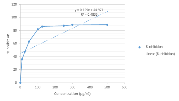
Figure 2: The % Inhibition of Standard Ascorbic Acid for the Calculation IC50 Value
The IC50 value of the standard ascorbic acid was calculated from the above graph using the equation y = 0.129x + 44.971, where y is 50 and x represents the IC50 value. From this, the IC50 value of the standard ascorbic Acid was calculated to be 38.9µg/ml. The IC50 values of all the other test extracts were calculated in a similar manner from their respective linear equations and summarized the IC50 values in Table 5.
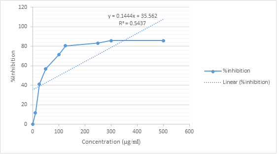
Figure 3: The % Inhibition of n-Hexane Extract of X. americana Stem Bark
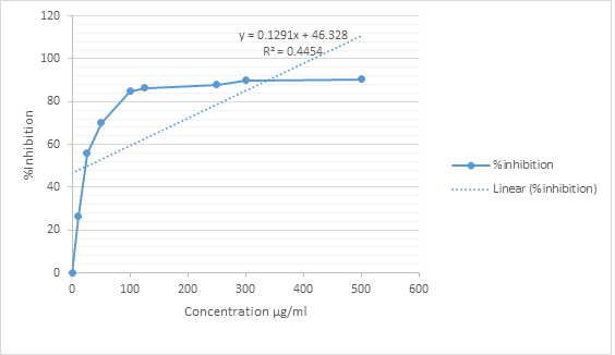
Figure 4: The % Inhibition of Ethyl Acetate extract of X. americana Stem Bark for the Calculation of IC50 value.
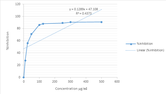
Fig. 5: The % Inhibition of Methanol Extract of X. americana Stem Bark
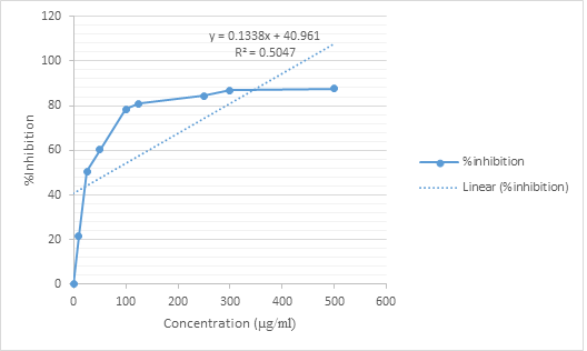
Fig. 6: The % Inhibition of Water Extract of X. americana Stem Bark
Table 5. The summary of the IC50 values of X. americana Stem Bark extracts as compared to standard ascorbic acid.
| IC50 (µg/ml) | |||
| S/No. | Extract/ Standard | ||
| 1 | Ascorbic acid | 38.9 | |
| 2 | n-hexane | 51.3 | |
| 3 | Ethyl acetate | 28.4 | |
| 4 | Methanol | 22.4 | |
| 5 | Water | 67.6 |
The results of the IC50 for antioxidant studies indicated that the methanol and ethyl acetate extracts of X. americana exhibited higher antioxidant activities (22.4 and 28.4µg/ml respectively) than the standard ascorbic acid (38.9 µg/ml). The methanol extract was almost twice (or 1.7 times) more potent free radical scavenger than the standard ascorbic acid. The n-hexane and water extracts exhibited lower activities (51.3 and 67.6µg/ml respectively). These results were taken to indicate that X. americana stem bark can be used as reliable source of antioxidants which are very important components of wound healing. The extent of antioxidant activities recorded for the X. americana stem bark is adequate to protect cells and tissues from destruction by free radicals. Furthermore, Lodhi et al (2016) reported that phytochemicals can partake in wound healing by exerting anti-inflammatory and/or analgesic and antioxidant activities.
RBC Membrane Stabilization
Membrane stabilization is a process of maintaining the integrity of biological membranes such as the erythrocyte and lysosomal membranes against osmotic and heat-induced lysis as reported by Sadique et al., (1989). It is believed that its stabilization indicates that the extract may capably stabilize lysosomal membranes. The stabilization of lysosomal membrane is important as it helps in limiting the inflammatory response by stopping the release of lysosomal constituents of activated neutrophils, such as enzymes like proteases and bacterial products which can be the cause of further tissue inflammation and harm upon extra cellular release. The lysosomal enzymes released during inflammation generate various disorders. The extra cellular activity of these enzymes are believed to be connected to acute or chronic inflammation. The Non-steroidal Anti-inflammatory Drugs (NSAIDs) produce their effects either by inhibiting the lysosomal enzymes or by stabilizing the lysosomal membranes (Rajendran et al., 2008). In this study the effects of the various extracts of X. americana extracts on RBC membrane stabilization against haemolysis induced by heat and hypotonicity were determined. The results of each test were expressed as mean ± SD using Graph Pad prism (version 4), employing a one-way analysis of variance (ANOVA). The statistical method applied in each analysis was described in each Table. Results were considered to be significant when p-values were less 0.05 (p<0.05)
Heat induced haemolysis.
The results of the effect of X. americana extracts on heat induced haemolysis of RBC are presented in (Table 6). The extracts were found to be effective in inhibiting the heat induced haemolysis at different concentrations. Diclofenac Sodium salt used as standard drug at 100µg/ml offered 90.66% protection, indicating a significant (p<0.05) protection against damaging effect of heat.
Table 6. The Effect of n-Hexane, Ethyl Acetate, Methanol and Water Extracts of X. americana on Heat Induced Haemolysis.
| Extract | Treatment | Absorbance at 560nm | % Inhibition |
| Negative Control (Normal Saline) | 1.264±0.0012a | 0 | |
| n-hexane | Positive Control (Diclofenac Sodium) | 0.1180±0.0000b | 90.66 |
| 100 | 0.1970±0.0006b | 84.41 | |
| 300 | 0.1833±0.0009b | 85.49 | |
| 500 | 0.1523±0.0003b | 87.95 | |
| Negative Control (Normal Saline) | 1.264±0.0012a | 0 | |
| Ethyl acetate | Positive Control (Diclofenac Sodium) | 0.1180±0.0000b | 90.66 |
| 100 | 0.3627±0.0009c | 71.30 | |
| 300 | 0.3227±0.0003c | 74.46 | |
| 500 | 0.2857±0.0003c | 77.39 | |
| Negative Control (Normal Saline) | 1.264±0.0012a | 0 | |
| Methanol | Positive Control (Diclofenac Sodium) | 0.1180±0.0000b | 90.66 |
| 100 | 0.7100±0.0021c | 43.82 | |
| 300 | 0.6840±0.0026c | 45.88 | |
| 100 | 0.4737±0.0003d | 62.52 | |
| Negative Control (Normal Saline) | 1.264±0.0012a | 0 | |
| Water | Positive Control (Diclofenac Sodium) | 0.1180±0.0000b | 90.66 |
| 100 | 0.6733±0.0012c | 46.73 | |
| 300 | 0.5673±0.0012c | 55.11 | |
| 500 | 0.2227±0.0041b | 82.38 |
Values are expressed in mean ± SD (n = 3). Data analyzed using one way ANOVA;
Values along same column differently superscripted differ significantly (P<0.05)
The results showed that all extracts of X. americana exhibited membrane stabilizing effects, but to varying degrees. The n-hexane extract showed the highest values (84.41-87.95%, p<0.05). This was followed by the ethyl acetate extract (71.3-77.4%, p<0.05). The lowest level of protection was shown by the methanol extract (43.82-62.52%, p<0.05). All the extracts exhibited concentration dependent increases in RBC membrane stabilization against heat induced haemolysis. It could be concluded that the n-hexane, ethyl acetate and water (at high concentrations) extracts of X. americana may serve as reliable sources of anti-inflammatory drugs/agents. However, further research work is needed to confirm this speculation.
Hypotonicity induced haemolysis of RBC.
The results generally showed that X. americana extracts at the concentration range of 100 – 500µg/ml showed significant protection (p<0.05) of RBC membrane. These results are presented in (Table 7). The membrane stabilizing effect was higher especially in the n-hexane extract where the % inhibition of RBC lysis ranged from 75.21 to 85.23% as concentration increases. This was followed by the ethyl acetate extracts at concentrations greater than 100 µg/ml (71.3 to 71,40%), Water extract provided more protection (59.98 to 67.08%) at 300 and 500 µg/ml concentrations which were higher than those exhibited by the methanol extract (48.15 to 58.77%). Diclofenac sodium (100 µg/ml), the standard drug used, offered a significant (p<0.05) protection (57.52%) against the damaging effect of hypotonic solution. The results showed that all the extracts exhibited higher levels of protection than the standard drug, Diclofenac sodium salt. These findings also suggest that the n-hexane and ethyl acetate extracts contain more potent agents that can protect the RBC from hypotonicity-induced haemolysis. This finding is similar to the conclusion drawn on the results of the heat induced haemolysis above, in both cases these two extracts showed higher RBC haemolysis and hence they could be used as sources of anti-inflammatory agents/drugs.
Table 7. Effect of n-Hexane, Ethyl Acetate, Methanol and Water Extracts of X. americana on Hypotonicity Induced Haemolysis
| Extract | Treatment(S) | Absorbance at 560nm | % Inhibition |
| Negative Control (Hyposaline) | 0.5690±0.0000a | 0 | |
| n-hexane | Positive Control (Diclofenac Sodium) | 0.2417±0.0003b | 57.52 |
| 100 | 0.1410±0.0130c | 75.21 | |
| 300 | 0.1390±0.0000c | 75.57 | |
| 500 | 0.0840±0.0000d | 85.23 | |
| Negative Control (Hyposaline) | 0.5690±0.0000a | 0 | |
| Ethyl acetate | Positive Control (Diclofenac Sodium) | 0.2417±0.0003b | 57.52 |
| 100 | 0.5457±0.0093a | 4.09 | |
| 300 | 0.1633±0.0063c | 71.30 | |
| 500 | 0.1627±0.0044c | 71.40 | |
| Negative Control (Hyposaline) | 0.5690±0.0000a | 0 | |
| Methanol | Positive Control (Diclofenac Sodium) | 0.2417±0.0003b | 57.52 |
| 100 | 0.2950±0.0000c | 48.15 | |
| 300 | 0.2593±0.0003b | 54.42 | |
| 500 | 0.2347±0.0003b | 58.75 | |
| Negative Control (Hyposaline) | 0.5690±0.0000a | 0 | |
| Water | Positive Control (Diclofenac Sodium) | 0.2417±0.0003b | 57.52 |
| 100 | 0.2277±0.0018b | 59.98 | |
| 300 | 0.2100±0.0000c | 63.09 | |
| 500 | 0.1873±0.0024c | 67.08 |
Values are expressed in mean ± SD (n = 3). Data analyzed using one way ANOVA;
Values along same column differently superscripted differ significantly (P<0.05)
Antibacterial activity Assays
The antibacterial effects of the standard drugs and the n-hexane, ethyl acetate, methanol and water extracts of X. americana were studied against isolate of two Gram positive (Staphylococcus aureus and Streptococcus pneumoniae) and two Gram negative (Escherichia coli and Salmonella Typhi) bacteria. The effects of the standard drugs (Gentamycin, Ofloxacin, Ciprofloxacin, Augmentin, Amoxicillin, Ceftriaxone, and Cefuroxime) are presented in (Table 8). The results of the in vitro antibiotic sensitivity tests showed that isolates of S. aureus, S. pneumoniae, S. Typhi and E. coli were generally susceptible to Ofloxacin, Ciprofloxacin, Augmentin and Amoxicilin. S. aureus, S. pneumoniae and E. coli were found to be resistant to Ceftriaxone and Cefuroxime but susceptible to Gentamycin while S. Typhi was found to be susceptible to the Ceftriaxone and Cefuroxime and resistant to Gentamycin.
Table 8. Antibacterial activity of Standard drugs
| Standard
Conc.100μg/disc |
(Zone of inhibition mm/dm) | |||
| Gram Positive | Gram Negative | |||
| S. aureus | S. pneumoniae | S. typhi | E. coli | |
| Gentamycin | 22 | 25 | 0 | 20 |
| Ofloxacin | 20 | 15 | 13 | 10 |
| Ciprofloxacin | 35 | 12 | 15 | 22 |
| Augmentin | 10 | 25 | 10 | 30 |
| Amoxicillin | 10 | 15 | 12 | 15 |
| Ceftriaxone | 0 | 0 | 15 | 0 |
| Cefuroxime | 0 | 0 | 10 | 0 |
Key: 0 = No Zone of Inhibition Detected
Table 9. Zone of inhibition (ZI) of extracts of Ximenia americana
| Zones of Inhibition (mm/dm) | |||||
| Extract | Concentration (µg/ml) | S. Typhi | S. aureus | E. coli | S. pneumoniae |
| n-Hexane | 100 | 0 | 7 | 8 | 9 |
| 200 | 0 | 1 | 10 | 12 | |
| 300 | 0 | 16 | 13 | 15 | |
| 400 | 0 | 22 | 16 | 17 | |
| 500 | 0 | 25 | 18 | 18 | |
| Ethyl acetate | 100 | 0 | 9 | 12 | 8 |
| 200 | 0 | 13 | 13 | 11 | |
| 300 | 0 | 15 | 15 | 14 | |
| 400 | 0 | 21 | 16 | 15 | |
| 500 | 0 | 25 | 19 | 17 | |
| Methanol | 100 | 0 | 12 | 15 | 12 |
| 200 | 0 | 15 | 17 | 15 | |
| 300 | 0 | 17 | 20 | 18 | |
| 400 | 0 | 25 | 23 | 22 | |
| 500 | 0 | 27 | 26 | 25 | |
| Water | 100 | 0 | 9 | 9 | 7 |
| 200 | 0 | 13 | 11 | 8 | |
| 300 | 0 | 15 | 14 | 11 | |
| 400 | 0 | 19 | 16 | 12 | |
| 500 | 0 | 22 | 17 | 16 | |
| Key: 0 = No activity | |||||
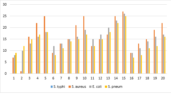
Figure 7. Antibacterial activity from X. americana extracts against S. Typhi, S. aureus, E. coli and S. pneumoniae.
The four extracts of X. americana were studied for their possible antibacterial activities. Results obtained (Table: 9 and Fig. 7) showed that all extracts inhibited the growth of S. aureus, S. pneumoniae and E. coli while S. Typhi was not inhibited by any of the extracts. The methanol extract showed the highest zones of inhibition against S. aureus (12-27 mm), S. pneumoniae (12-25 mm) and E. coli (12-26 mm), these results indicate that the extracts of X. americana, particularly the methanol extract, can be used to treat infections or ill-health conditions caused by S. aureus, E. coli and S. pneumoniae.
CONCLUSION
The present research work was carried out on phytochemical screening, antioxidant, antibacterial and anti-inflammatory activities of four extracts of X. americana stem bark, an indigenous plant of Yobe State, Nigeria. These extracts showed good antioxidant anti-inflammatory and antibacterial activities. The therapeutic potential of the stem barks of X. americana could be attributed to classes of active components present in the stem barks such as alkaloids, flavonoids, tannins and phenolic compounds etc. which may be acting in synergy or individually. The antioxidant, anti-inflammatory and anti-bacterial activities carried out in this study lend credence to the traditional claim about the wound healing property of the stem bark of X. americana.
Secondary metabolites such as alkaloids, flavonoids, phenols, tannins, terpenoids, etc. may be responsible for the positive antioxidant, anti-inflammatory and antibacterial activities reported in this study thus comparing with report of Nandagoapalan et al., (2016). Such activities as antimicrobial, anti-inflammatory, antioxidant, anticancer, among others, may be principal indicators of a plant’s value in medicine.
REFERENCES
- Amin, M. M., Sawhney, S. S. and Jassal, M. M. S. (2012). Qualitative and quantitative analysis of phytochemicals of Taraxacum officinale. Wudpecker Journal of Pharmacy and Pharmacology. 2(1): 001-005.
- Baiano, A. and Del Nobile, M. A. (2015). Antioxidant Compounds from Vegetable Matrices: Biosynthesis, Occurrence and Extraction Systems. Critical Review in Food Science and Nutrition. 56(12): 2053-68.
- Baris, O., Gulluce, M., Sahin, F., Ozer, H., Ozkan, H., Sokmen, M. and Ozbek, T. (2006). Biological activities of essential oil and methanol extract of Achillea Biebersteinii Afan. (Asteraceae). Turkish Journal of Biology. 30: 65-73
- Cai, Y., Luo, Q., Sun, M. and Corke, H. (2004). Antioxidant activity and phenolic compounds of 112 traditional Chinese medicinal plants associated with anticancer. Life Sciences. 74(17): 2157-84.
- Cheesbrough, M. (2002). Biochemical Test to Identify Bacteria. In: Laboratory Practice in Tropical Countries. Cambridge Edition. Pp. 63-70.
- Fang, Y. Z., Yang, S. and Wu, G. (2014). Free radicals, antioxidants and nutrition. Nutrition. 18: 872-879.
- Fawole, A. M. (2009). Pharmacological and Phytochemistry of South African Traditional Medicinal plants used as antimicrobials. M.sc Thesis. University of Kwazulu Natal, Pietermeritzburg.
- Gandhidasan, R., Thamaraichelvan, A. and Baburaj, S. (1991). Anti-inflammatory action of Lannea coromandelica by HRBC membrane stabilization. Fitoterapia. 12(1): 81-83.
- Gupta, M., Mazumdr, U. K., Sivakumar, T., Vamis M. L., Karkis, S., Sambathkumar, R. and Mainkndn, L. (2003).antioxidant and Anti-inflammatory activities of Acalypha fructicasa. Nig. J. Nat. Prd. Med. 25-29
- Hammer, K. A., Carson, C. F. and Riley, T. V. (1999). Antimicrobial activity of essential oils and other plants extracts. J. Appl. Microbiol. 86(6): 985.
- Hatano, T., Kagw, H., Yasuhara, T., and Okud, T., (1988). Two new flavonoid and other constituents in licorice root: their relative astringent and radical scavenging effects. Chem. Pharm. Bull. 36: 1090-2097.
- Kamil, M. (1993). Glimpses in plant research, Vol.XI, Today and Tomorrow̕s printers and publishers, Newyork. pp. 377-389.
- Khaleeliah, W. M. H (2001). Screening of anti-cancer activity of Palestinian plants. M.sc Thesis, An Najah National University, Palestine, pp: 1-10
- Kim, H., Son, K., Chang, H. and Kang, S. (2000). Effects of naturally occurring flavonoid on inflammatory response and their action mechanism. Natural Product Sciences; (6):170-178.
- Leelaprakash, G. and Mohan Dass, S. (2010). Invitro Anti-inflammatory Activity of Methanol Extract of Enicostemma axillare. International Journal of Drug Development & Research. 3(3): 189-196.
- Mamta, S., Jyoti, S., Rajeev, N., Dharmendra, S. and Abhishek, G. (2013). Phytochemistry of Medicinal Plants. Journal of Pharmacognosy and Phytochemistry. 1(6).
- Md. Lolo, C. M. (2009). Phytochemical analysis and selected biological activity of phyllanthus parvulus sond. Var. garipensis. M.sc thesis. University of Zululand, South Africa.
- Nandagoapalan V, Doss A, Marimuthu C. Phytochemical Analysis of Some Traditional Medicinal Plants. Bioscience Discovery, 2016:7(1):17-20.
- Okafor, T. and Mukhtar, M. D. (2002). Antibacterial Activity of Ethanolic Extract of Guiera senegalensis. International Journal of Pharmacology; 56: 213-216.
- Okwu, D. E. (2004) Evaluation of the chemical composition of Indigenous species and flavoring agents. Global Journal of Pure and Applied Science 7(3):455-459.
- Okwu, D. E. and Ekeke, O. (2003). Phytochemical Screening and mineral composition of chewing stick in south – eastern Nigeria. Global Journal of Pure and Applied Sciences (9):238 – 238.
- Park, K. H., Kwon, J. H. and Kim, S. B. (2011). Antioxidative and anti-inflammatory effects of phenolic compounds from the roots of Ulmus macrocarpa. Archieves of Pharmacal Research 34:1459.
- Rajendran, V. and Lakshmi, K. S. (2008). In vitro and in vivo anti-inflammatory activity of leaves of Symplocos cochinchinesis (Lour) Moore ssp Laurina. Bangladesh Journal of Pharmacology. 3, 121-124.
- Rubina, L. (2011). Evalution of antibacterial activity of plant extractson antibiotic suseptableand resistant staphylococcus aureus strains, Journal of Chemical and Pharmaceutical Research, Vol 3(4) p. 777-789.
- Sabri, F. Z., Belarbi, M., Sabri, S. and Alsaydi M. (2012). Phytochemical screening and identification of some compounds from Mallow ; J. Nat. Prod. Plant Reour., 2(4): 512-516.
- Sadique, J., Al-Rqobahs, W. A., Bughaith, and ElGindi, Ar. (1989). The bioactivity of certain medicinal plants on the stabilization of RBS membrane system. Fitoterapia. 60: 525-532.
- Santosh, C. H., Attitalla, I. H. and Mohan, M. M. (2013). Phytochemical analysis, antimicrobial and antioxidant activity of ethanolic extract of Vernonia anthelmintica. International Journal of Pharma and Bio Sciences. 4(1): 960-966.
- Shenoy, S., Shwetha, K., Prabhu, K., Maradi, R., Bairy, K. L. and Shanbhang, T. (2010). Evaluation of anti-inflammatory activity of Tephrosia purpurea in rats. Asian Pac J Trop Med. 3(3): 193-5.
- Sofowara A. Guideline for research promotion and development in traditional medicine. Nigerian Journal of pharmacy,1980:11:117–118.
- Sofowora A E. Medicinal plant and tradition medicine in Africa, John Wiley and sons, Ltd, Ife, Nigeria, 1993.
- Tanko, Y., Mohammad, A., Okasha, M. A., Shuaibu, A., Magaji, M. G. and Yaro, A. H. (2007). Analgesic and anti-inflammatory activities of ethanol seed extract of Nigella sativa 9black cumin) in mice and rats. European Journal of Scientific Research; 18:277-281.
- Tiwari, P., Bimlesh, K., Mandeep K., Gurpreet, K. and Harleen, K. (2011). Phytochemical screening and extraction: A review. Internationale Phrmaceuticasciencia, 1(1): 1-9.
- Van Wyk B. E and Gericke, N. (2000) People´s plants: A guide to useful plants of Southern Africa. Briza publications, Pretoria 226(3/4):245-247.
- Von koenem, E. (2001). Medicinal, poisonous and edible plants in Namibia. Windhoek, Namibia; Gottingen: Klaus Hess publisher, 190-195.
- Wugang. H (2008). Traditional Chinese medicinal plants and their endophytic fungi: isolation, identification and bio assay PhD thesis. University of Hong Kong. China, 1-17.