
Methanol Fraction of Ethanol Extract of Dialium guineense Stem Bark May Alter the Activity of Glucose 6-phosphatase/Aminotransferases and Levels of Lipids in Tissues of Diabetic Wistar Rats
- Abu, O.D.
- Okuo, A.V.
- Egili, S.
- Idehen, I.O.
- Esedebe, M.
- Etoroma, O.M.
- 523-532
- Jan 14, 2024
- Chemistry
Methanol Fraction of Ethanol Extract of Dialium guineense Stem Bark May Alter the Activity of Glucose 6-phosphatase/Aminotransferases and Levels of Lipids in Tissues of Diabetic Wistar Rats
Abu, O.D.1*, Okuo, A.V.2, Egili, S.3, Idehen, I.O.1, Esedebe, M.1 and Etoroma, O.M.3
1Department of Biochemistry, Faculty of Life Sciences, University of Benin, Benin City, Nigeria.
2Department of Chemistry, College of Arts and Sciences, University of Kentucky, USA
3Department of Pharmaceutical Chemistry, Faculty of Pharmacy, University of Benin, Benin City, Nigeria.
*Corresponding Author
DOI: https://doi.org/10.51244/IJRSI.2023.1012040
Received: 07 December 2023; Accepted: 15 December 2023; Published: 13 January 2024
ABSTRACT
The aim of the present study was to investigate the potential of methanol fraction of ethanol extract of Dialium guineense (MEDG) stem bark to alter the activity of glucose 6-phosphatase (G6Pase)/aminotransferases and levels of lipids in tissues of streptozotocin (STZ)-induced diabetic Wistar rats. Adult male Wistar rats (n = 25, mean weight = 215 ± 15 g) were randomly assigned to five groups (5 rats/group): normal control, diabetic, metformin, extract (200 mg/kg body weight, bwt) and extract (300 mg/kg bwt) groups. A single intraperitoneal injection of 50 mg/kg bwt STZ was used to induce diabetes mellitus (DM) in the experimental rats. The diabetic rats were subsequently treated for 21 days with 50 mg/kg bwt metformin (standard antidiabetic agent) or MEDG stem bark. Activities of G6Pase, alanine aminotransferase (ALT), aspartate aminotransferase (AST) and alkaline phosphatase (ALP) and levels of total cholesterol (TC), triacylglycerol (TG), very low-density lipoprotein cholesterol (VLDL-C), and nitric oxide (NO) were measured in plasma or hepatic/cardiac/renal tissues. The results showed that the G6Pase activity that was significantly increased in the plasma and hepatic/renal tissues of rats were markedly reduced after treatment with MEDG stem bark (p < 0.05). Similarly, STZ markedly elevated the activities of ALT and ALP in rat hepatic tissue (p < 0.05), but it did not alter the activity of AST in hepatic and cardiac tissues (p > 0.05). However, treatment of the diabetic Wistar rats with MEDG stem bark significantly reduced the hepatic activities of ALT and ALP (p < 0.05). Moreover, STZ-induced DM significantly elevated the levels of hepatic lipids (TC, TG and VLDL-C) and plasma NO, while MEDG administration significantly reversed the effect of the diabetogenic agent STZ by markedly reducing hepatic lipids and plasma NO levels (p < 0.05). These results suggest that the medicinal plant extract may alter the activity of G6Pase/aminotransferases and levels of lipids in tissues of STZ-induced diabetic rats.
Keywords: Gluconeogenesis, Glucose 6-phosphatase, Hyperglycemia, Lipids, Medicinal plant.
INTRODUCTION
Characterized by elevated blood glucose level (hyperglycemia) DM is caused by a relative or absolute deficiency of insulin [1]. The metabolic abnormalities of type-2 diabetes mellitus (T2DM) (hyperglycemia and hypertriacylglycerolemia) are the result of insulin resistance expressed primarily in liver, muscle, and adipose tissue [2]. Elevated levels of blood glucose and ketones are the hallmarks of untreated DM. Hyperglycemia is caused by increased hepatic production of glucose, combined with diminished peripheral use due to inability of muscle and adipose cells to take up glucose [3, 4]. Ketosis results from increased mobilization of fatty acids from adipose tissue, combined with accelerated hepatic synthesis of 3-hydroxybutyrate and acetoacetate [5 – 8]. Diabetic ketoacidosis occurs in 25 to 40 % of those newly diagnosed with type-1 DM (TIDM), and may recur if the patient becomes ill (most commonly with an infection) or does not comply with therapy. Ketoacidosis is treated by fluid/electrolytes replacement, followed by administration of low-dose insulin to gradually correct hyperglycemia without precipitating hypoglycemia [2]. Not all the fatty acids flooding the liver can be disposed of through oxidation or ketone body synthesis. The excess fatty acids are converted to triacylglycerols, which are packaged and secreted in VLDL-C [3]. Chylomicrons are synthesized from dietary lipids by intestinal mucosal cells following a meal. Because lipoprotein degradation catalyzed by lipoprotein lipase in adipose tissue is low in diabetics, plasma chylomicron and VLDL-C levels are elevated, resulting in hypertriacylglycerolemia [9].
Medicinal plants have long been recognized as important sources of therapeutically active compounds [11]. With special interest in the identification and characterization of bioactive compounds from natural sources, evidence-based research supports the medical and pharmacological uses of plant-derived compounds [12-14]. Dialium guineense is a medicinal plant used in Traditional Medicine for the treatment of different disease conditions, such as diarrhea, severe cough, bronchitis, wound, stomachaches, malaria, jaundice, ulcer and hemorrhoids [15-17]. The acute and subchronic toxicity of the plant stem bark have been reported [18, 19]. Extracts of D. guineense have been demonstrated to contain compounds with varied biological/pharmacological effects [20 – 32]. This study investigated the potential of MEDG stem bark to alter the activity of G6Pase/aminotransferases and levels of lipids in tissues of STZ-induced diabetic Wistar rats.
MATERIALS AND METHODS
Chemicals and Reagents
Metformin was a product of Micronova Laboratories (India), and STZ was purchased from British Drug House (BDH) Chemicals Ltd. (England). Solvents (methanol, ethanol and chloroform), and other materials/glass wares were bought from Bell, Sons & Co. (England), while formaldehyde was purchased from Thermo Fisher Scientific Ltd. (USA).
Glucose 6-phosphatase assay kit was obtained from Abcam (UK). All chemicals and solvents used in this study were of analytical grade.
Extract Preparation
Dialium guineense stem barks obtained from Auchi, Edo State, Nigeria, were authenticated at the University of Benin herbarium domiciled in the Department of Plant Biology and Biotechnology, University of Benin, Benin City, Nigeria. The prepared plant specimen was deposited in the herbarium of same department (No. UBHD330).
The plant’s stem bark was” washed and shade-dried for 4 weeks at room temperature, and thereafter ground into powder using a blender. A portion (500 g) of powdered plant material was steeped in 5,000 mL of absolute ethanol. The resulting extract was filtered through muslin cloth and freeze-dried with a lyophilizer. The ethanol extract was subsequently fractionated with 100% methanol [33 – 35].
Experimental Rats
Adult male Wistar rats (n = 25) weighing 200 to 230 g (mean weight = 215 ± 15 g) were bought from the Department of Anatomy, University of Benin, Nigeria and housed in wooden cages. They were acclimatized for fourteen (14) days before commencement of the study, and had free access to feed and clean water.
Experimental Design
The experimental design used in this study was a completely randomized block design. The rats were randomly assigned to five groups (5 rats/group): normal control, diabetic, metformin, 200 mg/kg bwt extract and 300 mg/kg bwt extract groups. Diabetes mellitus was induced in the rats using STZ (single intraperitoneal injection of 50 mg/kg bwt). The diabetic rats were then treated with metformin (50 mg/kg bwt) or MEDG stem bark (200 and 300 mg/ kg bwt), for 21 days.
Blood and Tissue Samples Collection and Preparation
At the end of the treatment period, the rats were subjected to mild chloroform anesthesia after an overnight fast. They were euthanized and blood was collected via retro-orbital sinus puncture and centrifuged for 10 min at 3000 rpm to obtain plasma. Rat liver, kidney and heart were excised, blotted dry and used to prepare 20 % tissue homogenate.
Biochemical Analyses
Activities of AST, ALT and ALP as well as levels of TP, TC, TG and VLDL-C were measured in hepatic, renal and cardiac tissues [36 – 41]. Glucose 6-phosphatase activity was determined in plasma, liver and kidney, while NO level was estimated in plasma only [42, 43].
Data Analysis
Data are expressed as mean ± standard error of mean (SEM, n = 5). Statistical analysis was performed using SPSS version 21. Statistical differences between means were compared using Duncan multiple range test. Statistical significance was assumed at p < 0.05.
RESULTS
Effect of MEDG Stem Bark on Weight and Blood Glucose of Rats
As shown in Table 1 and Figure 1, STZ-induced DM significantly increased the blood glucose concentrations of the rats, but it reduced the organ/body weight ratio (p < 0.05). However, treatment of the diabetic rats with the extract markedly reduced the fasting blood glucose (FBG) concentration and body weights of rats, but it increased the organ/body weight ratio (p < 0.05).
Table 1: Effect of MEDG Stem Bark on Weight and Blood Glucose Parameters
| Group | Weight Change (g) | % Weight Change | FBG (mg/dL) | Glycemic Change (mg/dL) | % Glycemic Change |
| Normal Control | – | – | – | – | |
| Diabetic | – | > 800 | – | – | |
| Metformin | 20.35 | 12.16 | > 800 | 399 | 49.88 |
| Extract (200 mg/kg bwt) | 12.26 | 7.87 | > 800 | 421 | 52.63 |
| Extract (300 mg/kg bwt) | 29.08 | 17.02 | 364 | 227 | 62.36 |
Data are weight and FBG parameters and are expressed as mean ± SEM (n = 5).

Figure 1: Organ Weight and Organ/Body Weight Ratio
Effect of MEDG Stem Bark on G6Pase Activity
Streptozotocin-induced DM significantly increased G6Pase activity in the plasma as well as hepatic and renal tissues of rats (p < 0.05). However, the activity of the enzyme was markedly reduced after treatment with MEDG stem bark (p < 0.05; Figure 2).
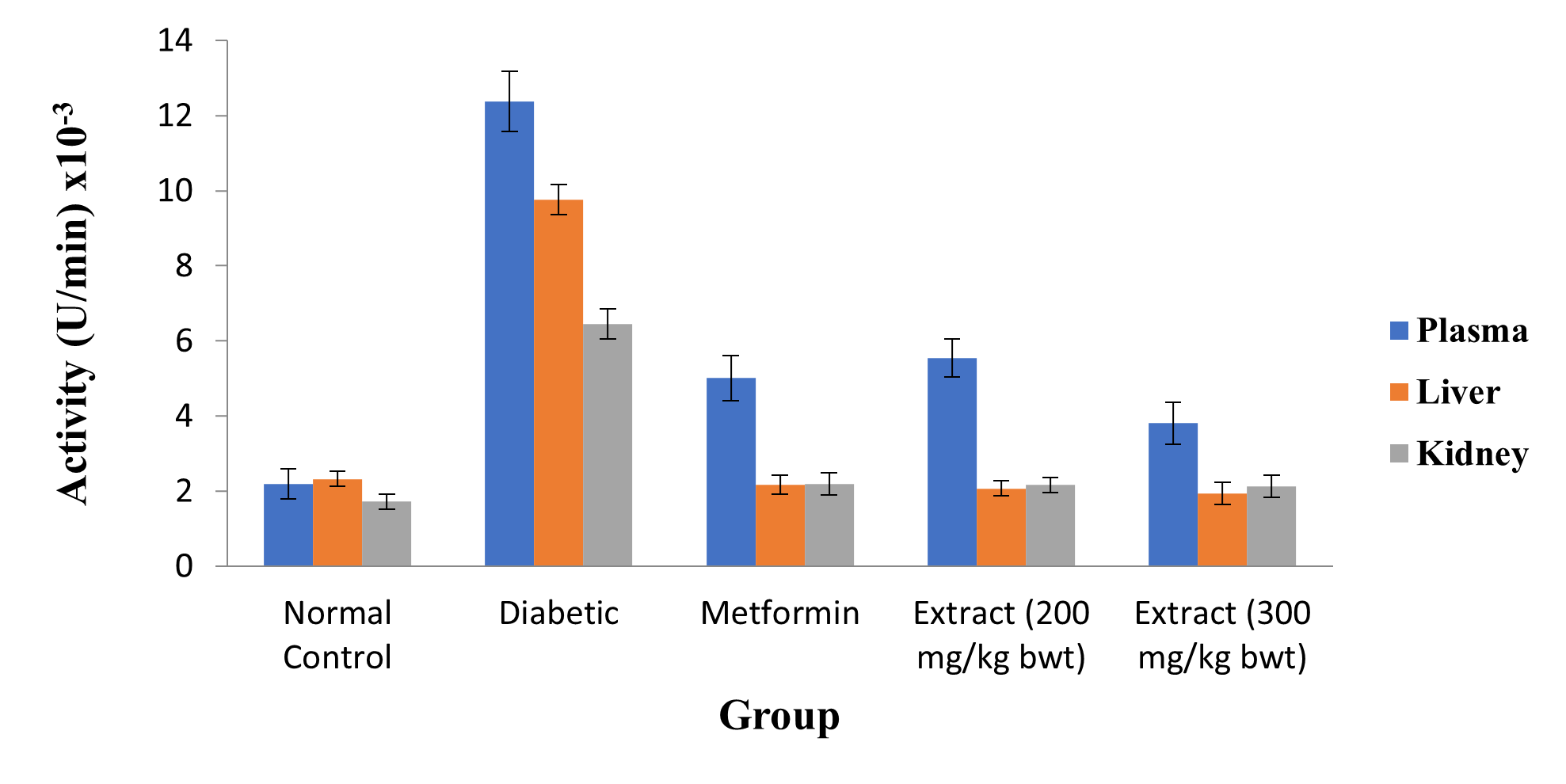
Figure 2: Activity of G6Pase in Plasma and Rat Hepatic and Renal Tissues
Data are plasma and tissue activities of G6Pase and are expressed as mean ± SEM (n = 5).
Effect of MEDG Stem Bark on Tissue Activity of AST, ALT and ALP
As shown in Figures 3 and 4, STZ markedly elevated the activities of ALT and ALP in rat hepatic tissue (p < 0.05), but it did not alter the activity of AST in hepatic and cardiac tissues (p > 0.05). On the contrary, treatment of the diabetic rats with MEDG stem bark significantly reduced the hepatic activities of ALT and ALP (p < 0.05).
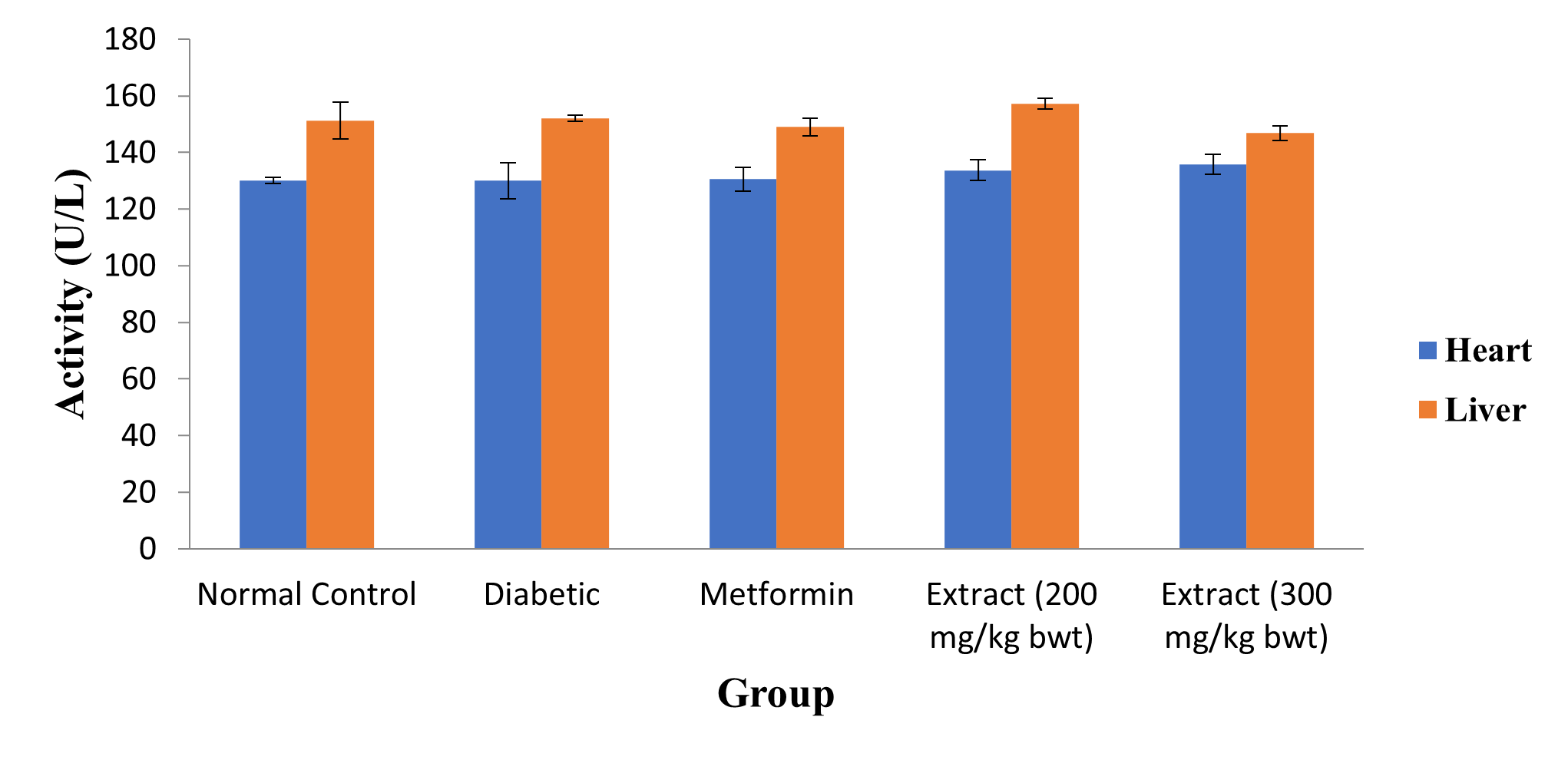
Figure 3: Activity of Cardiac and Hepatic AST
Data are tissue AST, and are expressed as mean ± SEM (n = 5).
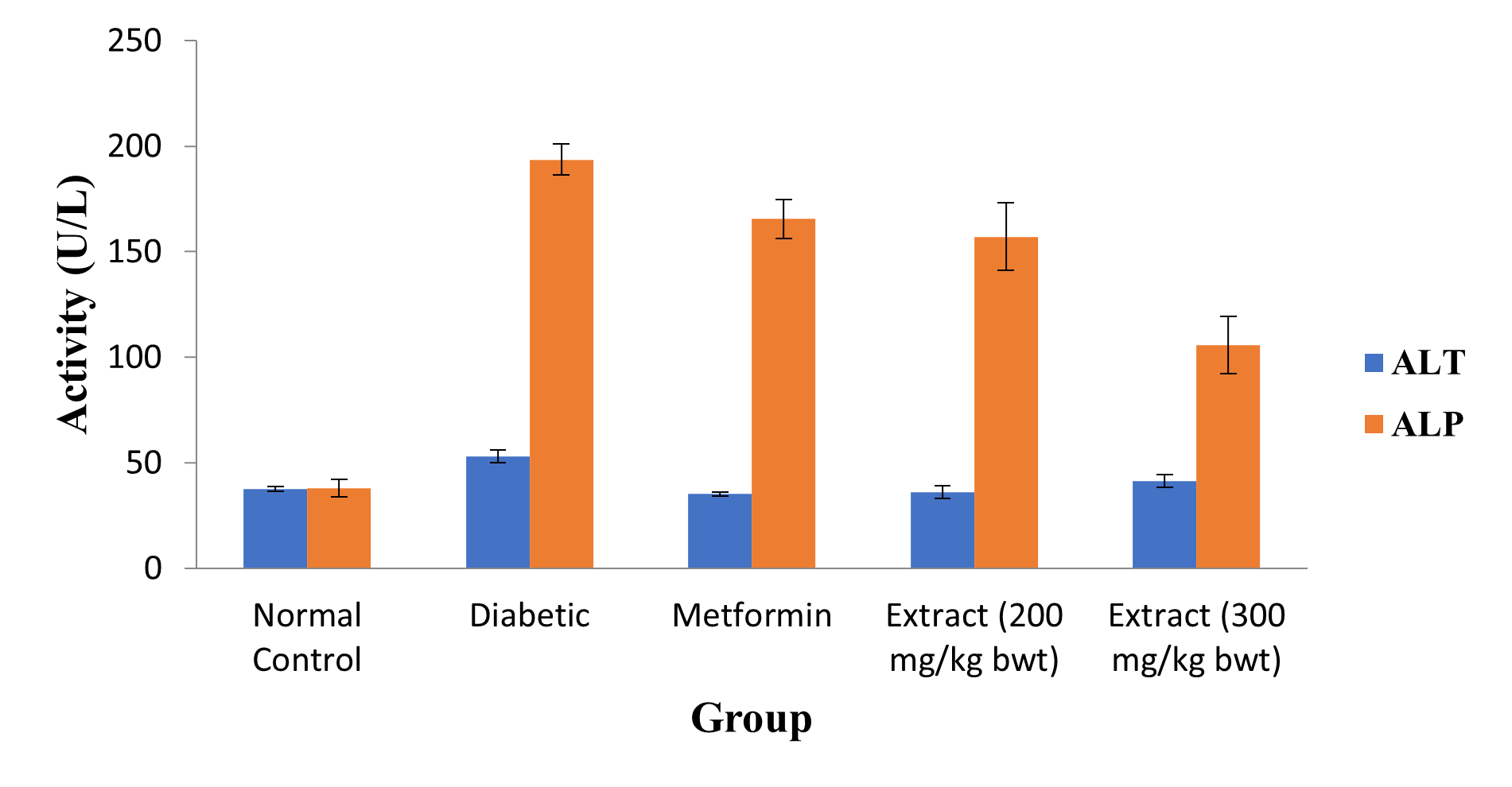
Figure 4: Activity of Hepatic ALT and ALP
Data are tissue ALT and ALP activity, and are expressed as mean ± SEM (n = 5).
Effect of MEDG Stem Bark on Tissue Lipids and NO Level
Induction of DM with STZ led to significant increase in the level of hepatic lipids (TC, TG and VLDL-C) and plasma NO, while treatment of the diabetic Wistar rats significantly reversed the effect of STZ by markedly reducing hepatic lipids and plasma NO levels (p < 0.05; Figures 5 and 6).
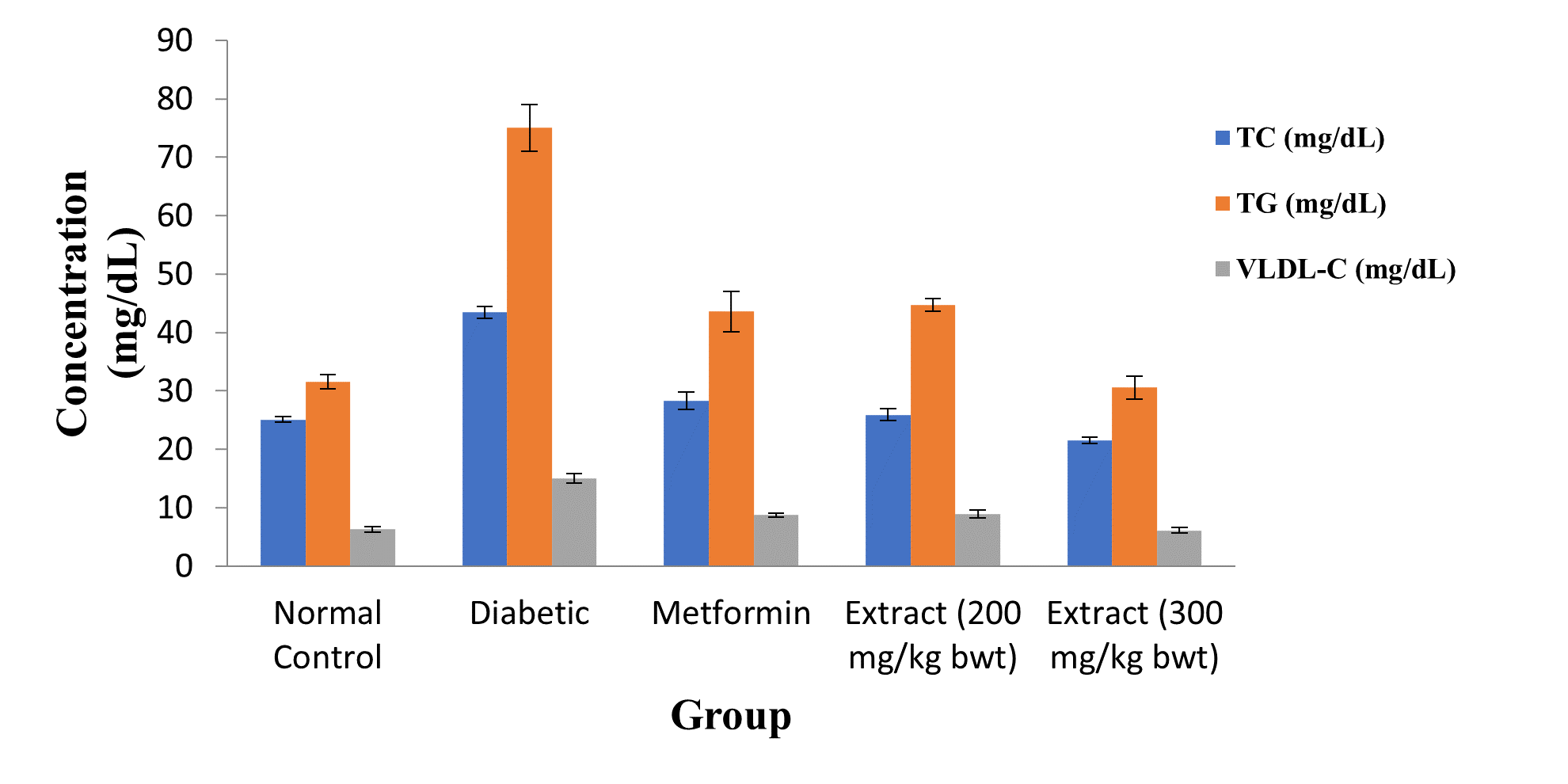
Figure 5: Comparison of Levels of Tissue Lipids
Data are levels of hepatic lipids and are expressed as mean ± SEM (n = 5).
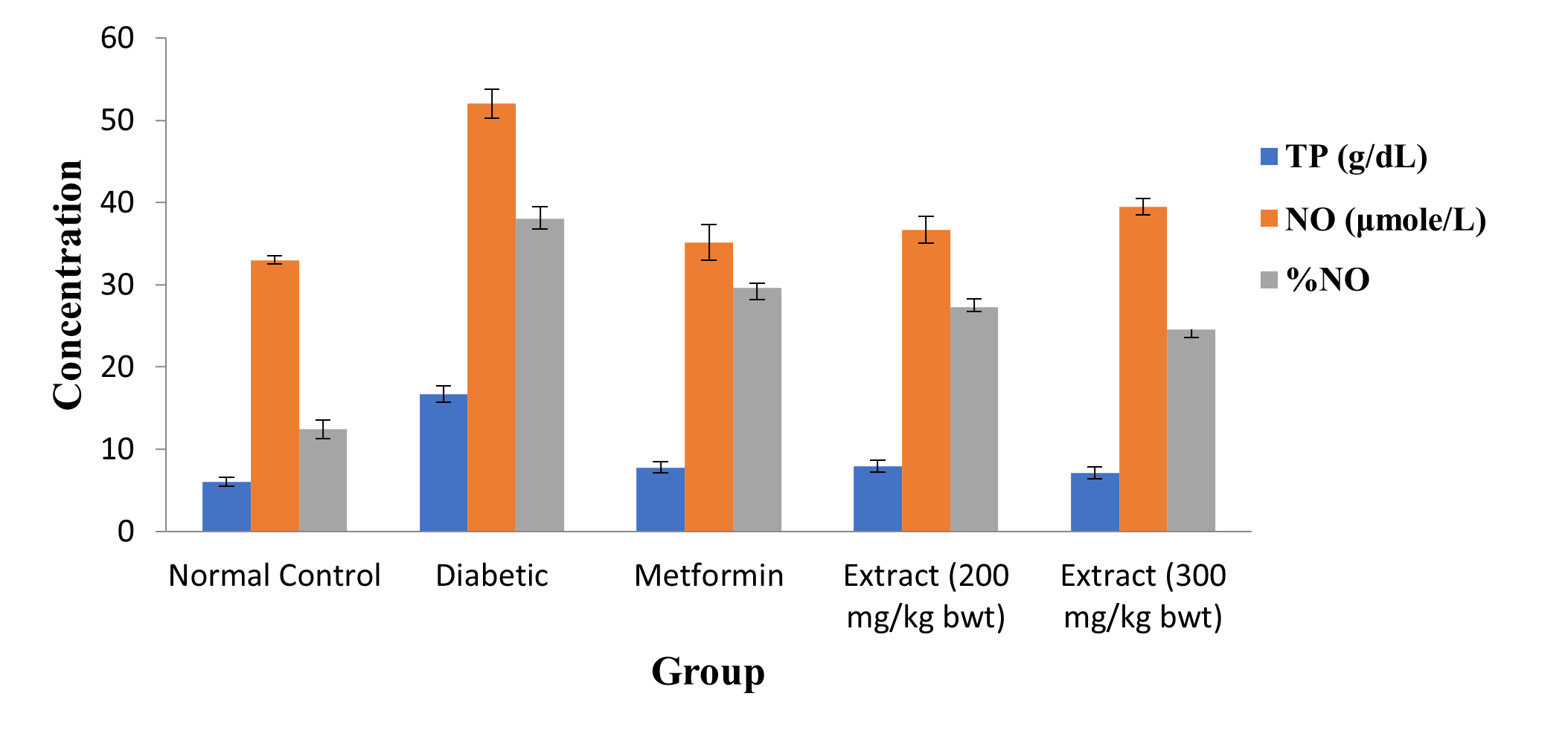
Figure 6: Plasma TP and NO Levels
Data are plasma TP and NO levels, and are expressed as mean ± SEM (n = 5).
DISCUSSION
Plants are at the center of Traditional Medicine. Their use in disease management is as old as man. Medicinal plants serve as cheap alternative to orthodox medicine since they are readily available [44, 45]. A variety of indigenous medicinal plants can effectively manage DM. One of the most significant benefits of medicinal plants is their ready availability and minimal side effects [46, 47].
Dialium guineense is a medicinal plant used locally to treat diverse kinds of diseases [48]. A substantial tropical fruit tree of the family Leguminosae, it bears tiny, frequently grape-sized edible fruits that are coated in brown, inedible shells. At the southernmost border of the Sahel in Africa, it grows in thick woods [15]. The Central African Republic, Sudan, and West Africa are the original home of this plant. In Nigeria, it is referred to by a variety of names, including “Icheku (Igbo), Awin (Yoruba), Tsamiyarkurm (Hausa), and Amughen (Bini) [16, 17]. According to reports, the plant’s extracts are rich in important phytochemicals [20, 21]. This study investigated the potential of MEDG stem bark to alter the activity of G6Pase/aminotransferases and levels of lipids in tissues of STZ-induced diabetic Wistar rats.
Hyperglycemia in DM results from increased hepatic gluconeogenesis and reduced glucose uptake in peripheral tissues. The chief and regulatory enzyme of gluconeogenesis is G6Pase. Along with elevated glycogenolysis during fasting, there is increased G6Pase activity in the liver [9]. Thus, glucose 6-phosphate generated from glycogenolysis is released from liver into the circulation for peripheral use. There does not appear to be G6Pase in skeletal muscle; hence, muscle glycogen is not a source of circulating glucose. Glucose-6-phosphatase deficiency (glycogen storage disease, GSD, type Ia) results in hypoglycemia and excessive intracellular accumulation of glucose-6-phosphate [9]. In this study, STZ-induced DM significantly increased G6Pase activity in the plasma as well as rat hepatic and renal tissues. However, the activity of the enzyme was markedly reduced after treatment with MEDG stem bark.
People with T2DM are at risk for both micro- and macrovascular complications [3, 49]. The toxicity produced by STZ impart negatively on organs such as liver, kidneys, heart and lung [50]. The exact molecular mechanism underlying the cytotoxic effect of STZ is not well-understood, however studies suggest that the cytotoxicity could be via production of reactive oxygen species (ROS) thus inducing oxidative stress, causing DNA damage with resultant necrosis due to the DNA methylating activity of the methyl nitroso urea moiety of the drug, release of NO which inhibits aconitase activity resulting in mitochondrial dysfunction, or via inhibition of O-linked β -N-acetylglucosaminase (O-GlcNAcase) [51]. In this study, the diabetogenic agent STZ markedly elevated the activities of ALT and ALP in rat hepatic tissue, but it did not alter the activity of AST in hepatic and cardiac tissues. On the contrary, treatment of the diabetic rats with MEDG stem bark significantly reduced the hepatic activities of ALT and ALP. In addition, induction of DM with STZ led to significant increases in the levels of hepatic lipids (TC, TG and VLDL-C) and plasma NO, while treatment of the diabetic Wistar rats markedly reversed the effect of STZ by significantly reducing hepatic lipids and plasma NO levels. These results are similarly to those obtained with ethanol extract of Cucumis sativus [52].
CONCLUSION
The results obtained in this study have shown that MEDG stem bark has the capacity to alter the activity of G6Pase/aminotransferases and levels of lipids in tissues of STZ-induced diabetic rats.
REFERENCES
- Centers for Disease Control and Prevention. Diabetes Report Card (2012). Atlanta, GA: Centers for Disease Control and Prevention, U.S Department of Health and Human Services.
- Onoagbe IO, Esekheigbe A (1999). Studies on the anti-diabetic properties of Uvaria Chamae in streptozotocin-induced diabetic rabbits. Biokemistri 9: 79 – 84.
- Centers for Disease Control (2011). National diabetes fact sheet: national estimates and general information on diabetes and prediabetes in the United States.
- Imperatore G, Cadwell BL, Geiss LS, Saaddine JB, Williams DE, Ford ES (2011). Thirty-year trends in cardiovascular risk factor levels among U.S adults with Am. J. Epidemiol 160: 531 – 539.
- World Health Organisation (2006). The World Health Report 2006: working together for Humans; Professional Competence.
- Abu OD, Ojo I, Ezike TV (2023). Methanol Fraction of Ethanol Extract of Dialium guineense Stem Bark Mitigates STZ-Induced Oxidative Stress in Rat Biomedical Journal of Scientific and Technical Research 51 (2): 42594 – 42600.
- Abu OD, Osime EC, Ngedaa OS (2023) Cardiac Oxidative Status in Diabetic Wistar Rats Exposed to Ethanol Extract of Cucumis sativus J. Diagnostics and Case Reports 4 (2): 1 – 5.
- Abu OD, Ojo I, Awhin EP (2023) Protective Property of Ethanol Extract of sativus on STZ-Induced Diabetic Rat Pancreas. Biomedical Journal of Scientific and Technical Research 52 (2): 43613-43618.
- Phibbs, B. (2007). The human heart: a basic guide to heart disease (2nd). Philadelphia: Lippincott Williams & Wilkins. Pp. 1.
- Hoffman, R.M., Boston, R.C., Stefanovski, D., Kronfeld, D.S. and Harris, P.A. (2003). Obesity and diet affect glucose dynamics and insulin sensitivity in Thoroughbred geldings. J Anim Sci. 81:2333 – 2342.
- Superling F. (1979). Introduction to Toxicity Evaluation Session. Environmental Health 32: 259.
- Walum E. (1998). Acute Oral Toxicity. Environmental Health Perspectives. 106(2):497-
- Handa SS, Khanuja SPS, Longo G, Rakesh DD. (2008). Extraction technologies for medicinal and aromatic plants. International Centre for Science and High Technology, Trieste, Italy, PP: 21-25.
- Sandoval M, Okuhama NN, Zhang XJ, Condezo LA, Lao J, Angeles FM (2002). Anti-inflammation and antioxidant activities of cat’s claw (Uncaria tomentos and Uncaria guinensis) are independent of their alkaloid content. Phytomedicine. 9(4): 325-337.
- Dalziel JM, Hutchison J. (1973). Flora of West Tropical Africa. Vol.1 (2nd Ed). The White friars Press Ltd. London PP: 561.
- Bero J, Ganfon H, Jonville MC, Frederich M, Gbaguidi F, De MP (2009) In vitro antiplasmodial activity of plants used in Benin in traditional medicine to treat malaria. Journal of Ethnopharmacology. 122(3): 439-444.
- Arogba SS, Ajiboro A, Odukwe I. (2006) A physicochemical study of Nigerianvelvet tamarind (Dialium guineense ) fruit. Journal of the Science of Food and Agriculture. 66(4): 533-534.
- Abu OD, Onoagbe IO. (2021). Acute toxicity of aqueous and ethanol extracts of Dialium guineense stem bark. Journal of Bioinnovation.10(2): 427-432.
- Abu, O.D., Adeogun, E.F. and Ebhohon S.O. (2019). Oral LD50 of total saponins and tannins isolated from Dialium guineense stem bark. European Journal of Experimental Biology. 9 (2): 11 – 13.
- Abu, O.D., Onoagbe, I.O. and Obahiagbon, O. (2020). In Vitro Antioxidant Activities of Extracts of Dialium Guineense Stem Bark. American Journal of Sciences and Engineering Research. 3 (4): 68 – 75.
- Abu, O.D., Onoagbe, I.O. and Obahiagbon, O. (2020). Phenolic contents of extracts of Dialium guineense stem bark. American Journal of Sciences and Engineering 3 (4): 92 – 96.
- Abu O.D., Onoagbe I.O. and Obahiagbon O. (2020). Analyses of metal and amino acid compositions of aqueous and ethanol stem bark extracts of Dialium guineense. Journal of Biogeneric Science and Research. 6 (4): 1 – 3.
- Abu, O.D., Onoagbe, I.O. and Obahiagbon, O. (2020). Vitamin contents of extracts of Dialium guineense stem bark. Biomed J Sci & Tech Res. 30 (2): 23263 – 23267.
- Abu, O.D., Onoagbe, I.O. and Obahiagbon, O. (2020). Qualitative phytochemical screening and proximate analysis of Dialium guineense stem bark. IAR Journal of Agriculture Research and Life Sciences. 1 (4): 108 – 112.
- Abu, O.D., Onoagbe, I.O., and Obahiagbon, O. (2020). Alpha amylase and alpha glucosidase inhibitory activities of extracts of Dialium guineense stem bark. International Journal of Clinical Biology and Biochemistry. 2 (1): 7 – 10.
- Abu, O.D., Onoagbe, I.O., and Osemwenoyenmwen, O. (2020). Alpha amylase and alpha glucosidase inhibitory activities of isolated total saponins and tannins of Dialium guineense stem bark. Journal of Cellular and Molecular Biolog Research. 1 (2): 1 – 3.
- Abu, O.D., Onoagbe, I.O. and Obahiagbon, O. (2020). In Vitro Antioxidant Activities of Isolated Total Saponins and Tannins of Dialium Guineense Stem Bark. IAR Journal of Medical Sciences. 1 (4): 193 – 199.
- Abu, O.D. and Onoagbe, I.O. (2019). Biochemical effect of aqueous extract of Dialium Guineense stem bark on oxidative status of normal Wistar rats. International Journal of Clinical Biology and Biochemistry. 1 (2): 15 – 18.
- Abu, O.D., Onoagbe, I.O., and Ojo, I. (2021). Graded and quantal dose response of total tannins isolated from the stem bark of Dialium guineense. Advanced Research Journal of Medicine and Clinical Science. 08 (10): 699 – 703.
- Abu, O.D., Onoagbe, I.O., and Ojo, I. (2022). Dose response study of aqueous extract of Dialium guineense stem bark. American Journal of Biomedical Science and 15 (2): 250 – 252.
- Abu, O.D., Onoagbe, I.O., and Ojo, I. (2022). Dose response of total saponins isolated from the stem bark of Dialium guineense. Journal of Advances in Plant Biology. 1 (4): 1 – 6.
- Abu, O.D., Onoagbe, I.O., and Ojo, I. (2021). Determination of effective dose for ethanol extract of Dialium guineense stem bark. Journal of Medical Research and Case 3 (2): 1 – 4.
- Abu, O.D. and Ikponmwosa-Eweka, O. (2022). Potential of Extracts of Dialium guineense Stem Bark in the Mitigation of Carbon Tetrachloride-induced Renal Oxidative Stress. BIU Journal of Basic and Applied Sciences. 7 (1): 62 – 69.
- Abu, O.D. and Ikponmwosa-Eweka, O. (2022). Evaluation of the Potential of Total saponins and Tannins of Dialium guineense Stem Bark in the Amelioration of Carbon Tetrachloride-Induced Renal Oxidative Stress. SAU Science-Tech. Journal. 7 (1): 42 – 50.
- Abu, O.D. and Ikponmwosa-Eweka, O. (2022). Potential of Total Saponins and Tannins Isolated from the stem bark of Dialium guineense in the Amelioration of Kidney Dysfunction Caused by CCl4. Journal of Basic and Applied Medical Sciences. 2 (1): 1 – 6.
- Reitman S, Frankel S (1957) A colorimetric method for the determination of serum glutamic oxalacetic and glutamic pyruvic transaminases Am J Clin Pathol 28(1): 56-63.
- Babson LA (1965) Phenolpthalein monophosphate, a new substrate for alkaline phosphatase. In: Abstracts of papers from scientific sessions: 17th National Meeting of the American Association of Clinical Chemists Chicago III.
- Henry RJ, Sobel C, Beckman S (1957) Determination of serum protein by the biuret reaction. Anal Chem 92(149): 1-5.
- Reiser R, Probstfield JL, Silvers A, Scott LW, Shorney ML, Wood RD, O’Brien BC, Gotto AM, Insull W (1985) Plasma lipid and lipoprotein response of humans to beef fat, coconut oil and safflower oil. Am J Clin Nutr 42 (2):190 – 197.
- Tietz NW, Finley PR, Pruden EL. (1990). Clinical Guide to Laboratory Tests. 2nd Edition, W.B. Saunders, Philadelphia. PP: 304-306.
- Friedewald WT, Levy RI, Fredrickson DS. (1972) Estimation of the concentration of low-density lipoprotein cholesterol in plasma, without use of the preparative Clin Chem 18 (6):499- 502.
- Garland RC, Cori, CF, Chang HW (1974) Relipidation of phospholipid-depleted microsomal particles with high glucose 6-phosphatase activity. Proc Nat Acad Sci 71 (10): 3805 – 3809.
- Marcocci L, Packer L, Droy Lefaix MT, Sekaki A (1994) Antioxidant action of Ginkgo biloba extract EGb 761. Methods in Enzymology 234: 462-475.
- Abu, O.D., Osagie, A.O. and Kolawole, O.M. (2022e). Ameliorative Effect of Extracts of Dialium guineense Stem Bark in CCL4–Induced Kidney Dysfunction in Wistar Rats. Biokemistri, 34 (2): 34310 – 34316.
- Abu, O.D., Awhin, E.P. and Ohikhuare, F. (2023c). Effect of Methanol Fraction of Ethanol Extract of Dialium guineense Stem Bark on Cardiovascular Disease Risk Factors in Diabetic Rats. Journal of Biology and Medicine, 4 (1): 128.
- Abu, O.D., Iyare, H.E. and Omoruyi, I.J. (2022h). Toxic Responses of the Blood of Rats Exposed to Aqueous Extract of Dialium guineense Stem Bark. FUDMA Journal of Science, 7 (2): 117 – 120.
- Abu OD, Imafidon KE, Obayuwana HO, Onodje S (2020) Quantitative phytochemical evaluation and phenolic contents of extracts of Citrullus lanatus seed. Int J Bioorg Chem Mol Biol 7: 31-35.
- Abu, O.D., Alegun, O. and Ifekwe, J.C. (2023). Renal Oxidative Status in Diabetic Wistar Rats Administered Methanol Fraction of Ethanol Extract of Dialium Medical and Clinical Case Reports Journal. 1(1): 1 – 13.
- Abu O.D., Awhin E.P. and Ozedu M.E. (2023). Evaluation of Cardiovascular Disease Risk Factors in Diabetic Rats Administered Ethanol Extract of Cucumis sativus Fruit. African Journal of Health, Safety and Environment. 4(1): 108 – 117.
- Abu O.D., Awhin E.P. and Iyare H.E. (2023). Assessment of Renal Function in Diabetic Wistar Rats Treated with Ethanol Extract of Cucumis sativus. African Journal of Health, Safety and Environment. 4(1): 101-107.
- Anabel, S.G-R., Shoshan, K-S., Jan, D.H., Alphons, P.L.V. and Dirk, W.B. (2010). Anomer equilibrated streptozotocin solution for the induction of experimental diabetes in mice (Musmusculus). Journal of the American Association for Laboratory Animal Science, 49 (1): 40 – 44.
- Abu, O.D., Obaze, G.E., Egili, S. and Idehen, I.O. (2023). Ethanol Extract of sativus Modulates the Activity of Glucose 6-phosphatase/ Aminotransferases and Levels of Lipids in Tissues of STZ-Induced Diabetic Rats. Biomedical Journal of Scientific and Technical Research. 53(4): 44989-44994.