
Amino Acid Profile, Total Protein and Histological Changes in Female Wistar Rats After Administration of Dichloromethane Extract of Heterotis Rotundifolia Leaves on Dermal Wound.
- Ogechukwu E. Ezim
- Jane A. Agwu
- 215-223
- Mar 9, 2024
- Education
Amino Acid Profile, Total Protein and Histological Changes in Female Wistar Rats After Administration of Dichloromethane Extract of Heterotis Rotundifolia Leaves on Dermal Wound.
Ogechukwu E. Ezim1*,Jane A. Agwu2.
1,2Department of Biochemistry, Faculty of Science, University of Port Harcourt, Choba, Rivers State, Nigeria.
*Corresponding Author
DOI: https://doi.org/10.51244/IJRSI.2024.1102017
Received: 06 February 2024; Accepted: 08 February 2024; Published: 06 March 2024
ABSTRACT
The study investigated the amino acid and wound healing potentials of the leaves of Heterotis rotundifolia. Amino acid analysis was performed using HPLC. Total protein level, body weight and histology were studied. Excision wound(2cm) was induced on the dorsal skin of 75 wistar rats (176-308g) divided into five groups of fifteen animals: control, 0.1g Vaseline, 0.1g gentamicin (reference drug), 0.1g extract+0.1g Vaseline (HRE1) and 0.2g extract+0.1g Vaseline (HRE2). At the end of days 1, 7, 14 and 21, rats were sacrificed and blood was collected for wound healing parameters while wound surface was taken for histology. The result revealed a higher total protein level of HRE1 with a significant difference (p<0.05) on day 21 compared to control and days 1-7 compared to gentamicin. Body weight in HRE2 significantly increased from days 1-21 with a significant difference (p<0.05) on days 1-7 compared to control and 1-14 compared to gentamicin. Histological examination revealed faster collagen deposition and angiogenesis on day 14 in HRE1 compared to control and gentamicin. Eighteen amino acids were detected with aspartate, methionine and arginine, (12.94, 9.38 and 8.74mg/100g respectively) having the highest concentrations. Heterotis rotundifolia leaves contains essential amiono acids and has wound healing potentials.
Keywords: Heterotis rotundifolia leaves, wound healing, amino acid and histology.
INTRODUCTION
Globally, herbal remedies derived from medicinal plant materials account for a significant portion of the medicinal market. Wound occurs when the skin is damaged anatomically or functionally. Wound healing is a complex process of tissue repair or remodeling (Ofusori et al., 2023). For centuries, plants and their constituents have been extensively used for the treatment and management of wounds. However, there is currently a great deal of research being done on different types of biopolymers for developing economical, sustainable, stable, and effective delivery systems for wound treatment (Sharma et al., 2021). The herbal remedies for wound healing are not only cheap and affordable but are also supposedly safe as highly sensitive reactions are barely encountered when these agents are being used. Through multiple mechanisms, these natural agents trigger healing and regeneration of the damaged tissue. However, scientific validation, standardization and safety evaluation of medicinal plants employed in traditional medicine is very imperative before they could be recommended as wound healing agents (Alam et al., 2011). Heterotis rotundifolia (Melastomataceae) known as pink lady is a perennial decumbent herb predominant in Tropical West Africa. H. rotundifolia leaves are reported to possess antibacterial, antioxidative, antitrypanosomal, antiplasmodial, antidiarrhoea, antisickling, antifungal and antiulcer potentials (Yeboah and Osafo, 2017).
There is no detailed scientific study on the use of the plant in wound healing although it is used in folk medicine. This study therefore examine the effect of dichloromethane extract of Heterotis rotundifolia leaves on the amino acid composition, total protein level, body weight and histology of dermal wound on female wistar albino rats.
MATERIALS AND METHODS
Samples
Heterotis rotundifolia fresh leaves were harvested from within Abuja campus, University of Port Harcourt. The vouche specimen of the same plant has been deposited at the Department of Plant Science and Biotechnology, University of Port Harcourt Herbarium (voucher number UPH/V/1323). The fresh leaves were carefully washed and air-dried at room temperature and grounded to powder using a mill.
Preparation of extract
Powdered leaves of Heterotis rotundifolia (477.12g) were dissolved in 1000ml of dichloromethane (70%) solvent, left for 3 days at room temperature and stirred 2-3 times a day. The mixture was filtered and the clear solution was concentrated using rotary evaporator at 45oC, it was transferred to evaporating dish placed on a water bath. The obtained extract was weighed, stored, labeled in screw-capped bottle and kept at room temperature.
Experimental animals and design
Female wistar albino rats weighing between 176-307g were purchased from the Department of Pharmacology and Toxicology animal house. The usage of the animals for this project was approved by University of Port Harcourt Ethical Committee with ethical number
UPH/CERAMAD/REC/MM60/040. Animals were housed and handled according to the appropriate guidelines principles for laboratory animal use and care. The rats were then divided into five groups of fifteen rats each, left for a week to acclimatize and they had access to normal rat chow and water ad libitum. Topical administration of animals in each group was as follows:
| Group | Treatment |
| Control | Untreated animals (natural healing) |
| Vaseline | Animals treated with Vaseline (vehicle control) |
| Gentamicin | Animals treated with gentamicin (reference drug) |
| HRE1 | Animals treated with 0.1g extract + 0.1g Vaseline |
| HRE2 | Animals treated with 0.2g extract + 0.1g Vaseline |
Wound creation
Partial thickness excision wound (2cm) was induced on the dorsal skin of each animal, according to Zaki et al., 2011 with little modifications.
Amino acid analysis (Elkin and Griffith, 1985)
Preparation of samples and standards
Prior to derivatization, sample proteins were hydrolyzed as follows. 0.1g of lyophilized sample was weighed and put in 16×125mm screw cap pyrex (Barcelona, Spain) tube, 15ml of 6N hydrochloric acid was added and the tube was thoroughly flushed with N2, quickly capped and put in an oven at 110oC for 24 hours. After hydrolysis, the tube contents were vacuum filtered (Whatmann #541, Maidstone, England) to remove solids. The filtrate was filled to 25ml with water, and aliquot of this solution was further filtered in a 0.5µm pore size membrane (Millipore, Madrid, Spain). A standard solution having 1.25µmol/ml per amino acid in 0.1N HCl was created.
Derivatization procedure
The procedure used was a modification of Elkin et al. (1985) method. A standard solution (5,10,15, or 20µL) or 50µL of sample solution was pipetted into a 10×5mm tube, dried at 65oC in vacuo. To the residue, 30µL of methanol-water-phenylisothiocyanate (2:2:1 [v/v]) was added and the tube was agitated and kept for 20 minutes at room temperature. Finally, the solvents were removed under a nitrogen stream, the tube sealed and stored at 4oC, pending analysis. Prior to injection, 150µL of diluents consisting of 5mM sodium phosphate with 5% acetonitrile was added to each tube.
Chromatographic procedure
Chromatography was performed at 30oC using a gradient elution as follows. Eluent A was an aqueous buffer prepared from the addition of 0.5mL/L Triethylamine to 0.14M sodium acetate, titrating with glacial acetic acid at pH 6.20; eluent B was acetonitrile-water (60:40 [v/v]). The gradient program given below:
Gradient program employed for PTC-Amino acids separation.
Time Flow rate
| (min) | (mL/min) | % Eluent A | % Eluent B |
| 0 | 1.0 | 90 | 10 |
| 12.0 | 1.0 | 70 | 30 |
| 20.0 | 1.0 | 52 | 48 |
| 22.0 | 1.0 | 0 | 100 |
| 24 | 1 | 0 | 100 |
| 30 | 1.5 | 0 | 100 |
| 37 | 1 | 90 | 10 |
Determination of Total Protein
Total protein level was determined using Colorimetric method-Biuret reagent.
Determination of Body weight
The body weights of the animals were taken before wound infliction and on days, 7, 14 and 21 of treatment.
Histology
At the end of days 1, 7, 14 and 21, animals from each group were sacrificed to obtain skin wound tissues by a scalpel for histological observation. The skin tissues were fixed with 10 % formalin. After fixation, samples were embedded in paraffin, cut into 3 mm frozen sections with a cryostat microtome, and stained with hematoxylin-eosin reagent. Keratin, blood vessel, fibroblast and granulation tissue of the rat skin were examined under a microscope separately.
STATISTICAL ANALYSIS
Values were expressed as means ± standard Error Mean (SEM). One way ANOVA was used to test for differences between treatment groups using statistical package for social sciences (SPSS) version 22. The results were considered significant at p< 0.05.
RESULTS
Amino acid composition
The result of the amino acid composition of H. rotundifolia leaves (Table 1) revealed the presence of 19 amino acids: nine essential amino acids and ten non essential amino acids. The leaves had a total essential amino acid composition of 40.21 and total non essential amino acid of 43.44.
Table 1: The amino acid composition of H. rotundifolia leaves
| Parameters | Concentration (mg/100g) |
| Essential Amino Acids (EAA) | |
| Valine | 3.39±0.01 |
| Lysine | 3.99 ± 0.00 |
| Leucine | 6.56± 0.03 |
| Isoleucine | 4.60 ± 0.00 |
| Methionine | 9.38 ± 0.01 |
| Threonine | 2.70 ± 0.01 |
| Tryptophan | 1.16 ± 0.04 |
| Phenylalanine | 5.23 ± 0.04 |
| Histidine | 3.20 ± 0.06 |
| Nonessential amino acid(NEAA) | |
| Aspartate | 12.94 ± 0.01 |
| Alanine | 2.92 ± 0.01 |
| Glutamate | 5.23 ± 0.01 |
| Arginine | 8.74 ± 0.02 |
| Cysteine | 1.50 ± 0.01 |
| Glycine | 3.84 ± 0.01 |
| Proline | 3.98 ± 0.00 |
| Serine | 1.92 ± 0.02 |
| Tyrosine | 2.37 ± 0.04 |
| Total EAA | 40.21 |
| Total NEAA | 43.44 |
| Total AA | 83.65 |
Total Protein level
The result of the total protein level is given on Table 2. Comparison of the total protein level of control group with Vaseline, gentamicin, HRE1 and HRE2 revealed a significant difference (p<0.05) only in gentamicin group on days 1 and 7, and a significant difference (p<0.05) in all groups on day 21. For comparison of vaseline, HRE1 and HRE2 with gentamicin, there was a significant difference (p<0.05) in all groups on day 1, significant difference (p<0.05) in Vaseline and HRE1 on day 7, no significant difference in any group on day 14 but a significant difference (p<0.05) only in Vaseline on day 21.
Table 2: Effect of dichloromethane extract of H. rotundifolia leaves on Total protein level (g/L) of wound treated wistar rats
| Group | Day 1 | Day 7 | Day 14 | Day 21 |
| Control | 59.67±1.45b | 67.67±1.45b | 64.33±0.88 | 59.67±0.88b |
| Vaseline | 61.00±1.15b | 72.00±3.06b | 65.33±0.88 | 80.67±0.88a,b |
| Gentamicin | 53.67±0.88a | 59.67±0.67a | 67.33±1.20 | 67.00±1.15a |
| HRE1 | 63.67±0.88b | 70.67±1.20b | 67.33±1.20 | 67.33±0.88a |
| HRE2 | 63.67±0.88b | 58.33±3.53 | 66.00±0.58 | 68.00±1.15a |
Data expressed as mean ± SEM (n=5). Values with different superscripts are statistically different at p<0.05. Values without superscripts are not statistically significant at p<0.05.Superscript a is statistically different (p<0.05) when control was compared with Vaseline, gentamicin and HRE 1 and HRE 2 on 1, 7, 14, and 21 days
Body weight
Comparison of the body weight of rats in vaseline, gentamicin, HRE 1 and HRE 2 with control indicated a significant difference (p<0.05) in all groups on day 1, in vaseline and HRE2 on day 7 and in vaseline alone on day 21. There was no statistical difference in any group on day 21. For comparison of Vaseline, HRE1 and HRE2 with gentamicin, there was a significant difference (p<0.05) in all groups on days 1 to 7 and significant difference (p<0.05) in Vaseline and HRE2 on days 14 and in Vaseline on day 21.
Table 3: Effect of dichloromethane extract of H. rotundifolia leaves on body weight (g) of wound treated wistar albino rats
| Group | Day 1 | Day 7 | Day 14 | Day 21 |
| Control | 214.88±1.02b | 214.52±4.41b | 226.65±6.55b | 236.55±11.95 |
| Vaseline | 221.10±2.34a,b | 219.84±3.59b | 224.29±0.897b | 217.30±5.50 |
| Gentamicin | 273.74±12.12a | 267.73±8.08a | 269.85±8.66a | 262.25±13.25 |
| HRE 1 | 236.48±1.58a,b | 230.34±5.99b | 249.08±8.40 | 242.20±16.50 |
| HRE 2 | 190.72±3.86a,b | 193.37±13.36a,b | 211.26±7.396b | 213.40±5.10 |
Superscript a is statistically different (p<0.05) when control was compared with Vaseline, gentamicin and HRE1 and HRE2 on 1, 7 14, and 21 days. Superscript b is statistically different (p<0.05) when gentamicin was compared with Vaseline, HRE1 and HRE2 on 1, 7, 14 and 21 days.
Table 4: Effect of dichloromethane extract of H. rotundifolia leaves body weight gain/loss (%) of wound treated wistar albino rat
| Group | After 7 days | After 14 days | After 21 days |
| Control | -0.17 | 5.48 | 10.08 |
| Vaseline | -0.57 | 1.44 | -1.72 |
| Gentamicin | -2.20 | -1.42 | -4.20 |
| HRE 1 | -2.60 | 5.33 | 2.42 |
| HRE 2 | 1.39 | 10.77 | 11.89 |
HISTOLOGY
Histological study revealed that all groups were yet to reach the skin structure and morphology of collagen sequence that matches the structure of the original skin.
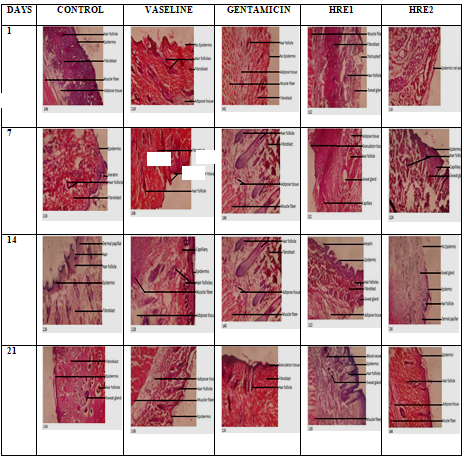
Photomicrograph (x100) of wound healing and epidermal/dermal from days 1 – 21 for wounds stained with H and E.
DISCUSSION
The result of the amino acid profile as presented in Table 1 revealed the presemce of all the essential amino acids as well as non-essential amino acids. Aspartate (12.94mg/100g), methionine (9.38mg/100g), Arginine (8.74mg/100g) were detected in substantive amount summing up to 31.06mg/100g of the total amino acid composition. The amino acid result of H. rotundifolia leaves revealed that Leucine, Lysine, Alanine, Methionine, Phenylalanine, Threonine, Serine and Valine were found in reduced concentrations except Histidine in comparison with the report of Shemishere et al. (2018) in dried leaves of M. cercropioides. Aspartate is essential for purine, pyrimidine, asparagine and inositol synthesis. It also participates in the detoxification and excretion of ammonia. Arginine is an indispensible amino acid in children because of its function in growth and development. Proteins and amino acids are relevant in the entire wound healing activity for instance methionine and cysteine participate in the biosynthesis of connective tissue and collagen while arginine aids in collagen proliferation and immune reaction.
Adequate supply of proteins is essential in wound healing. According to Candlish and Chandra (1996) the appearance of wound in animals leads to protein reduction in the blood and may change in a complex manner their protein metabolism. The significant increase in the total protein level as exhibited by the test animals (Table 2) in HRE1 as compared to control and reference drug (gentamicin) revealed the effectiveness of the plant extract in enhancing the synthesis of collagen content in excised tissue. The result also showed an enhanced nutritional condition of the rats which is vital for wound healing by improving cellular reproduction and migration to the wound tissue and fibroblast and granulation tissue formation. The observation from the histological examination of the wounded tissue also supports this result.
The percentage body weight gain recorded in HRE2 across the weeks, control and HRE1 after 2 and 3 weeks and Vaseline after 2 weeks. indicates that the extract had a positive outcome on their body weights. According to Demling and DeSanti, (2002), in spite of optimum nutrition and early wound closure, there could be notable involuntary weight loss which is majorly lean body mass. This weight loss is based on high rate of catabolic response leading to the use of body protein for energy. Also, Ma et al. (2015) suggestion on body weight loss in rats as seen in this work might be as a result of wound metabolic, circulatory and immunological imbalances.
Histological examination showed a normal skin for control on day 1 while other groups revealed a disrupted skin or skin without epidermis. On day 7, formation of new epithelial layer lined by thin layer of keratin, scab formation and fibroblast proliferation was revealed for control. Epithelial cells undergo mitosis to give off cells called keratinocytes, which form the superficial layers of the epidermis. Keratinized epithelium arranges a covering against abrasion, bacterial invasion and desic1cation. Vaseline and gentamicin revealed proliferation of fibroblast dominated by granulation tissue formation dominating the surface of the wound, formation of epidermis. HRE1 revealed new epithelial layer formation lined by a dense layer of keratin was seen at the skin surface. According to Pastar et al. (2014), keratinocytes are accountable for restoring the epidermis after injury through epithelialization process. HRE2 showed an incomplete epidermis that was beginning to form from the margin and less scab formation. On the 14th day, epithelial cells began to go into the proliferation phase that is typified by granulation tissue formation and collagen formation dominated by fibroblast, less scab formation and thin epidermis formation was detected for control. For Vaseline, proliferative phase continued with collagen deposition, angiogenesis and a thick epidermis was detected also. The epidermal thickness was analogous to intact epidermis. Gentamicin revealed granulation tissues formation by the fibroblast cells and production of thin epidermis. HRE1 revealed granulation tissues formation by fibroblast proliferation, collagen deposition and angiogenesis. Collagen formation and maturation may be based on the existence of flavanoids and steroids in Heterotis rotundifolia leaves (Soni and Singhai, 2012; Jain et al., 2018). A thin layer of epidermis can be seen formed on the outer surface, fibroblast cells and less scab formation in HRE2. Histological observation of wound tissue on the 21st day showed continuation of granulation tissue formation and deposition of collagen. This is the maturation and remodelling phase which begins day 8 through to one year. The collagen formed in granulation tissues is said to be thin. Broughton et al. (2006) suggests that the collagen primarily laid down is thinner than the collagen in uninjured skin (which has a basket weave pattern) and arranged parallel to the skin. He added that overtime, the initial collagen threads are reabsorbed and deposited thicker and organized along the stress lines. Vaseline and HRE1 revealed a repaired/regenerated skin showing an intact epidermis, dermis containing blood vessels, hair follicles, sweat glands and hypodermis containing muscle fibers. Gentamicin revealed continued formation of granulation tissue and thick epidermis while HRE2 revealed keratin formation, granulation tissue characterized by proliferation of fibroblast, scab formation and a thin epidermis formed on the surface.
CONCLUSION
In conclusion, the study reveals that H. rotundifolia leaves possess essential amino acids and wound healing potentials.
REFERENCES
- Abere, T.A., Okoto, P.E. and Agoreyo, F.O. (2010). Antidiarrhoea and toxicological evaluation of the leaf extract of Dissotis rotundifolia Triana (Melastomataceae). BMC Complementary and Alternative Medicin.e, 10:71
- Abere, T. A., Onwukaeme, D. N., Eboka, C. J. (2009). Pharmacognostic evaluation of the leaves of Dissotis rotundifolia Triana (Melastomataceae). African Journal of Biotechnology., 8(1): 113-115.
- Agwu, J. A., Uwakwe, A.A., Nzor, J. N. and Bobson, M.P. (2018). Effect of Heterotis rotundifolia leaf extract on Hemoglobin-S (HbS) Polymerization, Osmotic fragility and Fe2+/Fe3+ ratio. International Journal of Scientific and Engineering Research., 9(5):6-11.
- Aja, P.M, Alum, E.U, Ezeani, N.N, Ibiam, U.A and Egwu, C. (2015). Comparative phytochemical evaluation of Dissotis rotundifolia root and leaf. Global Veterinaria., 14(3):418-424.
- Amri, E and Kisangau, D.P. (2012). Ethnomedicinal study of plants used in villages around Kimboza forest reserve in Morogoro, Tanzania. Journal of Ethnobiology and Ethnomdicine., 8:1.
- Chandran, P. K and Kuttan, R. (2008). Effect of Calendula officinalis flower extract on acute phase proteins, antioxidant defense mechanism and granuloma formation during thermal burns. Journal of Clinical Biochemistry and Nutrition., 43:58-64.
- Demling, R.H. and DeSanti, L. (2002). Restoration of body weight, function and wound healing after severe burns using the anabolic agent oxandrolone is not age dependent. Ostomy Wound Management., 48(8):42-47.
- Dhumal, S.V. and Kulkarni, S.R. (2007). Antibacterial and wound healing activity of roots of Sesamum indicum. Indian Drugs., 44:937-944.
- Dielgelmann, R.F. and Evans, M.C. (2004). Wound healing: an overview of acute, fibrotic and delayed healing. Frontiers in Bioscience., 9: 283-289.
- Elkin, R.G. and Griffith, J.E. (1985). Amino acid analysis of feedstuff hydrolysates by cation exchange high performance liquid chromatography. Journal of Association of Official Analytical chemists. 68(5):1028-32.
- Firdous, S.M and Sautya, D. (2018). Medicinal plants with wound healing potential. Journal of Bangladesh Pharmacological Society (BDPS)., 13(1):41-52.
- Flanagan, M. (2000). The physiology of wound healing. Journal of Wound Care., 9(6):299-300.
- Hess, C. T. (2005). Clinical Guide: Wound care. 5th edition. USA: Lippnicott Williams and Wilkins Maryland.
- Ikeh, C.(2016). Protective role of aqueous leaf extract of Vernonia amygdalina Del. (Asteraceae) and Occium gratissium Linn (Lamiaceae) in cyclophosphamide induced urotixicity and myelosuppression (Doctoral dissertation).
- Kloth, L.C.,McGulloch, J.M., Feedar, J.A. (1990). Wound healing In: Kloth LC, McCulloch JM, Feedar JA, editors: Alternatives in management. Philadelphia (PA): FA Davis Co. Pp: 3-13.
- James, O and Friday, E.T. (2010). Phytochemical composition, bioactivity and wound healing potential of Euphorbia heterophylla (Euphorbiaceae) leaf extract. International Journal on Pharmaceutical and Biomedical Research. 1(1):54-63.
- Kumar, B., Vijayakumar, M., Govindarajan, R. and Pushpangadan, P. (2007). Ethnopharmacological approaches to wound healing-exploring medicinal plants of India. Journal of Ethnopharmacology, 114:103-113.
- Lin, T.S., Latiff, A.A., Hamid, A.A., Ngah, W.Z.W. and Mazlan, M. (2012). Evaluation of topical tocopherol cream on cutaneous wound healing in strptozotociin-induced diabetic rats. Evidenced-Based Complementary and Alternative Medicine.,Pp: 1-6.
- Ma, K., Du, M., Liao, M., Chen, S., Yin, G., Liu, Q., Wei, Q. and Qin, G. (2014). Evaluation of wound healing effect of Punica granatum peel extract on deep second-degree burns in rats. Tropical Journal of Pharmaceutical Research., 14(1):73-78.
- MacKay, D.J. and Miller, A.L. (2003). Nutritional support for wound healing. Alternative Medical Review., 8(4):359-377.
- Mann, A. and Ogbadoyi, E.O.( 2012). Evaluation of Medicinal Plants from Nupeland for Their in vivo Antitrypanosomal Activity. American Journal of Biochemistry., 2(1): 1-6.
- Noumi, E. and Yomi, A. (2001). Medicinal plants used in intestinal diseases in Mbalmayo Region. Fitoterapia., 72(3):246–254.
- Odimegwu, D.C., Ibezim, E.C., Esimone, C.O., Nworu, C.S., Okoye, F.B.C. (2008).Wound healing and antibacterial activities of the extract of Dissotis theifolia (Melastomataceae) stem formulated in a simple ointment base. Journal of Medicinal Plants Research., 2(1):11–16.
- Offor, C.E. (2015). Determination of vitamin composition of Dissotis rotundifolia leaves. International Journal of Current Microbiology and Applied Science., 4(1): 210-213.
- Ofusori, A.E., Raharjo, Y., Ofusori, D.A. and Adekunle, V.O. (2023). A comparative study of dichloromethane and ethylacetate root extract of Celosia trigyna: Phytochemical and wound healing effect analyses. Journal of Wound Management and Research., 19(2):87-95.
- Ogunka-Nnoka, C.U., Agwu, J.A., and Igwe, F.U. (2020) Nutrient and essential oil compositions of Heterotis rotundifolia leaves. American Journal of BioScience., 8(2):28-36.
- Parkar, H. (2016). Wound healing potential of Terminalia sericea. (Master of Science dissertation).
- Pérez-Urria, C.E. and García, A.A.(2009).Metabolismo secundario de plantas. REDUCA., 2: 119-145.
- Porembski, S.J., Szarzynski, J.P.M and Barrthlott, W. (1996). Biodiversity and vegetation of small-sized inselbergs in a West African rain forest (Tai, Ivory Coast). Journal of Biology., 23:47-55.
- Sabale, P., Bhimani, B., Prajapati, C. and Sabale, Y. (2012). An overview of medicinal plants as wound healers. Journal of Applied Pharmaceutical Sciences., 2(11):143-150.
- Shemishere, U.B., Taiwo, J.E., Erhunse, N. and Omoregie, E.S. (2018). Comparative study on the proximate analysis and nutritional composition of Musanga cercropiodes and Maesobotyra barteri leaves. Journal of Applied Sciences and Environmental Management., 22(2):287-291.
- Sorg, H., Tilkorn, D.j., Hager, S., Hauser, J. and Mirastschijiski, U. (2017). Skin wound healing: An update on the current knowledge and concepts.European Surgical Research., 58:81-94.
- Thakur, R., Jain, N., Pathak, R., and Sandhu, S.S. (2011). Practices in wound healing studies of plants. Evidence-Based Complementary and Alternative Medicine., 1-18.
- Thomas, J.C. (1997). Veternanj Pathology. 6th edition. USA: Williams and Wilkin Maryland. Pp:150-156.
- Thomas, J.C. and Howes, P.R. (1997). Effect of bacterial contamination on wound healing. Journal of Ethnopharmacology., 64:191-194.
- Trengove, N.J., Langton, S.R., Stacy, M.C. and Fracs, D.S. (1996). Biochemical analysis of wound fluid from non-healing and healing chronic leg ulcers. Journal of Wound Repair and Regeneration., 4(2): 234-239.
- Wicken, G.E. (1975). Melastomataceae. Flora of Tropical East Africa. Crown Agent of Overseas Government and Administration, 147:39.
- Wild, T., Rahbarnia, A., Kellner, M., Sobtka, L and Eberlein, T. (2010). Basics in nutrition and wound healing. Nutrition,. 26:862-866.
- Yeboah, O.K. and Osafo, N. (2017). Review of the ethnomedical, phytochemical, pharmacological and toxicological studies on Dissotis rotundifolia (Sm.) Triana.Journal of Complementary and Alternatice Medical Research., 2(3): 1-11.
- Zaki, M.Z.M, Hamid, A. and Latiff, M.A. (2011). Effects of Alocasia Sp. Stem juice on open wound healing in rats. Advances in Environmental Biology., 5(12):3734-3742.