
Antibacterial and Antioxidant Activity of Biosynthesized Silver Nanoparticle using Cassia Occidentalis
- Musa G.M.
- Modibbo A.A.
- Tanko M. M.
- 53-61
- Mar 29, 2024
- Chemistry
Antibacterial and Antioxidant Activity of Biosynthesized Silver Nanoparticle using Cassia Occidentalis
Musa G.M.1, Modibbo A.A.1 and Tanko M. M.2
1Department of Chemical Science Technology,
Federal Polytechnic, Mubi, Nigeria
2Department of Biomedical and Pharmaceutical Technology,
Federal Polytechnic, Mubi, Nigeria
DOI:https://doi.org/10.51244/IJRSI.2024.1103006
Received: 11 February 2024; Revised: 21 February 2024; Accepted: 26 February 2024; Published: 29 March 2024
ABSTRACT
Nanotechnology is a fascinating field of research that has applications in various areas such as material science, medical science, life science, physical and chemical sciences. A study was conducted to determine the antioxidant and antimicrobial properties of silver nanoparticles. The silver nanoparticles were biosynthesized from the leaves of Cassia occidentalis. The antimicrobial effectiveness of the biosynthesized silver nanoparticles was tested against Staphylococcus aureus and Pseudomonas Stutzeri, and in vitro antioxidant activities were measured using DPPH, FRAP, and Hydrogen peroxide. The microorganisms were treated with 10 and 20 µg/mL of the silver nanoparticles and 20 µg/mL of ciprofloxacin for 72 hours. After incubation, the silver nanoparticles significantly inhibited the growth of Staphylococcus aureus (17.00 ± 2.50), which was higher than the inhibition observed with ciprofloxacin (15.00 ± 0.05). The zone of inhibition of Pseudomonas Stutzeri was 15.00 ± 3.00, which was inhibited by the silver nanoparticles compared to ciprofloxacin (22.00 ± 2.00). The silver nanoparticles also showed significant antioxidant activity compared to the standard ascorbic acid. In conclusion, the study found that the biosynthesized silver nanoparticles exhibited antimicrobial and antioxidant properties against the tested microorganisms.
Keywords: Anti-microbial, silver nanoparticle, Cassia occidentalis, antioxidant,
INTRODUCTION
Nanotechnology deals with the manufacturing and applications of materials with Nano scale of 1–100 nm in size (Bhushan, 2017). Nanoparticles (NPs) have shown distinctive properties, which can be rightfully manipulated for several preferred applications due to their high surface area to volume ratio (Chadha et al., 2022). Nanoparticles (NPs) have wide range of applications in areas such as health care, cosmetics, food and feed, environmental health, biomedical sciences etc (Osama et al., 2020).
In general, NPs can be classified into metal, metal and metalloid oxides (inorganic) NPs (e.g. gold, silver, iron oxide and silicon dioxide), carbon-based NPs (e.g. carbon nanotubes and nanofibers) and organic NPs (e.g. dendrimers, liposomes and micelles) (Joudeh & Linke, 2022).
Silver nanoparticles (AgNPs) are applicable nanomaterial in medicine that are used as an antimicrobial production that shows antiseptic activity owing to silver toxicity that induces cell death by making changes in the enzymatic system, and cell permeation (Salleh et al., 2020). Several studies have shown the toxic effects of AgNP on numerous cell lines such as macrophages, embryonic kidney cells, skin keratinocytes, hepatocytes, neuroblastoma cells, etc (Glinski et al., 2021, Milić et al., 2015, Gao et al., 2021 and Zhang et al., 2016). Moreover, animal studies have demonstrated that AgNP administered to animals by different methods such as inhalation, ingestion, or Injections, these nanoparticles detect presence in the blood, and finally lead to toxicity of multiple organs such as the lungs, liver, kidneys, intestines, and brain (Ferdous & Nemmar, 2020, Bergin & Witzmann, 2013, and Nayek et al., 2021).
Silver nanoparticles (SNP) bind to the bacterial cell wall surface, which has a negative charge and cause stability interruption and permeability alteration in the envelope of the cell wall and the flow of silver ions inside the cell (Hassanien & Shaker, 2020). It has been reported that interaction of nanoparticles with cell protein and DNA results in interference of cell division and cell death (Sharmin et al., 2021).
This study was aim at determining the antioxidant and antimicrobial of silver nanoparticles
METHODOLOGY
Collection of Cassia occidentalis leaf
Fresh leaves of Cassia occidentalis was collected from Adamawa State University (ADSU) Mubi Botanical garden. Cassia occidentalis plant was botanically identified using the standard morphological characteristic features.
Aqueous extraction of Cassia occidentalis leaves
The fresh leaves of Cassia occidentalis was shade dried at room temperature for 11 days. The shade dried leaves of Cassia occidentalis was grounded into powder using a mortar and pestle (Raghasudha, 2016). The powdered leaves of Cassia occidentalis was stored in well labelled airtight container. Twenty grams of powdered leaves was weighed and transferred into 500 mL beaker containing 200 mL of deionized water. The mixture was boiled for 10 minutes allowed to cool and filtered through Whatman No.1 filter paper (Kumar et al., 2014).
Biosynthesis of silver nanoparticle:
Using the method of Keshari et al. (2016), 10 mL extract was added to the 180 mL silver nitrate solution (1mM) in 90 mL and stirred for 30 min using a magnetic stirrer at room temperature. The change in colour of the solution indicate the reduction of silver nitrate into the biosynthesized AgNPs. The precipitate formed was centrifuged and will behed with distilled water 3 times and be subjected to characterization as follows.
Separation and Purification of Silver Nanoparticles
After desired reaction period, the mixture containing product will be centrifuged at 110 X 100 rpm for 20 min. The process of centrifugation and re-dispersion in deionized water was repeated three times to ensure better removal of unreacted phytochemicals from the AgNPs (Premasudha et al., 2015).
Characterization of Synthesized Nanoparticles
The biosynthesized nanoparticles were characterized using the method according to Ghosh et al. (2015) UV visible spectrophotometer and Fourier Transmission Infar Red (FTIR) were used in characterizing the nanoparticle.
The Antimicrobial Activity of AgNPs
Agar well diffusion method was followed for the determination of Antimicrobial activity. The isolates were grown in Nutrient agar medium at 37 ºC for 24 hrs. Bacterial suspension (inoculum) was dilute with a sterile physiological solution to 108 cfu mL-1 with reference to the Mcfarland turbidometry. The bacterial suspension was added to each plate containing Muller Hinton Agar (MHA) by sterile cotton swab and allowed to remain in contact for 1 min. the AgNPs was dissolved by using Dimethyl sulphur oxide (DMSO) at concentration of 10 and 20 µg/mL, wells were made on the plates using a core borer with a diameter of 8 mm, these wells filled by addition 100 μL of each AgNPs concentration with the use of DMSO and Ciprofloxacin as a control. The plates were incubated at 37 ºC for 24 hours the inhibition zone around each well was measured in mm (Barapatre et al., 2016)
Free Radical-Scavenging Activity Using 2,2-Diphenyl-1-picrylhydrazyl (DPPH)
The antioxidant activity was monitored according to the DPPH method described by Martins et al. (2015) with modifications. This method was performed by using a SPECTRO star Nano microplate reader (BMG Labtech, Offenburg, Germany). The reaction mixture in each of the 96-wells consisted of 30 µL of sample solution (in an appropriated dilution, according to the reference range of the calibration curve) and a 0.004% methanolic solution of DPPH. The mixture was further incubated for 30 min in the dark, and the reduction of the DPPH radical was determined by measuring the absorption of the sample at 515 nm. Trolox was used as a standard reference and the results were expressed as Trolox equivalents (TE) per g of dry weight (mg TE/g dw TS).
Reducing power assay
The ferric reducing power of the Cassia occidentalis biosynthesized AgNPs were determined (Alavi and Karimi, 2017). The concentrations were used in the range of 1 – 5 µg/mL and the standard used as ascorbic acid. Based on the absorbance value, the ferric reducing power was expressed. IC50 values were calculated using the linear regression curve for all the antioxidant assays.
Hydrogen peroxide scavenging assay
The free radical scavenging activity was determined by hydrogen peroxide assay. Hydrogen peroxide (40 mM) solution was prepared in phosphate buffered saline (0.1 M, pH 7.4). 1 ml of the AgNPs containing samples of different concentration will be rapidly mixed with 600 μL of hydrogen peroxide solution. The absorbance was measured at 230 nm after 10 min of incubation at room temperature against a blank (without hydrogen peroxide). The percentage of scavenging of hydrogen peroxide will be calculated using the following formula. Quercetin was used as a positive control (Venkatesan et al., 2016).
Percentage scavenging H2O2 = Ao−A1/Ao×100.
Ao – Absorbance of control; A1 – Absorbance of sample.
Statistical Analysis
Statistical analysis was performed using SPSS version16 software (SPSS Inc., Chicago, IL) and the results were evaluated by one way ANOVA. Values were presented as mean ± S.EM of the three replicates of each experiment. The results with p < 0.05 were considered to be statistically significant
RESULTS
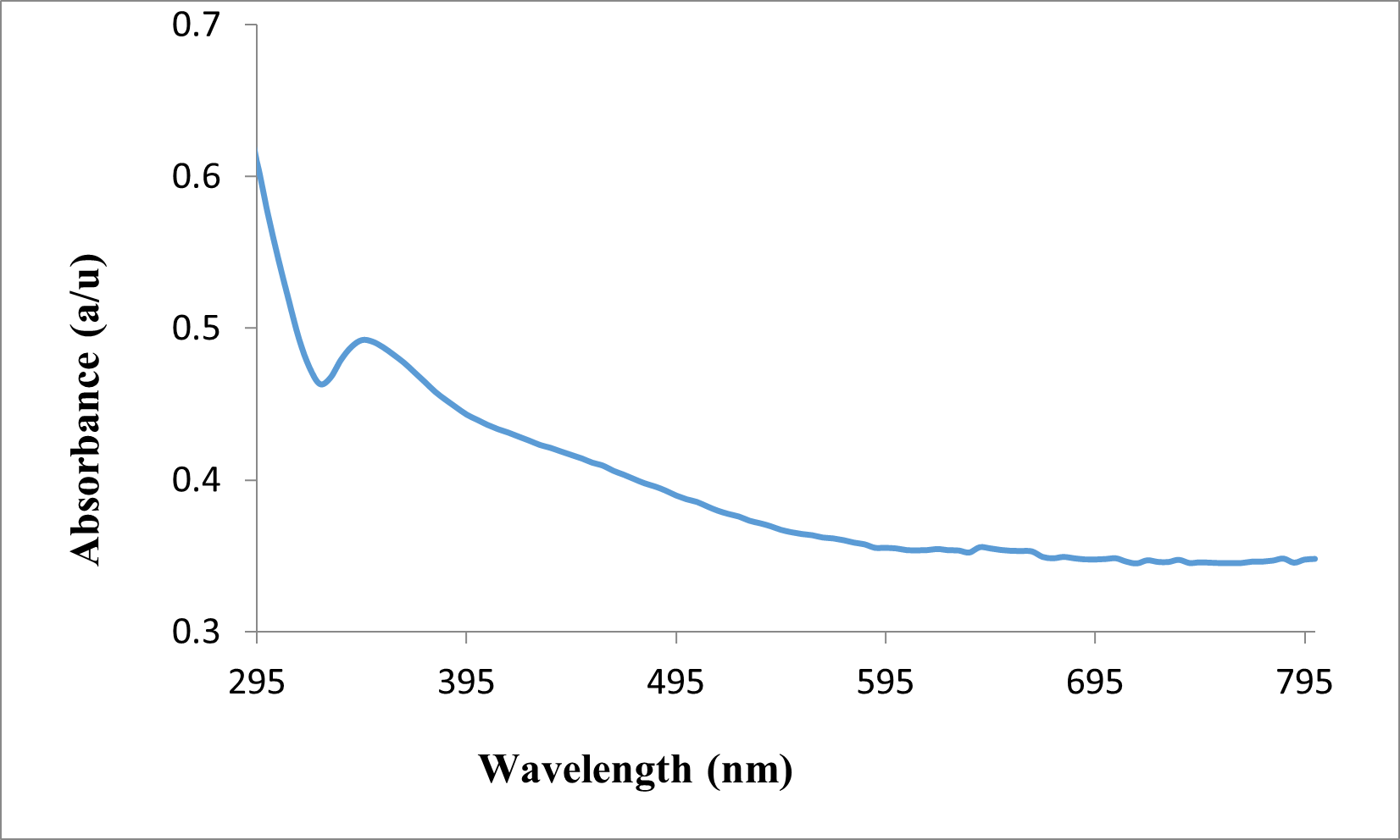
Figure 1: UV Visible spectrum of silver nanoparticle with absorption maximum at 350 nm
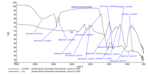
Figure 2: FTIR spectra of both silver nanoparticle and cassia occidentalis plant aqueous extract
Table 1: Zone of inhibition (mm) of antimicrobial activity silver nanoparticles of Cassia occidentalis leaf against Staphylococcus aureus and Pseudomonas stutzeri
| Culture | AgNPs
(10 µg/mL) |
AgNPs
(20 µg/mL) |
Ciprofloxacin
(20 µg/mL) |
| Staphylococcus aureus | 10.00 ± 1.00 | 17.50 ± 2.50 | 15.00 ± 3.00 |
| Pseudomonas stutzeri | 11.00 ± 1.00 | 15.00 ± 0.05 | 22.00 ± 2.00 |
All values are mean ± SD of three replicates
Table 2: Antioxidant activity of DPPH on silver nanoparticles and ascorbic acids
| S/No | Conc. (mg/mL) | AgNPs (%) | Ascorbic acid (%) |
| 1 | 5 | 76.5 ± 0.21 | 89 ± 0.12 |
| 2 | 4 | 44.4 ± 0.98 | 86 ± 0.53 |
| 3 | 3 | 42.3 ± 0.34 | 83 ± 1.01 |
| 4 | 2 | 33.0 ± 0.48 | 82 ± 0.56 |
| 5 | 1 | 21.6 ± 0.87 | 77 ± 0.78 |
| IC50 | 4.36 | 0.15 |
The values are mean ± SEM of three determinants
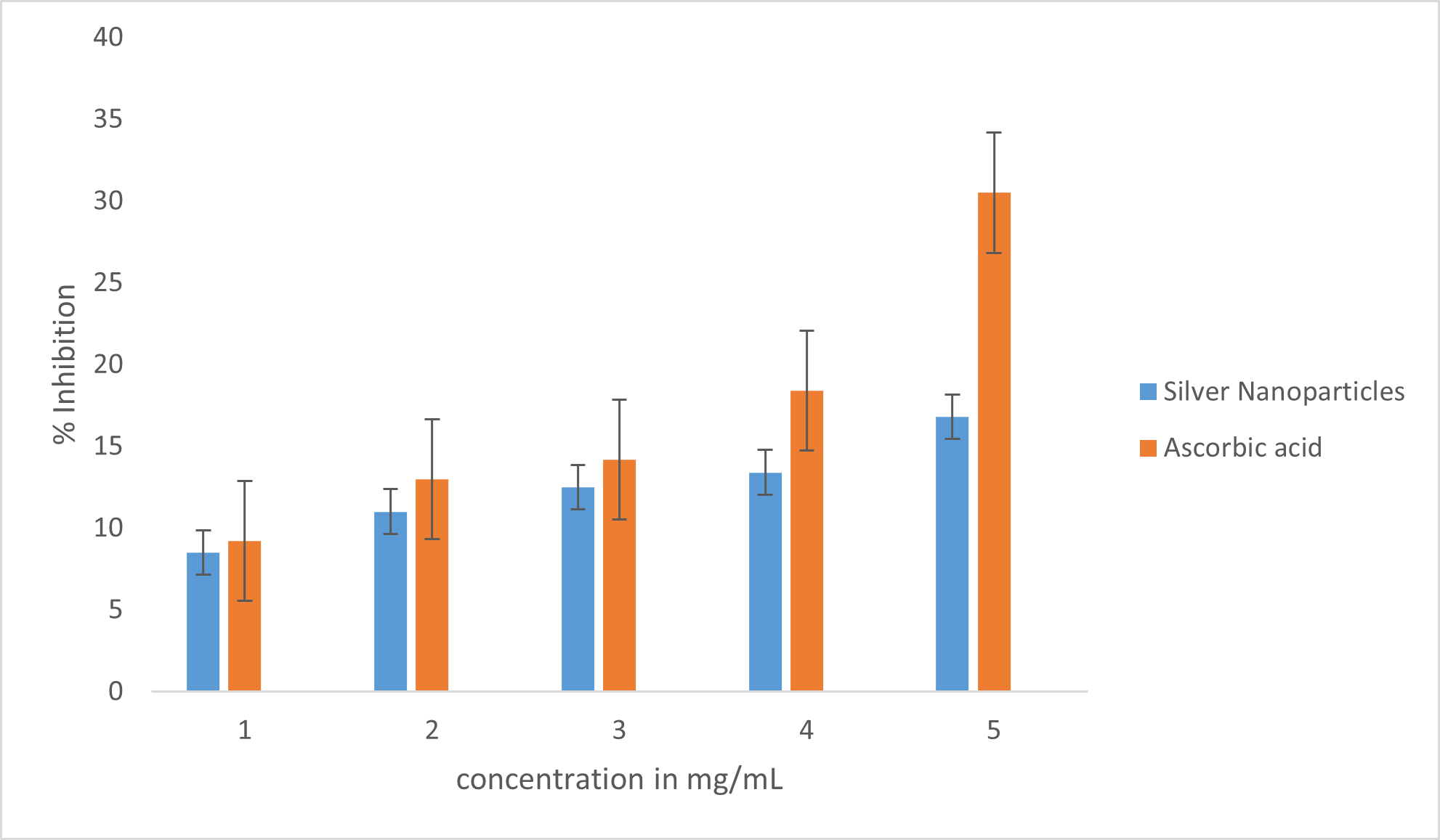
Figure 3: Antioxidant activity of AgNPs biosynthesized from Cassia occidentalis using FRAP
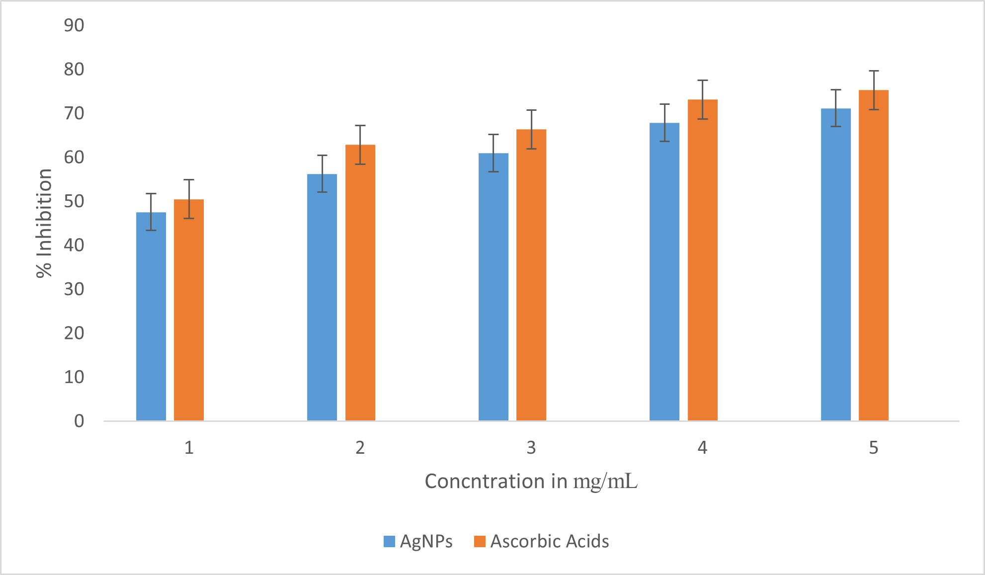
Figure 4: Antioxidant activity of AgNPs biosynthesized from Cassia occidentalis using H2O2
DISCUSSION
The UV visible spectroscopy analysis of the silver nanoparticle gave a sharp absorption maximum at 350 nm (figure 1) which is common to silver nanoparticles (Singh et al., 2012; Nuraqeelah et al., 2018). The narrow nature of the band and it singleness is an indication that the particles are same size, mono disperse and well aggregated (Tira et al., 2020). There has been report of silver nanoparticles with a wavelength maximum of 335.9 nm (Anith et al., 2022). Synthesized zinc oxide and observed that the UV-Vis absorbance spectrum shows strong absorption peaks occurring in the wavelength range of 220-380 nm for the unannealed samples and 190-235 nm for the annealed samples (Zikalala et al., 2022).
FTIR are employed in determining organic functional group present in organic compound or other substance. Eight different absorption peaks were observed in the plant extract as shown in Figure 2 at wavenumbers which corresponds to a particular organic functional group. The broad band observed at 3287.84 cm-1 corresponds to H–bonded O–H stretch alcohols. The peak at 2918.37 cm-1 is assign to C-H stretch of alkane. The absorption band at 2850.07 cm-1 corresponding to C-H stretch of alkane. The band at 1730.93 cm-1 was given CHO of saturated amide. The strong band at 1592.31 cm-1 reveals the presence of N-H bending of amide groups. Another absorption band was observed at 1406.97 cm-1 it was assigned to C-H bending of saturated aliphatic. The stretched absorption band at 1239.96 cm-1 indicate the C-O of ether while the absorption peak of 1024.02 cm-1 correspond to C-O stretched alcohol. This is an indication that the aqueous leaf extract of Cassia occidentalis may contain bioactive molecules like carboxylic acids, proteins, alcohol, alkaloid, flavonoid, etc. There is a shift in these absorption band when compared to the FTIR of the nanoparticle as seen in Figure 2. The band at 2918.37 cm-1 to 2329.20 cm-1, 2850.07 cm-1 to 2114.76 cm-1, 159.25 cm-1 shifted to 1524.68 cm-1, 1406.97 cm-1 to 1375.77 cm-1, 1730.93 cm-1 to 1991.69 cm-1, 1239.96 cm-1 to 1239.56 cm-1 and 1024.02 cm-1 to 958.91 cm-1 while the peak at 837.76 cm-1 presents in nanoparticle but absent in the plant extract was assigned to silver. Mohammad and Aneela, (2016) reported that during the biosynthesis of nanoparticle there is a shift in the peak position.
The bactericidal effect of metal nanoparticles has been attributed to their’ small size and high surface to volume ratio, which allows them to interact closely with microbial membranes and also due to the release of metal ions in solution (Gold et al., 2018). A cell wall is present around the outside of the bacterial cell membrane and it is essential for the survival of bacteria (Egan et al., 2017). The cell membrane is called peptidoglycan made up of polysaccharides and peptides (Garde et al., 2021). These can be thick cell wall containing many layers of peptidoglycan as in Gram positive or thin cell wall consisting of a few layers of peptidoglycan as in gram negative bacteria (Rohde, 2019). Surfaces of AgNPs interact directly with the bacterial outer membrane causing the membrane to rupture and killing bacteria (More et al., 2023).
Antimicrobial activity of the biosynthesized AgNPs based on size of zones of inhibition in millimetre (mm) is shown in table 1. Result revealed that the prepared nanoparticles Possessed antibacterial properties against both Staphylococcus aureus and Pseudomonas stutzeri. The screened nanoparticles exhibited higher activities of 17.54 ± 2.5 mm on Staphylococcus aureus test organisms at a concentration of 2.0 u/ml as compared with Pseudomonas stutzeri in which lower activity of 15 00 ± 0.01 mm was recorded. The result of Staphylococcus aureus in this study seem to be similar to that obtained by Taglialegna et al. (2019) but in contrast to their study, the inhibitory activity of the nanoparticles on Staphylococcus aureus was found to deceases as the concentration of the nanoparticles increases whereas in this study the inhibition activity increases as the concentration of the nanoparticles increases. This could possibly be due to the nature of the plant used in the synthesis of the nanoparticles (Jain & Mehata, 2017).
The antimicrobial activity of the nanoparticles could be due to the smaller size of the particles which leads to tightly adsorbed on the surface of the bacterial cells so as to disrupting the membrane which would lead to the leakage of intracellular component, there by killing the bacterial cells (Häffner & Malmsten, 2017). Another possible mechanism involves the association of AgNPs nanoparticles with oxygen and its reaction with sulfhydryl (-S-H) groups on the cell wall to form R-S-S-R bonds, thereby blocking respiration and causing cell death (Ezeuko et al., 2021). In this study Ciprofloxacin 20 ug/ml was used as positives control which has high zone of inhibition as compare with the nanoparticles in Pseudomonas stutzeri.
The antioxidant activity of Cassia occidentalis biosynthesized AgNPs was determined by using DPPH free radical and FRAP assay. DPPH is a stable compound which can be reduced by accepting the hydrogen or electrons and has been widely used to evaluate the antioxidant activity (Ivanova et al., 2020). The lower IC50 value indicates a stronger ability of the substance to act as a DPPH scavenger, while the higher IC50 value indicates a lower scavenging activity (Rahman et al., 2015). The results revealed that the synthesized AgNPs are free radical scavengers. The DPPH activity of the AgNPs was found to increase as the concentration increases ranging from 21.6 % to 76.5 % at concentrations 5 mg/mL, AgNPs showed a scavenging activity of 76.5 % with IC50 value 4.36. The antioxidant activity was far greater than that of standard ascorbic acid which is 0.15 at 5 mg/mL with highest inhibition of 89 %. In FRAP assay, the ability of AgNPs to reduce Fe3+ to Fe2+ was also found to increase as the concentration of the AgNPs in the analysis increases, again antioxidant activities of the nanoparticles were less than that of standard ascorbic acid (Dridi et al., 2022).
In conclusion, the AgNPs biosynthesised from Cassia occidentalis have the ability to inhibit the growth of bacteria.
ACKNOWLEDGMENT
I wish to acknowledge the Tertiary Education Fund Nigeria (Tetfund) for funding this research under the Institutional Based Research (IBR) intervention. Furthermore, my sincere appreciation also goes to the Management of Federal Polytechnic Mubi for the timely approval and onward submission in the course of accessing the funding.
REFERENCES
- Alavi M, Karimi N. (2017). Characterization, antibacterial, total antioxidant, scavenging, reducing power and ion chelating activities of green synthesized silver, copper and titanium dioxide nanoparticles using Artemisia haussknechtii leaf extract. Artif Cells Nanomedicine Biotechnol. 46:2066–81.
- Anith, R. J., Devina, D. M., Arulananth, T. S. and Nagaraju, S. (2022) Characterization Analysis of Silver Nanoparticles Synthesized from Chaetoceros calcitrans Journal of Nanomaterials Volume 2022, Article ID 4056551, 15
- Barapatre A., Aadil K.R.and Jha H. (2016). “Synergistic antibacterial and antibiofilm activity of silver nanoparticles biosynthesized by lignindegrading fungus”. Bioresour Bioprocess. 3(8): 1-13
- Bergin, I. L., & Witzmann, F. A. (2013). Nanoparticle toxicity by the gastrointestinal route: evidence and knowledge gaps. International journal of biomedical nanoscience and nanotechnology, 3(1-2), 163-210.
- Bhushan, B. (2017). Introduction to nanotechnology. Springer handbook of nanotechnology, 1-19.
- Chadha, U., Selvaraj, S. K., Ashokan, H., Hariharan, S. P., Mathew Paul, V., Venkatarangan, V., & Paramasivam, V. (2022). Complex nanomaterials in catalysis for chemically significant applications: from synthesis and hydrocarbon processing to renewable energy applications. Advances in Materials Science and Engineering, 2022, 1-72.
- Dridi, R., Essghaier, B., Hannachi, H., Khedher, G. B., Chaffei, C., & Zid, M. F. (2022). Biosynthesized silver nanoparticles using Anagallis monelli: Evaluation of antioxidant activity, antibacterial and antifungal effects. Journal of Molecular Structure, 1251, 132076.
- Egan, A. J., Cleverley, R. M., Peters, K., Lewis, R. J., & Vollmer, W. (2017). Regulation of bacterial cell wall growth. The FEBS journal, 284(6), 851-867.
- Ezeuko, A. S., Ojemaye, M. O., Okoh, O. O., & Okoh, A. I. (2021). Potentials of metallic nanoparticles for the removal of antibiotic resistant bacteria and antibiotic resistance genes from wastewater: A critical review. Journal of Water Process Engineering, 41, 102041.
- Ferdous, Z., & Nemmar, A. (2020). Health impact of silver nanoparticles: a review of the biodistribution and toxicity following various routes of exposure. International journal of molecular sciences, 21(7), 2375.
- Gao, X., Li, R., Sprando, R. L., & Yourick, J. J. (2021). Concentration-dependent toxicogenomic changes of silver nanoparticles in hepa t ocyte-like cells derived from human induced pluripotent stem cells. Cell Biology and Toxicology, 37, 245-259.
- Garde, S., Chodisetti, P. K., & Reddy, M. (2021). Peptidoglycan: structure, synthesis, and regulation. EcoSal Plus, 9(2).
- Ghosh, P. K., Bikash, M., Susanta, G., & Basumallick, a. I. ( 2015). Synthesis and Study of Corrosion Protection Efficiency of Silica Nanoparticles on Aluminium. British Journal of Applied Science & Technology 5(2), 198-209.
- Glinski, A., de Souza, T. L., da Luz, J. Z., Junior, A. G. B., de Oliveira, C. C., de Oliveira Ribeiro, C. A., & Neto, F. F. (2021). Toxicological effects of silver nanoparticles and cadmium chloride in macrophage cell line (RAW 264.7): An in vitro approach. Journal of Trace Elements in Medicine and Biology, 68, 126854.
- Gold, K., Slay, B., Knackstedt, M., & Gaharwar, A. K. (2018). Antimicrobial activity of metal and metal‐oxide based nanoparticles. Advanced Therapeutics, 1(3), 1700033.
- Häffner, S. M., & Malmsten, M. (2017). Membrane interactions and antimicrobial effects of inorganic nanoparticles. Advances in colloid and interface science, 248, 105-128.
- Hassanien, A. A., & Shaker, E. M. (2020). Investigation of the effect of chitosan and silver nanoparticles on the antibiotic resistance of Escherichia coliO157: H7 isolated from some milk products and diarrheal patients in Sohag city, Egypt. Veterinary World, 13(8), 1647.
- Ivanova, A., Gerasimova, E., & Gazizullina, E. (2020). Study of antioxidant properties of agents from the perspective of their action mechanisms. Molecules, 25(18), 4251.
- Jain, S., & Mehata, M. S. (2017). Medicinal plant leaf extract and pure flavonoid mediated green synthesis of silver nanoparticles and their enhanced antibacterial property. Scientific reports, 7(1), 15867.
- Joudeh, N., & Linke, D. (2022). Nanoparticle classification, physicochemical properties, characterization, and applications: a comprehensive review for biologists. Journal of Nanobiotechnology, 20(1), 262.
- Martins, N.; Barros, L.; Dueñas, M.; Santos-Buelga, C.; Ferreira, I.C.F.R. (2015). Characterization of phenolic compounds and antioxidant properties of Glycyrrhiza glabra L. rhizomes and roots. R. Soc. Chem. Adv. 5, 26991–26997.
- Milić, M., Leitinger, G., Pavičić, I., Zebić Avdičević, M., Dobrović, S., Goessler, W., & Vinković Vrček, I. (2015). Cellular uptake and toxicity effects of silver nanoparticles in mammalian kidney cells. Journal of Applied Toxicology, 35(6), 581-592.
- More, P. R., Pandit, S., Filippis, A. D., Franci, G., Mijakovic, I., & Galdiero, M. (2023). Silver nanoparticles: bactericidal and mechanistic approach against drug resistant pathogens. Microorganisms, 11(2), 369.
- Muhammad, I. D., & Aneela, R. (2016). Recent Advances in the Synthesis and Stabilization of Nickel and Nickel Oxide Nanoparticles: A Greener Adeptness. International Journal of Analytical Chemistry Volume 2016, 1-14.
- Nayek, S., De Silva, I. W., Aguilar, R., Lund, A. K., & Verbeck, G. F. (2021). Toxicological alterations induced by subacute exposure of silver nanoparticles in Wistar rats. Journal of Applied Toxicology, 41(6), 972-986.
- Nuraqeelah, M. S., Boon, S. W., Suk, F. C. and Kuan, Y. K. (2018) Synthesis and Characterization of Zinc Oxide Nanoparticles with Small Particle Size Distribution Acta Chim. Slov. 2018, 65, 578–585
- Osama, E., El-Sheikh, S. M., Khairy, M. H., & Galal, A. A. (2020). Nanoparticles and their potential applications in veterinary medicine. Journal of Advanced Veterinary Research, 10(4), 268-273.
- Rahman, M. M., Islam, M. B., Biswas, M., & Khurshid Alam, A. H. M. (2015). In vitro antioxidant and free radical scavenging activity of different parts of Tabebuia pallida growing in Bangladesh. BMC research notes, 8(1), 1-9.
- Rohde, M. (2019). The Gram-positive bacterial cell wall. Microbiology Spectrum, 7(3), 10-1128.
- Salleh, A., Naomi, R., Utami, N. D., Mohammad, A. W., Mahmoudi, E., Mustafa, N., & Fauzi, M. B. (2020). The potential of silver nanoparticles for antiviral and antibacterial applications: A mechanism of action. Nanomaterials, 10(8), 1566.
- Sharmin, S., Rahaman, M. M., Sarkar, C., Atolani, O., Islam, M. T., & Adeyemi, O. S. (2021). Nanoparticles as antimicrobial and antiviral agents: A literature-based perspective study. Heliyon, 7(3).
- Singh, D. S., Pandey, D. K., Yadav, R. R. and Devraj, S. (2012) A study of nanosized zinc oxide and its nanofluid PRAMANA Indian Academy of Sciences journal of physics 78 (5):759–766
- Taglialegna, A., Varela, M. C., Rosato, R. R., & Rosato, A. E. (2019). VraSR and virulence trait modulation during daptomycin resistance in methicillin-resistant Staphylococcus aureus infection. MSphere, 4(1), 10-1128.
- Tira, A., Windri, H., Cuk, I. (2020) The UV visible spectrum
- Venkatesan J, Kim S-K, Shim M. (2016). Antimicrobial, Antioxidant, and Anticancer Activities of Biosynthesized Silver Nanoparticles Using Marine Algae Ecklonia cava. Nanomaterials. 6:1–18.
- Zhang, X. F., Shen, W., & Gurunathan, S. (2016). Silver nanoparticle-mediated cellular responses in various cell lines: an in vitro model. International journal of molecular sciences, 17(10), 1603.
- Zikalala, N. E., Azizi, S., Zikalala, S. A., Kamika, I., Maaza, M., Zinatizadeh, A. A., … & Kaviyarasu, K. (2022). An Evaluation of the Biocatalyst for the Synthesis and Application of Zinc Oxide Nanoparticles for Water Remediation—A Review. Catalysts, 12(11), 1442.