
Determination of Radiological Variations of Crista Galli in Humans
- Okoseimiema Sonny Clement
- Ekpenyong Aniedi Moses
- 534-541
- Apr 18, 2025
- Education
Determination of Radiological Variations of Crista Galli in Humans
Okoseimiema Sonny Clement1, Ekpenyong Aniedi Moses2*
1Department of Anatomy, Faculty of Basic Medical Sciences, University of Port Harcourt, Port Harcourt, Nigeria,
2Department of Mathematics, Rivers State University, Port Harcourt, Nigeria
*Corresponding Author
DOI: https://doi.org/10.51244/IJRSI.2025.121500049P
Received: 25 March 2024; Accepted: 01 April 2025; Published: 18 April 2025
ABSTRACT
A cross sectional study that was ethically approved and conducted at the Rivers State University Teaching Hospital and Intercontinental Diagnostic Center in Rivers State. The crista galli is a bony prominence within the anterior cranial fossa that exhibits significant anatomical variations that have clinical implications for neurosurgical and endoscopic sinus procedures. This review examines the diverse morphological presentations of the crista galli, including variations in size, shape, position, and pneumatization. Radiological imaging techniques, particularly computed tomography (CT), have provided valuable insights into these variations. Understanding these anatomical nuances is essential for surgeons to navigate the complex anatomy of the anterior skull base and minimize potential complications. The computed tomography images of 251 adults were evaluated for the types of Crista galli, based on its relation to the cribiform plate, and the Crista galli was also accessed for the presence of pneumatization. The generated data were analyzed using SPSS; the frequencies of the variants were presented in percentages. The K test was used to evaluate the differences in types of Crista galli, and the independent t test was used to evaluate sex differences. The type II Crista galli was the most common kind in both males and females, accounting for 71.2% and 72.3% of cases, respectively. Additionally, the data demonstrated a statistically significant association between the gender and the of crista galli as (P=0.812). However, the Statistical Significance of the P-value 0.135 indicates that there is a statistically significant difference in the Crista Galli types.
Keywords: Crista Galli, Types, Teaching Hospital, Rivers State.
INTRODUCTION
The field of medicine known as radiology uses medical imaging to identify illnesses and direct treatment in both human and animal bodies. Its name has a root that refers to radiation because it started with radiography. Today, it encompasses all imaging modalities, including those that use electromagnetic radiation (like computed tomography (CT), fluoroscopy, and nuclear medicine, including positron emission tomography (PET), as well as those that don’t (like magnetic resonance imaging and ultrasonography).
Several healthcare professionals collaborate in the current practice of radiography. A radiologist is a medical professional who has finished the necessary post-graduate training. They interpret medical images, report or verbally discuss their findings with other medical professionals, and use imaging to perform minimally invasive procedures [3]. In addition to administering medication, keeping an eye on vital signs, and monitoring sedated patients, the nurse also assists with patient care both before and after imaging or treatments. A specially trained healthcare professional, the radiographer—also referred to as a “radiologic technologist” in some countries, including the United States and Canada—uses advanced technology and positioning techniques to create medical images that the radiologist can interpret [13].
A wedge-shaped, vertical, midline upward continuation of the perpendicular plate of the skull’s ethmoid bone, the crista gali (or “crest of the rooster”) protrudes above the cribriform plate into the cranial cavity. The membranes encircling the brain attach to it [4].
It divides the olfactory bulbs, which are located in the cribriform plate’s olfactory fossae on either side of it. It acts as the falx cerebri’s anterior connection. The nasal slits, where the anterior ethmoidal nerves descend into the nasal cavity’s roof, are located directly lateral to it. Because it connects to the falx cerebri posteriorly, it is a helpful anatomical site in anterior skull base surgery [2]. From the ethmoid bone, Crista galli grows embryologically [15]. On computed tomography (CT), pneumatization of the crista galli is a known incidental finding that often has minimal bearing on the clinical picture. Nonetheless, there are several noteworthy exceptions: nasal dermoids and other congenital midline nasal abnormalities have been shown to pass through or close to the crista galli. Additionally, mucocele development has been observed.
The crista galli is not always a compact bone; occasionally, it is pneumatized or filled with spongy bone. There are three sizes of pneumatized crista galli based on the proportions of pneumatization: small, moderate, and large. In clinical practice, pneumatized crista galli can be useful as a morphologic barrier in neurosurgical approaches to certain cancers of the front skull base, or as an inflamed sinus or other pathologies (sinusitis cristae galli, mucocele). During endoscopic sinus surgery, the sinus crista galli may be mistaken for an ethmoidal cell, which increases the risk of an unintentional invasion of the skull base. The thick, triangular crista galli bone extends perpendicularly into the anterior cerebral fossa from the superior surface of the ethmoid bone. The falx cerebri of the dura mater is anchored by its thin, somewhat curved posterior edge [12]. The mesethmoid cartilage, which forms the anterior skull base, including the cartilaginous crista galli, is formed by the presphenoidal cartilage during the eighth week of intrauterine life. Crista galli ossification begins at two months after birth and progresses quickly until 14 months. By the second year of life, ossification has slowed down and stopped ([16], [11]), It is known as sinus crista galli because the crista galli may become pneumatized once air cells from nearby paranasal sinuses extend [10]. Due to its ethmoid bone origin, displaced ethmoidal air cells may aerate the crista galli. Additionally, pneumatization of the crista galli may result from the frontal sinus’s aeration extending over the frontal bone’s boundaries. Clinicians should be aware of this variation since it can cause chronic sinusitis that is resistant to medication if its drainage is blocked [17]. Additionally, because it serves as a crucial landmark during endoscopic sinus and skull base procedures, the crista galli is significant to surgeons. Its proximity to olfactory nerves that enter the orbital content, the anterior cerebral fossa, and the cribriform foramina raises the possibility of iatrogenic consequences, including loss of vision or olfaction and cerebrospinal fluid leaking [1]. Additionally, the crista galli’s mucoceles may extend intracranially, raising the possibility of unintentional intracranial penetration during surgery.
Development and applied importance of the Crista Galli
The mesethmoid cartilage, which forms the anterior skull base, including the cartilaginous crista galli, is formed by the presphenoidal cartilage during the eighth week of intrauterine life. Crista galli ossification begins at two months after birth and progresses quickly until 14 months. By the second year of life, ossification has slowed down and stopped [16]. It is known as sinus crista galli because the crista galli may become pneumatized once air cells from nearby paranasal sinuses extend [10]. Due to its ethmoid bone origin, displaced ethmoidal air cells may aerate the crista galli. Additionally, pneumatization of the crista galli may result from the frontal sinus’s aeration extending past the frontal bone’s edges. Clinicians should be aware of this variation since it can cause chronic sinusitis that is resistant to medication if its drainage is blocked [17].
The orbit and sinonasal compartments below are divided from the cranial cavity above by the anterior skull base. Two bones create this boundary: the orbital plates of the frontal bone laterally and the cribriform plate centrally. The olfactory bulbs rest on the cribriform plate, which is the portion of the ethmoid bone that has two parallel grooves. These are divided by a midline triangular structure known as the crista galli, which gets its name from its similarity to a rooster’s comb. For the falx cerebri, the crista galli is an attachment point. Each groove’s floor contains several small holes that let olfactory nerves reach the nasal mucosa. In front of the cribriform plate are the frontal sinuses. Coronal CT images clearly show the thin cribriform plate. This structure can also be seen on T1-weighted and T2-weighted MRI sequences as a linear hypointense signal. Tumors often affect it, and trauma or surgery can break it. The frontal bone’s orbital plates provide a strong and substantial barrier against the transmission of illness. The medial orbital wall that divides the orbit from the ethmoid sinus is called the lamina papyracea. It gets its name from the fact that it looks like paper and that tumors can readily break or destroy it. Similar to the cribriform plate, its structure is easily accessed by CT. The periosteum that lines the bony orbit’s walls and to which it is tangentially attached is known as the periorbita. Through the superior orbital fissure, it is continuous with the dura mater posteriorly and the periosteum of the face bones anteriorly. In healthy patients, this structure is indistinguishable from the surrounding orbital wall; nevertheless, when it is raised off the underlying bone by a tumor, blood, or pus, it appears as a hypointense line on T1-weighted and T2-weighted MRI.
Additionally, because it serves as a crucial landmark during endoscopic sinus and skull base procedures, the crista galli is significant to surgeons. Its proximity to olfactory nerves that enter the orbital content, the anterior cerebral fossa, and the cribriform foramina raises the possibility of iatrogenic consequences, including loss of vision or olfaction and cerebrospinal fluid leaking [1]. Additionally, the crista galli’s mucoceles may extend intracranially, raising the possibility of unintentional intracranial penetration during surgery.Crista galli bases come in different heights [5]. A nasal dermoid cyst’s intracranial expansion may be indicated by a bifid crista galli.
Types of Radiographic Diagnosis
Computed Tomography
CT imaging creates images of the body by using X-rays and computer algorithms [6]. In CT, a computer-generated cross-sectional image (tomogram) is created by rotating an X-ray tube across from an X-ray detector (or detectors) in ring-shaped equipment around a patient [7]. CT is obtained in the axial plane, and computer reconstruction is used to create the sagittal and coronal pictures. CT is frequently used with radiocontrast drugs to improve anatomical delineation. CT can identify more subtle changes in X-ray attenuation (greater contrast resolution), even though radiographs offer higher spatial resolution. CT exposes the patient to significantly more ionizing radiation than a radiograph.
A spiral multidetector CT scan uses 16, 64, 254, or more detectors while the subject moves continuously across the radiation beam to capture fine-grained images quickly. These fine-grained images can be converted into three-dimensional (3D) images of the carotid, cerebral, coronary, or other arteries by quickly administering intravenous contrast during the CT scan. Early in the 1970s, computed tomography was introduced, revolutionizing diagnostic radiology by giving front-line doctors three-dimensional, detailed views of anatomic structures. Cerebral hemorrhage, pulmonary embolism (clots in the lungs’ arteries), aortic dissection (tearing of the aorta wall), diverticulitis, appendicitis, and obstructive kidney stones are among the urgent and emergent disorders for which CT scanning has emerged as the preferred test. Before the invention of CT imaging, the only reliable method of determining the source of acute abdominal discomfort that could not be determined by external observation was frequently through invasive and painful exploratory surgery [8]. The accuracy and utility of CT scanning have significantly risen due to ongoing advancements in technology, such as quicker scanning times and better resolution, which may help explain why it is being used more frequently for medical diagnosis.
Magnetic Resonance Imaging (MRI)
In MRI, atomic nuclei (often hydrogen protons) are aligned within human tissues using powerful magnetic fields. A radio signal is then used to disrupt the nuclei’s axis of rotation, and the radio frequency signal produced as the nuclei return to their baseline states is observed. Small antennae, known as coils, are positioned close to the region of interest to gather the radio signals. The capacity of MRI to easily create pictures in axial, coronal, sagittal, and numerous oblique planes is one of its advantages. Out of all the imaging modalities, MRI scans provide the best soft tissue contrast. MRI has grown in importance as a tool in neuroradiology and musculoskeletal radiology due to advancements in computer 3D algorithms and technology, as well as increases in scanning speed and spatial resolution.
One drawback is that throughout the imaging procedure, the patient must remain still for extended periods in a crowded, noisy environment. Up to 5% of patients are known to have claustrophobia, or a dread of enclosed spaces, severe enough to end the MRI examination. For claustrophobic patients, recent advancements in magnet design, such as more open magnet designs, shorter exam times, larger, shorter magnet bores, and higher magnetic fields (3 teslas), have provided some respite. However, image quality and open design are frequently traded off for magnets with comparable field strengths. MRI is very useful for imaging the musculoskeletal system, spine, and brain. Due to the strong fluctuating radio signals and powerful magnetic fields that the body is exposed to, MRI is currently not recommended for patients with pacemakers, cochlear implants, some indwelling medication pumps, certain types of cerebral aneurysm clips, metal fragments in the eyes, and some metallic hardware. Potential advancements include cardiovascular MRI, MRI-guided treatment, and functional imaging. On MRI, the pyramidal low-intensity cortex entirely encloses the crista galli. It should not be mistaken for a lipoma or dermoid.
X-ray
High-energy electromagnetic radiation includes X-rays and, less frequently, X-radiation. After the German scientist Wilhelm Conrad Röntgen discovered it in 1895 and called it X-radiation to denote an unidentified form of radiation, it is known in various languages as Röntgen radiation [14].
The wavelengths of X-rays are longer than those of gamma rays and shorter than those of ultraviolet radiation. The boundaries of the X-ray band are not strictly defined or agreed upon by everyone. The wavelength of X-rays is approximately between 10 nanometers and 10 picometers, which corresponds to frequencies between 30 petahertz and 30 exahertz (3×10¹⁶ Hz and 3×10¹⁹ Hz) and photon energies between 100 eV and 100 keV, respectively.
X-rays can penetrate many solid substances such as construction materials and living tissue, so X-ray radiography is widely used in medical diagnostics (e.g., checking for broken bones) and material science (e.g., identification of some chemical elements and detecting weak points in construction materials. However X-rays are ionizing radiation and exposure to high intensities can be hazardous to health, causing damage to DNA, cancer, and at high dosages, burns and radiation sickness. Their generation and use are strictly controlled by public health authorities.
X-ray photons carry enough energy to ionize atoms and disrupt molecular bonds. This makes it a type of ionizing radiation, and therefore harmful to living tissue. While lesser doses can raise the risk of radiation-induced cancer, very high doses of radiation over short periods can cause burns and radiation sickness. In medical imaging, the advantages of the test typically exceed this elevated cancer risk. In cancer treatment, X-rays’ ionizing power can be utilized to destroy cancerous cells using radiation therapy. X-ray spectroscopy is also utilized for material characterization. Medical imaging has made use of X-rays ever since Röntgen discovered that they could detect bone formations. Less than a month after his paper on the topic, it was used for the first time in medicine. Radiation therapy is the term for the use of X-rays as a treatment; it is mostly used for the management (including palliation) of cancer and necessitates larger radiation doses than imaging alone. According to [9], lower-energy X-ray beams are used to treat skin cancers, whereas higher-energy beams are utilized to treat cancers in the brain, lung, prostate, and breast.
Aim
The objective of this study was to determine whether certain morphologies occur, compare the differences between males and females, and examine the variation in pneumatization and crista galli morphology.
Scope of the Study
The 14-day retrospective study was conducted at Intercontinental Diagnostic Center and the Radiology Department of the River State University Teaching Hospital (UPTH) in Nigeria.
Study Design
This study used a prospective cross-sectional descriptive survey research design, which is a method of gathering data in which no particular control over any of the variables of interest is exerted.
Study Population
For this investigation, 251 participants—both male and female—who had undergone a skull CT scan were used. These participants did not have an age range.
Materials
- CT monitor
- CD plate
METHODS
The Radiology Department of the Rivers State University Teaching Hospital and Intercontinental Laboratory served as the site for the retrospective investigation. A 64-slice CT scanner was used to get the brain CT images of the 70 participants, which were then saved in picture archiving and communication software. Pictures that showed signs of face injuries, diseases of the sinuses, or procedures on the delicate sinuses were not included. The subjects’ anthropometric information was documented. The coronal portion was used to identify the crista galli, and its location was categorized according to how it related to the cribriform plate. The following were the classifications:
Type I: is when the base of the crista galli is at the cribriform plate’s level
Type II: is when less than half of the crista galli’s height is below the plate’s level.
Type III: The crista galli is situated below the cribriform plate by more than 50% of its height.
This can be shown as
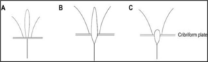
Figure 1: Showing the 3 types of Crista galli
Methods of Data Collection
The archived findings of patients who had undergone a skull CT scan were retrieved in order to collect data for this investigation.
Inclusion criteria for Data collection
The results must come from a patient who has had a head CT scan and lives in Port Harcourt.
The radiological film or CD must be free of artifacts and sufficiently clear to show the fissure and foramina.
Exclusion criteria
The outcome cannot come from another state or city. Patients who have had any kind of head surgery were not to be taken into consideration. The patient’s results must not indicate the presence of any craniofacial fractures. Every CT scan was carried out using the standard protocol of a 16-slice multidetector CT scanner, which is available in the Intercontinental Diagnostic Center and the Radiology Department of the River State University Teaching Hospital (UPTH). After volumetric imaging data gathering, post-reconstruction procedures were used to create smaller slices of 0.625 mm from the axial and coronal slices created in the DICOM viewer’s bone window. The size of the fissures and foramina was measured using the electronic caliper on the bone window.
Data Analysis
SPSS version 23 was used for data analysis, gender classification, and frequency summarization. A gender difference in the prevalence of these variations was assessed using t-test. Additionally, a P value above 0.05 was regarded as statistically significant.
RESULTS
Table 1: length of crista galli base on sex
| TYPE | Male (N=139) (%) | Female (N=112) (%) |
| TYPE 1 | 26(18.7) | 24(21.4) |
| TYPE 2 | 99(71.2) | 81(72.3) |
| TYPE 3 | 14(10.1) | 7(6.3) |
| P _ Value | 0.812 |

Figure 2: Pie Chart of the Sex
Table 1 and Figure 2 above shows the result of the analysis of crista galli based on sex. From the result, it can be seen that in male 18.7% are type 1, 71.2% fall under type 2 and 10.1% fall under type 3 while in female 21.4% are type 1, 72.3% fall under type 2 while 6.3% fall under type 3.
Table 2: length of crista galli base on type
| Units | TYPE 1(N=50) (%) | TYPE 2 (N=180) (%) | TYPE 3(N=21) (%) |
| MALE | 26(52) | 99(55) | 14(66.7) |
| FEMALE | 24(48) | 81(45) | 7(33.3) |
| P-Value | 0.135 |

Figure 3: Pie Chart of the Grista Galli Types
The results of the type-based study of Crista Galli are displayed in Table 2 and Figure 3. It is evident from the statistics that type 1 comprises 48% females and 52% males. Type 2 is made up of 55% male and 45% females, whereas Type 3 is made up of 66.7% male and 33.3% females.
DISCUSSION
In this study, 251 patients—139 men (55.4%) and 112 women (44.6%)—had their CT scan images reviewed. Crista Galli’s sex-based analytical results are shown in Table 1, and her type-based results are shown in Table 2.
According to Table 1, type II Crista galli was the most common kind in both males and females, accounting for 71.2% and 72.3% of cases, respectively. Type I comes next, accounting for 18.7% of males and 21.4% of females, respectively. With 10.1% of males and 6.3% of females, type III is the least common. Additionally, the data demonstrated a statistically significant correlation between gender and the type of crista galli (P=0.812).
The distribution of men and females within each Crista galli type, Type 1 Crista galli was detected in 48% of females and 52% of males. Type 2 (N=180): Males accounted for 55% and females for 45% of type 2 Crista galli. Type 3 (N=21): Males accounted for 67% and females for 33% of type 3 Crista galli.
The Statistical Significance of the P-value 0.135 indicates that there is a statistically significant difference in the distribution of males and females across the different Crista Galli types.
In the current investigation, type II crista galli were the most common, followed by type I and type III. The prevalence of the various forms of Crista galli varies throughout research; however, this can be explained by variations in sample size, technique, race, and ethnicity [1].
Summary
The findings indicate that sex has a major impact on the distribution of Crista Galli types. It has been confirmed that there is a statistically significant difference in the types of Crista galli, and there seems to be a little male predominance in type 3. Additionally, this study’s prevalence of the various crista galli types was different from certain findings in the literature. Thus, radiologists, otorhinolaryngologists, and neurosurgeons must be able to identify these variations when evaluating CT scans in our community. According to this study, radiologists, neurosurgeons, and otorhinolaryngologists can evaluate CT images more effectively before surgery if they are aware of the morphological variations of the crista galli. It is also acknowledged that knowledge of the morphology of the crista galli is beneficial in several fields, such as Neurosurgery: To navigate the intricate skull base and prevent harming vital structures during surgical procedures, neurosurgeons must have a thorough understanding of the anatomy of the crista galli. Anatomy and Embryology: Researchers can better comprehend the embryological origins of the skull and the development of the brain by examining the crista galli’s growth and form. Forensic Anthropology: Since the size and shape of crista galli can differ from person to person, understanding their morphology can help identify human remains. Imaging and Radiology: Radiologists must be familiar with the morphology of the crista galli to correctly interpret imaging tests, such as CT and MRI scans, and detect disorders affecting the base of the skull. Comparative Anatomy: Researching the crista galli in various animals can reveal information on how the brain and skull have evolved. In conclusion, forensic anthropology, radiography, neurosurgery, anatomy, and comparative anatomy are among the disciplines that require an understanding of crista galli morphology.
REFERENCES
- Acar, G., Cicekcibasi, AE., Koplay, M., Kelesoglu, KS, The relationship between the pneumatization patterns of the frontal sinus, crista galli and nasal septum. A tomography study. Turkish Neurosurgery; (2020); 30(4):532-41.
- Akiyama, O,. Kondo, A., Classification of crista galli pneumatization and clinical considerations for anterior skull base surgery. Journal of Clin Neuroscience, (2020); 82(Pt B): 225-230.
- American Medical Association; “Radiology — Diagnostic Specialty Description, Retrieved 19 October 2020
- Fehrenbach, Margaret J., Herring, Susan W., Illustrated Anatomy of the Head and Neck (5th ed.), St. Louis: Elsevier. (2017); p. 57. ISBN 978-0-323-39634-9
- Hajiioannou, J., Owens, D., Whittet, HB., “Evaluation of anatomical variation of the crista galli using computed tomography” Clinical Anatomy. (2010); 23 (4): 370–373.
- Herman, GT., Fundamentals of Computerized Tomography: Image Reconstruction from Projections. Springer. ISBN 978-1-84628-723-7.
- Hermena, Shady., Young, Michael., “CT-scan Image Production Procedures”, StatPearls, Treasure Island (FL): StatPearls Publishing, 2022; PMID 34662062, retrieved 2023-11-24.
- Hillman, B J., Goldsmith, J C., The Sorcerer’s Apprentice: How Medical Imaging Is Changing Health Care. Oxford and New York: Oxford University Press. (2011); pp. 83–85. ISBN 9780195386967. Retrieved 30 July 2023
- Hill, R., Healy, B., Holloway, L., Kuncic, Z., Thwaites, D., Baldock, C., “Advances in kilovoltage x-ray beam dosimetry”, Physics in Medicine and Biology. 59 (6): R183–R231.
- Kamala, E., Vijaya, SU., Gugapriya, TS., kumar, NV., A computerized tomographic study of morphology and pneumatization of crista galli. International Journal of Anatomy and Research; 2016;4(2):2429-33.
- Kim, JJ., Cho, JH., Choi, JW., Lim, HW., Song, YJ., Choi, SJ., Morphologic analysis of crista galli using computed tomography, Journal of Rhinology; 2012; 19(2):91-5.
- Metin-Tellioglu, A., Polat, Y., The incidence of sinus crista galli in children and adults. A computerized tomography study, International Journal of Morphology.;2019;37(2):735-8. [doi:10.4067/S0717-95022019000200735]
- Murphy, A., Ekpo, E., Steffens, T., Neep, MJ., “Radiographic image interpretation by Australian radiographers: a systematic review”, Journal of Medical Radiation Sciences, 2019; 66 (4): 269–283.
- Novelline, Robert., Squire’s Fundamentals of Radiology, Harvard University Press, 5th edition
- Papadopoulou, AM., Chrysikos, D., Samolis, A., Tsakotos, G., Troupis, T., Anatomical variations of the nasal cavities and paranasal sinuses: A systematic review. Cureus 2021;,13(1): e12727.
- Som, PM., Park, EE., Naidich, TP., Lawson, W., Crista galli pneumatization is an extension of the adjacent frontal sinuses. American Journal of Neuroradiology; 2009;30(1):31-33.
- Tetiker, H., Kosar, MI., Çullu, N., Sahan, M., Gençer, CU., Derin, S., Pneumatization of crista galli in pre-adult and adult stages, International Journal of Morphology; 2016, 34(2):541-4.