
Review on MALDI Imaging for Direct Tissue Imaging and its Application in Pharmaceutical Research
- Oluwatosin Ogundairo
- Oluwatoyin Ayo-Farai
- Chinedu Paschal Maduka
- Chiamaka Chinaemelum Okongwu
- Abdulraheem Olaide Babarinde
- Olamide Tolulope Sodamade
- 130-141
- Jan 5, 2024
- Social Welfare
Review on MALDI Imaging for Direct Tissue Imaging and its Application in Pharmaceutical Research
Oluwatosin Ogundairo1*, Oluwatoyin Ayo-Farai2,Chinedu Paschal Maduka3,Chiamaka Chinaemelum Okongwu4, Abdulraheem Olaide Babarinde5, Olamide Tolulope Sodamade6
1Independent Researcher
2Jiann-Ping Hsu College of Public Health, Georgia Southern University.
3Institute of Human Virology, Nigeria
4Faculty of Community Health and Primary Care, Department of Public Health, University of Lagos
5The Heller School for Social Policy and Management, Brandeis University, Waltham Massachusetts.
6Africa Voices HQ, Nigeria
*Corresponding Author
DOI: https://doi.org/10.51244/IJRSI.2023.1012011
Received: 01 December 2023; Accepted: 11 December 2023; Published: 04 January 2024
ABSTRACT
Matrix-Assisted Laser Desorption/Ionization (MALDI) imaging has emerged as a powerful analytical technique, enabling direct tissue imaging in pharmaceutical research. This review provides an in-depth exploration of the principles, methodologies, and applications of MALDI imaging in the context of studying drug distribution and molecular changes within tissues. provides a structured approach to exploring the principles, methodologies, applications, challenges, and future directions of MALDI Imaging in pharmaceutical research. Each section aims to contribute to a comprehensive understanding of the technique’s significance and potential for transformative contributions to the field. The study begins by elucidating the underlying principles of MALDI imaging, highlighting the role of matrix-assisted laser desorption/ionization in generating spatially resolved mass spectra from biological samples. Special emphasis is placed on advancements in instrumentation and sample preparation techniques that have enhanced the spatial resolution and sensitivity of MALDI imaging. The application of MALDI imaging in pharmaceutical research is comprehensively explored, focusing on its pivotal role in drug development, pharmacokinetic studies, and toxicity assessments. Case studies and examples illustrate how MALDI imaging facilitates the visualization of drug distribution within tissues, offering valuable insights into drug metabolism, biodistribution, and pharmacodynamics.
Keyword: MALDI, Imaging, Tissue Imaging, Pharmaceutical, review
INTRODUCTION
Matrix-Assisted Laser Desorption/Ionization Imaging Mass Spectrometry (MALDI-IMS) stands at the forefront of technological advancements in analytical chemistry, particularly in its application to the field of pharmaceutical research (Fournelle et al., 2023). This innovative imaging technique enables direct visualization and spatial mapping of molecular distributions within biological tissues, revolutionizing the way researchers investigate drug compounds, biomarkers, and their interactions in situ (Moore, and Charkoftaki, 2023).
The conventional methodologies in pharmaceutical research often involve the analysis of homogenized tissue samples, potentially losing critical information about the spatial localization of molecules (Roseboom et al., 2020). MALDI-IMS, on the other hand, allows researchers to preserve the spatial context of molecular distributions directly within intact tissue sections. This breakthrough capability has profound implications for understanding the pharmacokinetics, biodistribution, and efficacy of pharmaceutical agents.
At its core, MALDI-IMS combines the principles of mass spectrometry with imaging capabilities. A matrix, typically an organic compound, is applied to the tissue, facilitating the desorption and ionization of molecules upon laser irradiation. The resulting mass spectra provide a molecular fingerprint, and by scanning across the tissue, a spatially resolved map of molecular distributions is generated.
Various MALDI-IMS methodologies exist, including traditional MALDI (Wang et al., 2023), MALDI-TOF (Time-of-Flight) (Gopal, and Muthu, 2023, Pan et al., 2023), and MALDI-Q-TOF (Quadrupole-Time-of-Flight) (Huang et al., 2023). Each methodology offers specific advantages in terms of resolution, sensitivity, and mass range. Coupled with advancements in instrumentation, these techniques enable high-throughput and high-resolution imaging, allowing researchers to investigate complex biological samples with unprecedented detail.
MALDI-IMS has found diverse applications in pharmaceutical research. It plays a pivotal role in drug discovery by elucidating the spatial distribution of pharmaceutical compounds and their metabolites within tissues. Researchers can assess drug penetration, metabolism, and potential toxicity directly in the anatomical context, facilitating informed decision-making in preclinical studies.
The spatially resolved information provided by MALDI-IMS is instrumental in biomarker discovery and validation. By correlating molecular signatures with specific tissue regions, researchers can identify potential biomarkers associated with diseases or drug responses, contributing to the development of personalized medicine.
While MALDI-IMS offers unparalleled advantages, challenges such as data complexity, standardization, and integration with other imaging modalities remain. The ongoing evolution of MALDI-IMS technologies and methodologies, coupled with collaborative efforts across disciplines, holds the promise of addressing these challenges and unlocking new dimensions in pharmaceutical research.
This review explores the transformative impact of MALDI-IMS on pharmaceutical research. By providing a direct and spatially resolved view of molecular distributions within tissues, MALDI-IMS significantly enhances our understanding of drug behavior and molecular interactions. The subsequent sections delve into the principles, methodologies, applications, and challenges of MALDI-IMS, illuminating its pivotal role in advancing pharmaceutical research and paving the way for more targeted and effective therapeutic interventions.
MALDI IMAGING FOR DIRECT TISSUE IMAGING
Matrix-Assisted Laser Desorption/Ionization Imaging Mass Spectrometry (MALDI-IMS) has emerged as a cutting-edge technology for direct tissue imaging, revolutionizing the way researchers investigate the spatial distribution of molecules within biological tissues. This innovative technique allows for the visualization of molecular information directly from intact tissue sections, providing valuable insights into the complex and heterogeneous nature of biological samples.
MALDI Imaging for direct tissue imaging represents a paradigm shift in the way researchers study biological samples. Its ability to provide spatially resolved molecular information within intact tissues opens up new possibilities in drug development, disease understanding, and personalized medicine. While challenges exist, ongoing technological advancements and collaborative efforts promise to further enhance the capabilities and applications of MALDI-IMS in the realm of direct tissue imaging.
Matrix-assisted laser desorption/ionization (MALDI) imaging mass spectrometry (IMS) is a method that allows the investigation of the molecular content of tissues within its morphological context (Balluff, et al., 2011). MALDI-IMS has been successfully used for the molecular assessment of tissue samples mainly in biomedical research and also in other scientific fields. In direct tissue analysis by MALDI-IMS, matrix is applied over the entire tissue section either manually, by robotic spotting, nebulization or sublimation, and laser shots are subsequently performed across the tissue sample (Mainini et al., 2015).
Matrix-Assisted Laser Desorption/Ionization Imaging Mass Spectrometry (MALDI-IMS) has emerged as a powerful analytical technique, enabling the direct analysis of molecular distributions within biological tissues. Its applications in pharmaceutical research have significantly advanced our understanding of drug pharmacokinetics, biodistribution, and disease pathology.
MALDI-IMS allows researchers to visualize how pharmaceutical compounds are distributed within tissues over time. Temporal mapping of drug concentrations and identification of metabolites provide crucial insights into drug absorption, distribution, metabolism, and excretion (ADME). MALDI-IMS provides spatial information on drug localization, aiding in assessing the efficacy of drug delivery systems and guiding formulation strategies. Evaluating the effectiveness of pharmaceutical interventions at the molecular level. Spatial correlation between drug distribution and therapeutic responses helps assess target engagement and overall treatment efficacy. Identifying and validating molecular markers associated with diseases or treatment responses. MALDI-IMS facilitates the discovery of spatially distinct molecular patterns, aiding in the identification of potential biomarkers for early disease diagnosis and personalized medicine.
MALDI-IMS allows researchers to decipher the complex molecular landscape of tissues, providing a holistic view of the interactions between drugs and biological systems. This molecular insight is crucial for optimizing drug formulations, understanding drug mechanisms of action, and identifying potential off-target effects. MALDI-IMS contributes to streamlining drug development processes by providing spatially resolved data early in the drug development pipeline. Accelerating decision-making in drug development, reducing costs, and improving the success rate of candidate compounds.
MALDI-IMS enables the development of personalized medicine approaches by linking individual molecular profiles with treatment responses. Tailoring therapeutic strategies based on the unique molecular characteristics of patients improves treatment outcomes and reduces adverse effects. MALDI-IMS aids in unraveling the molecular basis of diseases, contributing to a deeper understanding of disease pathology. Insights gained from direct tissue analysis enhance our knowledge of disease mechanisms, potentially leading to the identification of novel therapeutic targets.
Despite its significant contributions, challenges such as data complexity, standardization, and integration with other imaging modalities persist. Future directions involve technological advancements for improved resolution and sensitivity, as well as fostering collaborative research initiatives to maximize the potential of MALDI-IMS in pharmaceutical research.
MALDI-IMS stands as a transformative technology in pharmaceutical research, offering a unique window into the molecular complexities of biological tissues. Its applications in pharmacokinetics, biodistribution, efficacy assessment, and biomarker discovery underscore its importance in advancing drug development and personalized medicine. As technological innovations continue and collaborative efforts expand, MALDI-IMS is poised to play an increasingly pivotal role in shaping the future of pharmaceutical research.
2.1. Principles of Matrix-Assisted Laser Desorption/Ionization Imaging Mass Spectrometry: Matrix-Assisted Laser Desorption/Ionization Imaging Mass Spectrometry (MALDI-IMS) is a powerful analytical technique that combines mass spectrometry with spatial information, allowing researchers to visualize the distribution of molecules directly within biological tissues. The principles of MALDI-IMS encompass several key steps. A matrix, typically an organic compound, is applied to the tissue surface. The matrix serves multiple purposes, including absorbing the laser energy, facilitating the desorption of analytes, and promoting ionization. Secondly, Laser Desorption/Ionization. The tissue section coated with the matrix is subjected to laser irradiation. For the desorption/ionization process, the laser energy is absorbed by the matrix, leading to its vaporization and desorption of analyte molecules from the tissue. This process also ionizes the desorbed molecules. Mass Spectrometry Analysis vis-a-viz ionization of analytes. The desorbed molecules become ionized in the gas phase. The resulting ions are accelerated into the mass spectrometer, where they are separated based on their mass-to-charge ratio (m/z). The ions are detected, and their abundance at specific m/z values is recorded, generating a mass spectrum.
Scanning Across the Tissue in which the tissue section is systematically scanned, and mass spectra are acquired at each position. The mass spectra from different positions are integrated to create a spatially resolved map of molecular distributions within the tissue. The resulting images depict the spatial distribution of specific molecules, providing insights into their localization within the biological sample.
2.1.1. Molecular Fingerprinting: Molecular fingerprinting is a technique that allows the investigation of the molecular content of tissues within its morphological context (Capecchi, Probst, and Reymond, 2020). It is used in biomedical research and other scientific fields to assess the molecular content of tissue samples. Matrix-assisted laser desorption/ionization (MALDI) imaging mass spectrometry (IMS) is a method that allows the investigation of the molecular content of tissues within its morphological context. In direct tissue analysis by MALDI-IMS, matrix is applied over the entire tissue section either manually, by robotic spotting, nebulization or sublimation, and laser shots are subsequently performed across the tissue sample. Each type of molecule produces a characteristic mass spectrum, creating a molecular fingerprint. Researchers can identify and analyze specific molecules based on their unique mass spectra. The abundance of molecules at different locations within the tissue can be quantified.
2.1.2. Advancements in Resolution and Sensitivity: Advancements in spectrophotometry techniques have improved sensitivity and accuracy, enabling researchers to achieve more precise and reliable results (Zhang, Zhang, and Liu, 2013). One of the key areas of advancement in spectrophotometry techniques lies in the improvement of optical components and detectors. Optical components, such as light sources and monochromators, have become more efficient and precise, allowing for better control and manipulation of light wavelengths. This ensures optimal spectral resolution and accuracy in measurements. Detectors, including photodiodes and photomultiplier tubes, have also undergone significant improvements. These advancements have resulted in increased sensitivity and wider dynamic range, enabling the detection and quantification of lower concentration analytes. High-performance detectors facilitate the measurement of absorbance over a broader spectrum, providing a more comprehensive analysis of samples (Das, and Agrawal, 2011). Ongoing technological advancements aim to enhance the spatial resolution of MALDI-IMS, allowing for detailed imaging at the subcellular level. Improvements in sensitivity enable the detection of low-abundance molecules, expanding the range of analytes that can be studied.
The principles of MALDI-IMS, rooted in the combination of matrix-assisted laser desorption/ionization and mass spectrometry, enable the visualization and analysis of molecular distributions within biological tissues. This innovative technique has diverse applications in fields such as drug discovery, biomarker research, and understanding disease pathology. Ongoing advancements continue to refine and expand the capabilities of MALDI-IMS for comprehensive spatial analysis in complex biological systems. Figure 1 shows the various stages of drug development. It begins with the drug discovery which entails target identification.
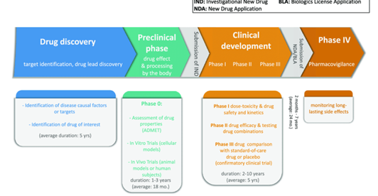
Figure 1. Drug development stages (Réda, Kaufmann, and Delahaye-Duriez, 2020)
2.2. Methodologies and Instrumentation in MALDI-IMS: Matrix-Assisted Laser Desorption/Ionization Imaging Mass Spectrometry (MALDI-IMS) utilizes various methodologies and instrumentation approaches to enhance its capabilities for spatially resolved molecular analysis. These include traditional methods and advanced technologies (Aminlashgari, and Hakkarainen, 2012).
2.2.1. Traditional MALDI: Traditional matrix-assisted laser desorption/ionization (MALDI) is a technique that allows the investigation of the molecular content of tissues within its morphological context (Keller et al., 2018). In direct tissue analysis by MALDI, matrix is applied over the entire tissue section either manually, by robotic spotting, nebulization or sublimation, and laser shots are subsequently performed across the tissue sample (Lu and Cai, 2012).
The foundational approach involves applying a matrix to the tissue and subjecting it to laser irradiation, generating mass spectra at each spatial position. Widely used for fundamental studies and initial applications in direct tissue imaging.
2.2.2. MALDI-TOF (Time-of-Flight): Time-of-flight mass analyzers measure the time it takes for ions to travel a fixed distance. This method provides mass spectra with improved resolution. Enhanced mass accuracy and resolution, allowing for more precise identification of molecules. Commonly applied in MALDI-IMS for detailed molecular profiling (Wu, Shaler, and Becker, 1994). Matrix-assisted laser desorption/ionization time-of-flight (MALDI-TOF) is a soft ionization technique used in mass spectrometry that produces rapid and efficient ionization of a wide variety of molecules (Hosseini, et al., 2017). MALDI-TOF mass spectrometry is a versatile analytical technique to detect and characterize mixtures of organic molecules. In microbiology, it is used as a rapid, accurate, and cost-effective method for identifying microorganisms (bacteria, fungi, and viruses) (Croxatto, Prod’hom, and Greub, 2012). Identification of the organisms by MALDI-TOF is based on assessing protein profiles and database comparison. A typical experiment consists of the growth of the organism (e.g., bacteria), colony selection and placement on a target, addition of matrix, and analysis with MALDI-TOF MS. The MALDI TOF process is a two-phase procedure; Ionization Phase and Time of Flight Phase (Jiang et al., 2019). In the ionization phase, samples are fixed in a crystalline matrix on a target plate and are bombarded by a laser. The sample molecules vaporize into the vacuum while being ionized at the same time. High voltage is then applied to accelerate the charged particles. The second step is the time-of-flight mass spectrometry phase. In the linear mode, particles will impinge upon the linear detector within a few nanoseconds after ionization. Higher mass molecules will arrive later than lighter ones. Flight time measurement makes it possible to determine molecule masses directly. Each peak in the spectrum corresponds to the specific mass of the particle along the time axis, starting with the ionization moment (Dichtl et al., 2023, Dingle, and Butler-Wu, 2013).
2.2.3. MALDI-Q-TOF (Quadrupole-Time-of-Flight): It combines the selective filtering capabilities of a quadrupole mass analyzer with the high-resolution capabilities of a time-of-flight analyzer. Improved selectivity and resolution, enabling more accurate and comprehensive analysis. Particularly valuable for complex samples and detailed molecular imaging studies.
Quadrupole-Time-of-Flight (Q-TOF) is a hybrid mass spectrometry technique that combines the features of quadrupole and time-of-flight mass analyzers (Allen, and McWhinney, 2019). The Q-TOF MS uses a quadrupole mass analyzer to select ions of a specific mass-to-charge ratio (m/z) and then passes them through a collision cell where they are fragmented (Kind et al., 2018). The resulting fragments are then passed into a time-of-flight mass analyzer, which measures the time it takes for the fragments to travel a known distance. The mass-to-charge ratio of the fragments is then calculated from the time-of-flight data. Q-TOF MS systems have MS-MS capabilities and can be coupled to GC or LC systems.
2.2.4. Advancements in Instrumentation: Advancements in spectrophotometry techniques have improved sensitivity and accuracy, enabling researchers to achieve more precise and reliable results (Berkhout, and Ram, 2019). One of the key areas of advancement in spectrophotometry techniques lies in the improvement of optical components and detectors. Optical components, such as light sources and monochromators, have become more efficient and precise, allowing for better control and manipulation of light wavelengths (Khanafer, and Shirmohammadi, 2020). This ensures optimal spectral resolution and accuracy in measurements. Detectors, including photodiodes and photomultiplier tubes, have also undergone significant improvements (Gross, Lockwood, and Spence, 2017). These advancements have resulted in increased sensitivity and wider dynamic range, enabling the detection and quantification of lower concentration analytes. High-performance detectors facilitate the measurement of absorbance over a broader spectrum, providing a more comprehensive analysis of samples. Instrumentation advancements aim to achieve finer spatial resolution in imaging. Allows for imaging at the subcellular level, providing unprecedented detail. Essential for studying cellular structures and interactions within tissues. Instrumentation designed for rapid data acquisition, enabling the analysis of large sample sets. Accelerates data generation, making MALDI-IMS more applicable in high-throughput studies. Particularly useful in drug discovery and biomarker research, where large datasets need to be analyzed efficiently.
These methodological and instrumental variations cater to the diverse needs of researchers engaged in MALDI-IMS studies. The choice of methodology depends on the specific requirements of the study, such as the desired resolution, sensitivity, and throughput. Advancements in instrumentation continually expand the capabilities of MALDI-IMS, making it a versatile and powerful tool for molecular imaging in various biological contexts.
2.3 Applications in Pharmaceutical Research: AI and machine learning have become the breakthrough technology most anticipated to have a transformative effect on pharmaceutical research and development (R&D) (Bhattamisra et al., 2023). AI/ML techniques are appealing to the pharmaceutical industry due to their automated nature, predictive capabilities, and the consequent expected increase in efficiency. ML approaches have been used in drug discovery over the past 15–20 years with increasing sophistication (Wang, 2006). The most recent aspect of drug development where positive disruption from AI/ML is starting to occur, is in clinical trial design, conduct, and analysis (Kellogg, 2006). Computer applications in pharmaceutical research and development include bioinformatics, data mining, predicting human response to drugs, and high-throughput screening (Kolluri et al., 2022). Figure 2 shows the schematic visualization of the patient measurement.
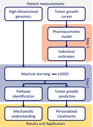
Figure 2. A schematic visualization of the patient measurements (Zwep, 2023)
2.3.1. Drug Discovery and Development: Drug discovery and development is a complex and resource-intensive process that involves the identification of new chemical or biological substances and transforming them into products approved for use by patients (Southey, and Brunavs, 2023). The process typically involves several stages, including discovery, preclinical research, clinical research, FDA review, and post-market safety monitoring.
Advancements in spectrophotometry techniques have improved sensitivity and accuracy, enabling researchers to achieve more precise and reliable results. One of the key areas of advancement in spectrophotometry techniques lies in the improvement of optical components and detectors. Optical components, such as light sources and monochromators, have become more efficient and precise, allowing for better control and manipulation of light wavelengths. This ensures optimal spectral resolution and accuracy in measurements. Detectors, including photodiodes and photomultiplier tubes, have also undergone significant improvements. These advancements have resulted in increased sensitivity and wider dynamic range, enabling the detection and quantification of lower concentration analytes. High-performance detectors facilitate the measurement of absorbance over a broader spectrum, providing a more comprehensive analysis of samples (Hughes et al., 2011). AI and machine learning have become the breakthrough technology most anticipated to have a transformative effect on pharmaceutical research and development (R&D) (Vamathevan et al., 2019). AI/ML techniques are appealing to the pharmaceutical industry due to their automated nature, predictive capabilities, and the consequent expected increase in efficiency. ML approaches have been used in drug discovery over the past 15–20 years with increasing sophistication (Kolluri, et al., 2022). The most recent aspect of drug development where positive disruption from AI/ML is starting to occur, is in clinical trial design, conduct, and analysis. Computer applications in pharmaceutical research and development include bioinformatics, data mining, predicting human response to drugs, and high-throughput screening.
2.3.2 Applications of MALDI-IMS in Pharmaceutical Research: Matrix-Assisted Laser Desorption/Ionization Imaging Mass Spectrometry (MALDI-IMS) plays a crucial role in advancing pharmaceutical research by providing valuable insights into the spatial distribution of molecules within biological tissues. Its applications are diverse, contributing to various aspects of drug discovery, development, and personalized medicine.
MALDI-IMS enables the study of how pharmaceutical compounds distribute and change over time within the body. Visualizing the temporal changes in drug concentrations. Identifying and mapping drug metabolites. Understanding the distribution of drugs and their metabolites in different organs and tissues. Mapping the presence and concentration of drugs in specific organs. Assessing how well drugs penetrate tissues at the site of action.
Evaluating the effectiveness of pharmaceutical interventions within the context of tissue morphology. Assessing if drugs reach their intended target sites. Correlating drug distribution with therapeutic responses. Investigating the spatial distribution of molecules in diseased tissues to identify potential biomarkers. Identifying regions of altered molecular expression in disease. Establishing links between molecular patterns and pathological conditions. Tailoring treatment strategies based on individual molecular profiles. Using molecular information to predict individual responses to specific treatments. Developing personalized treatment plans based on spatial molecular information.
These applications showcase how MALDI-IMS contributes to the optimization of drug development processes and the advancement of personalized medicine. By providing spatially resolved molecular information, MALDI-IMS enhances our understanding of drug behavior, efficacy, and the molecular basis of diseases, ultimately facilitating more targeted and effective therapeutic interventions.
2.4 Challenges and Considerations in MALDI-IMS: Matrix-Assisted Laser Desorption/Ionization Imaging Mass Spectrometry (MALDI-IMS) presents several challenges and considerations that researchers must address to maximize its potential and ensure the reliability of results. The generation of spatially resolved data results in large datasets that can be complex to manage and analyze. Implementing efficient storage solutions for vast amounts of spatial data. Ensuring access to sufficient computational resources for data processing.
Extracting meaningful information from the complex datasets requires sophisticated bioinformatics approaches. Advancing algorithms for effective data processing and interpretation. Fostering collaboration between analytical chemists and bioinformaticians for comprehensive analyses.
Lack of standardized protocols can lead to variations in sample preparation and data acquisition. Developing standardized protocols for sample preparation and instrument calibration. Encouraging the adoption of community-driven standards to enhance reproducibility. Inconsistencies in experimental conditions may result in irreproducible results. Implementing robust quality control measures to ensure reproducibility. Conducting validation studies to verify the reliability of findings.
Integrating MALDI-IMS with other imaging modalities for comprehensive data interpretation. Ensuring compatibility between MALDI-IMS and other imaging instruments. Developing methodologies for merging data from different imaging modalities. Maximizing the synergies between MALDI-IMS and existing imaging technologies. Encouraging collaboration between researchers specializing in different imaging techniques. Exploring opportunities for developing hybrid imaging systems.
Addressing these challenges and considerations is essential for advancing the field of MALDI-IMS and ensuring the reliability and applicability of its results. By focusing on data management, standardization, and integration with complementary technologies, researchers can unlock the full potential of MALDI-IMS in unraveling the spatial molecular complexities within biological tissues.
2.5 Future Directions and Innovations in MALDI-IMS: Matrix-Assisted Laser Desorption/Ionization Imaging Mass Spectrometry (MALDI-IMS) holds immense potential for further development and innovation. Future directions are likely to focus on technological advancements and collaborative research initiatives to enhance the capabilities and applications of this powerful analytical technique. Enhancing spatial resolution for more detailed imaging at the cellular and subcellular levels. Development of higher-resolution mass spectrometers. Innovations in matrix application techniques to achieve finer spatial resolution. Improving sensitivity to detect low-abundance molecules and trace compounds. Integration of advanced laser technologies for improved ionization. Refinement of matrix compositions to enhance sensitivity.
Fostering collaborations between researchers from diverse disciplines. Collaboration between analytical chemists and biologists to understand complex biological systems. Engaging data scientists and bioinformaticians to develop advanced algorithms for data interpretation. Facilitating collaborative efforts on a global scale. Creation of shared databases for standardized data sets and methodologies. Collaborative projects involving researchers from different countries to address global challenges.
These future directions aim to propel MALDI-IMS into new frontiers of analytical capabilities, making it an even more indispensable tool in various scientific fields. By pushing the boundaries of resolution, sensitivity, and collaboration, researchers can unlock novel applications and gain deeper insights into the spatial distribution of molecules within biological tissues. The collaborative nature of these advancements reflects the interdisciplinary and global effort required to fully realize the potential of MALDI-IMS in advancing scientific understanding and discovery.
CONCLUSION
Matrix-Assisted Laser Desorption/Ionization Imaging Mass Spectrometry (MALDI-IMS) has emerged as a transformative tool in pharmaceutical research, offering unprecedented insights into the spatial distribution of molecules within biological tissues. This review has explored the principles, methodologies, applications, challenges, and future directions of MALDI-IMS in the context of direct tissue imaging and its implications for advancing pharmaceutical research.
MALDI-IMS provides a unique capability to visualize and analyze molecular distributions directly within intact tissues. This spatial information is invaluable for understanding the localization of pharmaceutical compounds, metabolites, and biomolecules. MALDI-IMS has demonstrated versatile applications in drug discovery and development. From studying pharmacokinetics and biodistribution to assessing drug efficacy, MALDI-IMS contributes critical data for optimizing pharmaceutical interventions.
The technique contributes significantly to biomarker discovery and validation by allowing researchers to correlate molecular patterns with disease states. This has implications for early diagnosis, personalized medicine, and the development of targeted therapies. The review has identified challenges such as data complexity, standardization issues, and the need for integration with other imaging modalities. Addressing these challenges is crucial for ensuring the reliability and reproducibility of MALDI-IMS results.
The future of MALDI-IMS lies in continuous technological innovations. Improvements in resolution and sensitivity are anticipated, enabling researchers to explore biological tissues at finer scales and detect even trace amounts of molecules. The collaborative nature of future research initiatives is emphasized. Interdisciplinary collaborations between chemists, biologists, data scientists, and international research communities will be pivotal in pushing the boundaries of MALDI-IMS applications.
RECOMMENDATION
Continued research and exploration of MALDI-IMS in diverse biological contexts and disease models. Enhanced integration with other imaging modalities for a comprehensive understanding of biological systems. Continued efforts towards establishing standardized protocols and data-sharing practices to enhance reproducibility and comparability across studies.
In conclusion, MALDI-IMS has revolutionized our approach to studying biological tissues in the context of pharmaceutical research. Its unique ability to provide spatially resolved molecular information opens new avenues for drug discovery, disease understanding, and personalized medicine. As we move forward, a collective effort from the scientific community is essential to overcome challenges, drive technological advancements, and fully harness the potential of MALDI-IMS in shaping the future of pharmaceutical research.
REFERENCE
- Allen, D.R. and McWhinney, B.C., 2019. Quadrupole time-of-flight mass spectrometry: a paradigm shift in toxicology screening applications. The Clinical Biochemist Reviews, 40(3), p.135.
- Aminlashgari, N. and Hakkarainen, M., 2012. Surface assisted laser desorption ionization-mass spectrometry (SALDI-MS) for analysis of polyester degradation products. Journal of The American Society for Mass Spectrometry, 23(6), pp.1071-1076.
- Atanasov, A.G., Zotchev, S.B., Dirsch, V.M. and Supuran, C.T., 2021. Natural products in drug discovery: Advances and opportunities. Nature reviews Drug discovery, 20(3), pp.200-216.
- Balluff, B., Schöne, C., Höfler, H. and Walch, A., 2011. MALDI imaging mass spectrometry for direct tissue analysis: technological advancements and recent applications. Histochemistry and cell biology, 136, pp.227-244.
- Capecchi, A., Probst, D. and Reymond, J.L., 2020. One molecular fingerprint to rule them all: drugs, biomolecules, and the metabolome. Journal of cheminformatics, 12(1), pp.1-15.
- Croxatto, A., Prod’hom, G. and Greub, G., 2012. Applications of MALDI-TOF mass spectrometry in clinical diagnostic microbiology. FEMS microbiology reviews, 36(2), pp.380-407.
- Berkhout, J.H. and Ram, A., 2019. Recent advancements in spectrophotometric pKa determinations: a review. Indian J. Pharm. Educ. Res, 53(4), pp.S475-S480.
- Bhattamisra, S.K., Banerjee, P., Gupta, P., Mayuren, J., Patra, S. and Candasamy, M., 2023. Artificial Intelligence in Pharmaceutical and Healthcare Research. Big Data and Cognitive Computing, 7(1), p.10.
- Das, R.S. and Agrawal, Y.K., 2011. Raman spectroscopy: Recent advancements, techniques and applications. Vibrational spectroscopy, 57(2), pp.163-176.
- Dichtl, K., Klugherz, I., Greimel, H., Luxner, J., Köberl, J., Friedl, S., Steinmetz, I. and Leitner, E., 2023. A head-to-head comparison of three MALDI-TOF mass spectrometry systems with 16S rRNA gene sequencing. Journal of Clinical Microbiology, 61(10), pp.e01913-22.
- Dingle, T.C. and Butler-Wu, S.M., 2013. MALDI-TOF mass spectrometry for microorganism identification. Clinics in laboratory medicine, 33(3), pp.589-609.
- Fournelle, F., Lauzon, N., Yang, E. and Chaurand, P., 2023. Metal-assisted laser desorption ionization imaging mass spectrometry for biological and forensic applications. Microchemical Journal, 185, p.108294.
- Gopal, J. and Muthu, M., 2023. Matrix-assisted laser desorption ionization-time of flight mass spectrometry (MALDI-TOF MS) for the analysis of mushroom polysaccharides of clinical, nutritional, and medicinal significance-contemplating the achievements and eliciting future trends. TrAC Trends in Analytical Chemistry, p.117095.
- Gross, B., Lockwood, S.Y. and Spence, D.M., 2017. Recent advances in analytical chemistry by 3D printing. Analytical chemistry, 89(1), pp.57-70.
- Huang, L., Nie, L., Dong, J., Yao, L., Kang, S., Dai, Z., Wei, F. and Ma, S., 2023. Differential distribution of phytochemicals in Scutellariae Radix and Scutellariae Amoenae Radix using microscopic mass spectrometry imaging. Arabian Journal of Chemistry, 16(5), p.104590.
- Hughes, J.P., Rees, S., Kalindjian, S.B. and Philpott, K.L., 2011. Principles of early drug discovery. British journal of pharmacology, 162(6), pp.1239-1249.
- Hosseini, S., Martinez-Chapa, S.O., Hosseini, S. and Martinez-Chapa, S.O., 2017. Principles and mechanism of MALDI-ToF-MS analysis. Fundamentals of MALDI-ToF-MS Analysis: Applications in Bio-diagnosis, Tissue Engineering and Drug Delivery, pp.1-19.
- Jiang, B., Na, J., Wang, L., Li, D., Liu, C. and Feng, Z., 2019. Separation and enrichment of antioxidant peptides from whey protein isolate hydrolysate by aqueous two-phase extraction and aqueous two-phase flotation. Foods, 8(1), p.34.
- Keller, C., Maeda, J., Jayaraman, D., Chakraborty, S., Sussman, M.R., Harris, J.M., Ané, J.M. and Li, L., 2018. Comparison of vacuum MALDI and AP-MALDI platforms for the mass spectrometry imaging of metabolites involved in salt stress in Medicago truncatula. Frontiers in plant science, 9, p.1238.
- Khanafer, M. and Shirmohammadi, S., 2020. Applied AI in instrumentation and measurement: The deep learning revolution. IEEE Instrumentation & Measurement Magazine, 23(6), pp.10-17.
- Kind, T., Tsugawa, H., Cajka, T., Ma, Y., Lai, Z., Mehta, S.S., Wohlgemuth, G., Barupal, D.K., Showalter, M.R., Arita, M. and Fiehn, O., 2018. Identification of small molecules using accurate mass MS/MS search. Mass spectrometry reviews, 37(4), pp.513-532.
- Kolluri, S., Lin, J., Liu, R., Zhang, Y. and Zhang, W., 2022. Machine learning and artificial intelligence in pharmaceutical research and development: a review. The AAPS Journal, 24, pp.1-10.
- Lu, M. and Cai, Z., 2012. Advances of MALDI-TOF MS in the analysis of traditional Chinese medicines. Applications of MALDI-TOF Spectroscopy, pp.143-164.
- Marchenko, O.V. and Katenka, N.V. eds., 2020. Quantitative methods in pharmaceutical research and development: Concepts and applications.
- Mainini, V., Lalowski, M., Gotsopoulos, A., Bitsika, V., Baumann, M. and Magni, F., 2015. MALDI-imaging mass spectrometry on tissues. Clinical Proteomics: Methods and Protocols, pp.139-164.
- Moore, J.L. and Charkoftaki, G., 2023. A Guide to MALDI Imaging Mass Spectrometry for Tissues. Journal of Proteome Research.
- Pan, F., Zhao, N., Zhao, W., Wang, C., Sun, Y., Zhang, H., Qin, J., Liu, Q. and Zhang, H., 2023. Performance of Two Matrix-Assisted Laser Desorption Ionization–Time-of-Flight Mass Spectrometry (MALDI-TOF MS) Systems for Identification of the Viridans Group Streptococci. Infection and Drug Resistance, pp.2901-2909.
- Réda, C., Kaufmann, E. and Delahaye-Duriez, A., 2020. Machine learning applications in drug development. Computational and structural biotechnology journal, 18, pp.241-252.
- Roseboom, I.C., Rosing, H., Beijnen, J.H. and Dorlo, T.P., 2020. Skin tissue sample collection, sample homogenization, and analyte extraction strategies for liquid chromatographic mass spectrometry quantification of pharmaceutical compounds. Journal of Pharmaceutical and Biomedical Analysis, 191, p.113590.
- Southey, M. and Brunavs, M., (2023). Introduction to Small Molecule Drug Discovery and Preclinical Development. Frontiers in Drug Discovery, 3, p.1314077.
- Topić Popović, N., Kazazić, S.P., Bojanić, K., Strunjak‐Perović, I. and Čož‐Rakovac, R., 2023. Sample preparation and culture condition effects on MALDI‐TOF MS identification of bacteria: A review. Mass Spectrometry Reviews, 42(5), pp.1589-1603.
- Vamathevan, J., Clark, D., Czodrowski, P., Dunham, I., Ferran, E., Lee, G., Li, B., Madabhushi, A., Shah, P., Spitzer, M. and Zhao, S., 2019. Applications of machine learning in drug discovery and development. Nature reviews Drug discovery, 18(6), pp.463-477.
- Wang, J., Zhao, J., Nie, S., Xie, M. and Li, S., 2023. MALDI mass spectrometry in food carbohydrates analysis: A review of recent researches. Food Chemistry, 399, p.133968.
- Wu, K.J., Shaler, T.A. and Becker, C.H., 1994. Time-of-flight mass spectrometry of underivatized single-stranded DNA oligomers by matrix-assisted laser desorption. Analytical chemistry, 66(10), pp.1637-1645.
- Zhang, Z., Zhang, F. and Liu, Y., 2013. Recent advances in enhancing the sensitivity and resolution of capillary electrophoresis. Journal of chromatographic science, 51(7), pp.666-683.
- Zwep, L.B., 2023. Statistical learning for complex data to enable precision medicine strategies (Doctoral dissertation, Leiden University).