
The Cushioning Effect of Extracts of Amaranthus Viridis Linn (Green Amaranth) on Acetaminophen-Induced Hepatotoxicity.
- Enemali, Michael Okey
- Asogwa Kingsley Kelechi
- Ezeigwe Christian Obiajulu
- Ezenwelu Obinna Chijioke
- 1117-1128
- Jun 25, 2024
- Chemistry
The Cushioning Effect of Extracts of Amaranthus Viridis Linn (Green Amaranth) on Acetaminophen-Induced Hepatotoxicity.
Enemali, Michael Okey, *Asogwa Kingsley Kelechi, Ezeigwe Christian Obiajulu and Ezenwelu Obinna Chijioke
Department of Applied Biochemistry, Nnamdi Azikiwe University, Awka, Anambra State, Nigeria
*Corresponding Author
DOI: https://doi.org/10.51244/IJRSI.2024.1105075
Received: 24 April 2024; Revised 20 May 2024; Accepted: 24 May 2024; Published: 25 June 2023
ABSTRACT
Vegetables serve as indispensable constituents of the human diet, supplying the body with minerals, vitamins and certain hormone precursors, in addition to protein and energy. Huge quantities of Amarranthus viridis (Green amaranth), is consumed all over Nigeria vegetable for various dishes. In the current study, the hepatoprotective properties of aqueous and ethanol extract of A. viridis was studied. Thirty (30) male Wister albino rats weighing between 120-140g were used for the study. The animals were divided into five (5) groups of six animals each. These include three (3), control groups; Normal Control (Water and Feeds only), Negative Control (Water, Feed and 2g/kgbw of Acetaminophen) and Positive control (Water, Feed, Sylimarin and 2g/kgbw of Acetaminophen). The other groups were two groups administered with the aqueous and ethanol leaf extracts of the plant, respectively. The results obtained shows that oral administration of a single 2g/kgbw dose of acetaminophen led to a marked increase in the serum activities of the ALT, AST and ALP of the animals in the negative control group compared to the normal control group .The groups pre-treated with 400mg/kgbw of aqueous leaf extracts of the plant showed significantly (p<0.05) decreased activity of ALT and AST but insignificant change in the serum activity of ALP of the animals. It can therefore, be suggested that Amaranthus viridis produced effects which may contribute to the preservation of cellular GSH levels in the acetaminophen-intoxicated rats, which further provides cellular defense both as a hydroxyl radical scavenger and also a detoxifying agent against the toxic intermediate of acetaminophen in a GST-catalysed reaction. Therefore, it can be a good source of raw material for the production of drugs used for the prevention and treatment of liver diseases and other pathological conditions associated with oxidative stress. This encourages and supports its use as edible vegetables and for therapy respectively.
INTRODUCTION
Vegetables serve as indispensable constituents of the human diet, supplying the body with minerals, vitamins and certain hormone precursors, in addition to protein and energy, (Aja et al., 2010). Leafy vegetables have been found to boost the concentration of Red Blood cell and significantly increase the serum activity of AST in experimental animals, (Ezekwe et al., 2013). With the increasing focus on plant research all over the world, a large body of evidence has been collected to show immense potential of medicinal plants for treatment purposes or for the production of drugs (Dahanukar et al., 2001; Olamide and Mathew, 2013; Udochukwu, et al., 2015). The use of plants in ethnomedicine for the management of ailments stem from the presence of phytochemicals, (Aja, et al., 2010). It has been suggested that ethanol extracts of vegetables could be used in treating diseases caused by microorganisms (Ladipo, et al., 2010). Polyphenols and flavonoids in plant extracts had been implicated for their antioxidant potentials by improving the functionality of the antioxidant system of test rats, (Edeoga, et al., 2005; Sudha et al., 2011; Imaga and Bamigbetan 2013; Alamgir et al., 2016; Enemali et al., 2018). Many remedies from plant origin have been evaluated for their potential antioxidant and hepatoprotective effect in experimental animal model (Enemali and Udedi 2018; Enemali, et al., 2019).
Acetaminophen (APAP) is an analgesic and antipyretic substance used in the production of the drug paracetamol. Although safe at therapeutic doses of 1000mg/75kgbw, APAP had been found to cause severe liver injury (Erica and Emily. 2014) at higher doses (Enemali et al., 2018). Mitchell, et al. (1973), reported that Acetaminophen (APAP) overdose is the predominant cause of acute liver failure in the United States and that toxicity begins with a reactive metabolite that binds to proteins. These findings indicated that acetaminophen was metabolically activated by cytochrome P450 enzymes to a reactive metabolite that depleted glutathione (GSH) and covalently bonded to protein. It has also been shown by James, et al. (2009) that repletion of glutathione (GSH) prevented the toxicity. Depletion of GSH which is an intrinsic antioxidant is capable of introducing peroxidation of cell membrane lipids, regeneration of reactive oxygen free radicals and hepatocellular fatty regeneration with centriolobular necrosis of the liver.
Reactive oxygen species (ROS) are produced in the cells by cellular metabolism and other exogeneous environmental agents. They are generated by a process known as redox cycling and are catalysed by transition metals such as Fe2+ and Cu2+ (Halliwell and Guteridge, 1999). Over production of ROS can damage cellular biomolecules like nucleic acids, proteins, lipids, carbohydrates and enzymes resulting in several diseases. Living systems have specific pathways to overcome the adverse effects of various damages. However, sometimes these repair mechanism fail to keep pace with such deleterious effects (Nilsson et al., 2004). In chronic liver diseases caused by oxidative stress (alcoholic and non-alcoholic fatty liver diseases, drug- and chemical-induced hepatic toxicity), conventional therapeutics options available for managing liver hepatotoxicity include a commonly used drug called sylibon 140, whose active ingredient is sylimarin. (Feher and Lengyey, 2017). Sylimarin is the active ingridient in the branded drug, Sylibon 140 (a known hepatoprotective drug), manufactured by Micro Laboratory Ltd, India. Silymarin has cytoprotective activity mediated by its antioxidative and radical-scavenging properties, (Křena and Walterovab, 2005). As an antioxidant, silymarin scavenges for free radicals that can damage cells exposed to toxins. Silymarin has been said to be at least ten times more potent in antioxidant activity than vitamin E.
In a poor resource country like Nigeria, access to quality drugs continue to be a big challenge to rural dwellers and Those in extreme poverty. In such countries, leafy vegetables contribute greatly to the nutritional and medicinal needs of local populations where they are used as staples, flavours, condiments, spices, drinks and beverages in most developing countries (Mbang et al., 2008). It is generally known that the consumption of a variety of local herbs and vegetables by man contributes significantly to the improvement of human health, in terms of prevention and cure of diseases because plants have long served as a useful and natural source of therapeutic agents (Chevellier, 1996). Living systems have specific pathways to overcome the adverse effects of various damages, but sometimes, these repair mechanism fail to keep pace with such deleterious effects (Stegmark and Akesson, 2004; Nilsson et al, 2004). Therefore, there is need to seek an alternative to augment the effort of the internal protection and repair mechanisms.
Townsend (1985) typically described the plant, Amaranthus viridis as an annual herbaceous plant, of the family Amaranthaceae, genius Amaranthus and specie Amaranthus viridis L (USDA, 2008). A.viridis grows in heavy organic to very sandy soil, including muck soil after the water has gone down for the season. Amaranthus viridis is grown and utilized in many areas of the world as both a wild and cultivated pot herb (Uphof, 1968). The plant is rich in calcium and iron and it is a good source of vitamin B and C (Morton, 1981). Amaranthus viridis’ common names include: green amaranth, slender amaranth, African spinach, wild amaranth, (English), inine (Igbo), tete (Yoruba).
Huge quantities of Amarranthus viridis (Green amaranth), are consumed all over Nigeria for flavouring and spicing of various types of food. Available information on the medicinal property is scanty, therefore, it is expedient to establish the hepatoprotective potentials of aqueous and the ethanol leaf extracts of this plant
MATERIALS AND METHODS
Experimental Animals
Thirty (30) male Wister albino rats weighing between 120-140g were used for the study. These rats were purchased from the animal house of the National Veterinary Research Institute (NVRI), Vom, in Plateau state. They were housed in clean, well ventilated metal cages in the animal house of the Department of Biological Sciences (Zoology unit), Nasarawa State University Keffi. The animals were kept under 24 hours light/dark cycling. They were allowed access to unlimited food and water supply and allowed to acclimatize for two (2) weeks before the commencement of the study.
Plant Material
The leaf of the plant under study were harvested from a farm at Angwan Lambu in Keffi, Nasarawa State (North Central Region of Nigeria). They were taken to the Department of Plant Science and Biotechnology for identification and assignment of voucher number (NSUKPSB112V), before some samples were deposited in the Herbarium.
Methods
Plant Sample preparation
They were rinsed in water to remove dust and sand particles, and then dried under room temperature for fourteen (14) days. The dried leaves were then ground into powder using electric blender. Part of the ground leaf materials were used to determine the proximate composition while the other was used for preparation of aqueous and ethanol extracts.
Proximate Analysis of the Plant Material
Standard methods of the Association of Analytical Chemist (AOAC), 2006 was used to determine the moisture content, crude protein, crude fat, total ash, crude fiber and carbohydrate content of the materials.
Preparation of Leaf Extracts
Ground plant material were divided into two; one was extracted with ethanol by soaking 200g of the ground samples in 1000ml of 98% ethanol (i.e. ratio 1:5; weight to volume) for 48hrs. The extracts were filtered using muslin cloth and then concentrated by heating in a water bath and stored in airtight containers.
The other part was extracted by soaking 200g of the powdered samples in 1000ml of distilled water (i.e. ratio 1:5; weight to volume) for 24hrs. The extracts were filtered using muslin cloth and then concentrated by heating to evaporate the water and stored in airtight containers.
Phytochemical Screening of the Leaf Extracts:
Portions of the concentrated extracts were used for phytochemical screening using standard procedures of the Association of Analytical Chemist to identify the constituents as described by Odebiyi and Sofowora(1990), Fadeyi (1983), Sofowara (1993), Trease and Evans (1989) and Harborne (1973).
Animal Handling
The animals were divided into five (5) groups of six animals each. These include three (3), control groups; Normal Control (Water and Feeds only), Negative Control (Water, Feed and 2g/kgbw of Acetaminophen) and Positive Control (Water, Feed, Sylimarin and 2g/kgbw of Acetaminophen). The other groups were two groups administered with the aqueous and ethanol leaf extracts of the plant, respectively prior to administration of acetaminophen (2g/kgBW) through oral gavage.
Preparation of Biological Samples
Collection of Blood Samples
At the end of the experimental period, (after 9 hours of intoxication), the animals were made unconscious by exposure to chloroform in an enclosed container, according to the method described by Ekor, et al. (2006) Incisions were quickly made into the animals’ cervical region with the aid of sterile blades. Blood samples were collected by cervical decapitation into plain tubes. Serum was collected by centrifuging the clotted blood in a HSC (1000-4000rpm) bench centrifuge at 3000 rpm for 10 minutes.
Preparation of Liver Homogenate:
The liver homogenate was prepared according to the method described by Enemali et al. (2019). The sample was stored at the temperature of -4oC and later used for the assay of the activities of the antioxidant enzymes (Super Oxide Dismutase, Catalase and Glutathione Peroxidase).
Determination of the Liver Function
The prepared serum was analyzed for various biochemical parameters – Aspartate Aminotransferase (AST) (King, 1965a), Alanine aminotransferase (ALT) (Lowry et al., 1951) and Alkaline phosphatase (ALP) (King, 1965b); Total and Direct Bilirubin were estimated by the method of Malloy-Evelyn, (1937). Albumin concentration was determined using Randox kits using the method outlined in the manual for albumin determination.
Histopathological Study on the Liver Tissues
Histopathological study on the liver tissues was carried out at Federal College of Veterinary and Medical Laboratory Technology, Vom, Plateau State, using the method described by Bancroft et al., (2013).
Statistical Analysis
Statistical analysis was conducted, for the enzyme assay and lipid profile values using the one-way ANOVA and data were expressed as mean ± standard deviation (SD) of six values. The differences between the mean values of the test groups were analysed for statistical significance using one-way ANOVA. The differences between groups were considered to be significant if p<0.05.
RESULTS
The Proximate Compositions of the Leaves of Amaranthus viridis:
The leaves of Amaranthus viridis were found to be rich in protein, having percentage compositions of 25.09 ± 0.07%. The leaf also has high ash content of above 11% and protein content of above 20% as shown in Table 1
Table 1: The proximate Compositions of Amaranthus viridis the leaf
| Proximate Parameters | Composition |
| Crude protein (%) | 25.09 ± 0.07 |
| Crude Fibre (%) | 9.54 ± 0.10 |
| Ash (%) | 17.77 ± 0.07 |
| Total Lipids (%) | 11.97 ± 0.05 |
| Moisture (%) | 9.80 ± 0.06 |
| Total Carbohydrate (%) | 4.88 ± 0.10 |
Values are mean ± SD of triplicate (X3) results
The Qualitative Phytochemical Compositions of Aqueous and Ethanol Leaf Extracts of the Plants
The results from the phytochemical analysis of the aqueous and ethanol leaf extracts of the plant showed the presence of Phenols, Tannins, Terpenoids and Cardiac glycosides both aqueous and ethanol extracts. While other phytochemicals were differently distributed in the aqueous and ethanol leaf extracts of the plant. The water extracted higher quantities of tannin and saponin from all the plant and ethanol leaf extract contained more Phenol and alkaloids, see Table 2
Table 2: The qualitative phytochemical compositions of aqueous and ethanol leaf extracts of Amaranthus viridis
| Phytochemical | Aqueous Extract | Ethanol Extract |
| Alkaloid | – | + |
| Tannin | + | + |
| Total Phenol | + | + |
| Cardiac Glycoside | + | + |
| Triterpenoid | – | + |
| Sterol | – | + |
| Saponin | + | – |
| Balsam | + | + |
| Cardenolides | + | – |
| Resin | + | – |
| Glycoside | + | – |
| + indicates presence; – indicates absence | ||
The Effect of Pre-Treatment with 400gm/Kgbw Of Leaf Extracts of Amaranthus Viridis on the Serum Activities of AST, ALT And ALP of Rats Intoxicated with 2g/Kgbw Single Dose of Acetaminophen.
The oral administration of a single 2g/kgbw dose of acetaminophen led to a marked increase in the serum activities of the ALT, AST and ALP of the animals in the negative control group compared to the normal control group .The groups pre-treated with 400mg/kgbw of aqueous leaf extracts of the plant showed significantly (p<0.05) decreased activity of ALT and AST but insignificant change in the serum activity of ALP of the animals (Table 3).
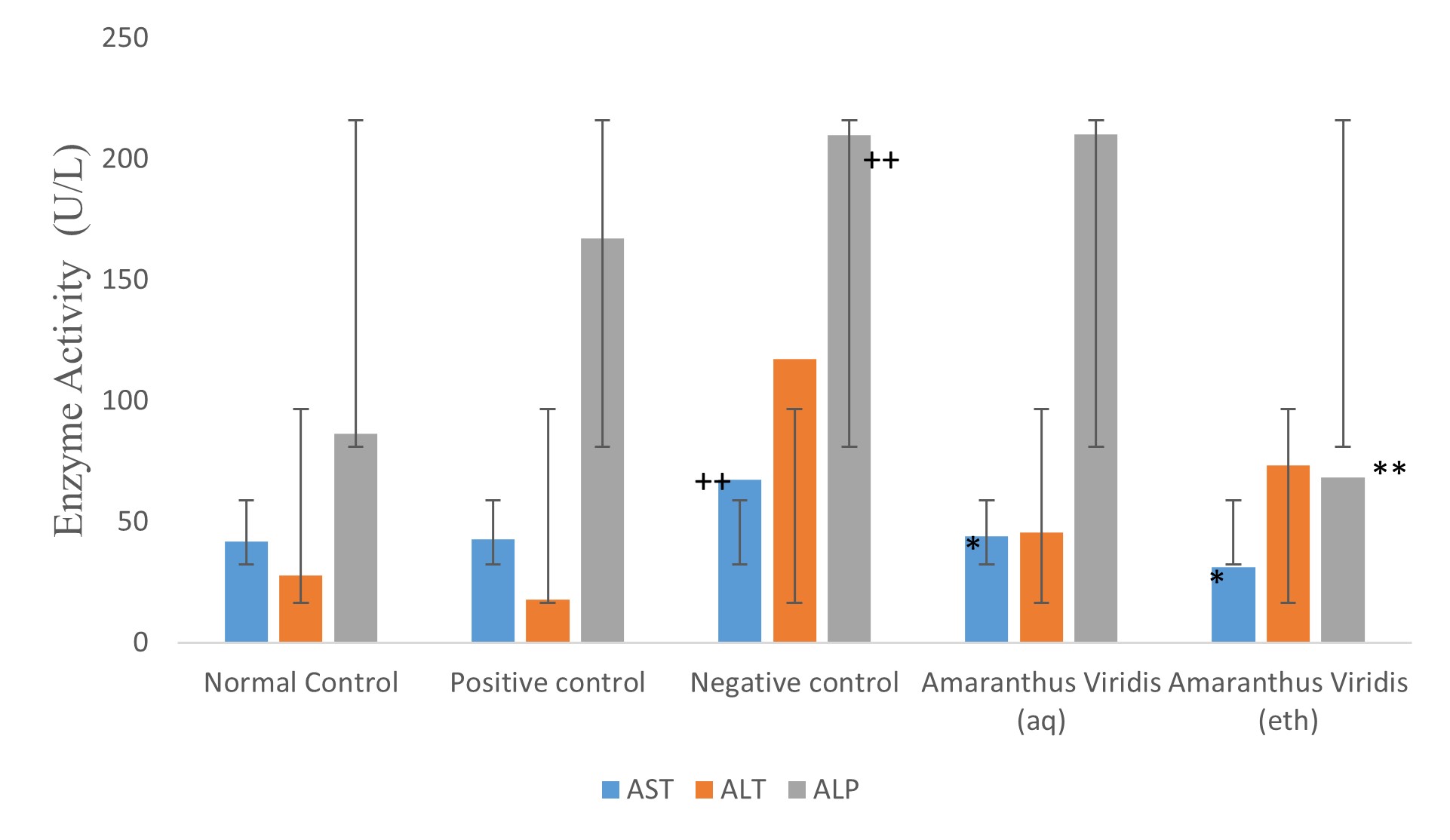
**Fig 1: The effect of pre-treatment with 400gm/kgbw leaf extracts of Amaranthus viridis on the serum activities of AST, ALT and ALP of rats intoxicated with 2g/kgbw single dose of acetaminophen.
Values are mean ± SD of six (6) results, * and ** show values with significant increase and decrease respectively, compared to the Negative control while ++ and — indicate values with significant increase and decreases respectively compared to the Normal control. AST = Aspartate aminotransferase, ALT = Alanine aminotransferase, ALP = Alkaline phoshatase
The Effect of Pre-Treatment with 400gm/Kgbw Aqueous and Ethanol Leaf Extracts of Amaranthus Viridis on the Serum Concentrations of Total Protein, Albumin, and Bilirubin of Rats Intoxicated with 2g/Kgbw Single Dose of Acetaminophen.
The oral administration of a single 2g/kgbw dose of acetaminophen to the rats elicited a significant (p< 0.05) decrease on the serum albumin concentration of the rats. However this intoxication caused insignificant (p>0.05), change on the serum concentrations of total protein, total bilirubin, direct bilirubin and indirect bilirubin (fig 2). The 7day pre-treatment of the animals with both aqueous and ethanol leaf extracts of the plant elicited significant (p< 0.05) increase on the serum concentrations of the albumin of some of the animals, see Table 4.
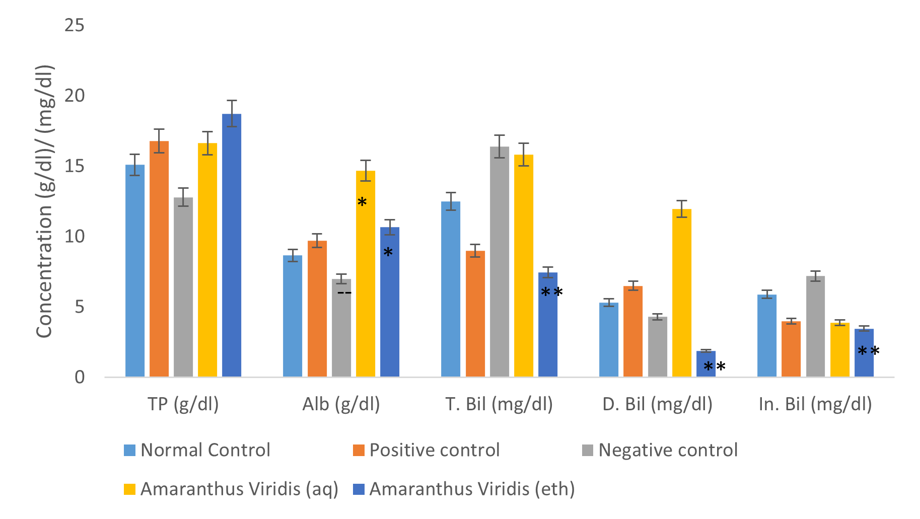
Fig 2: The effect of pre-treatment with 400gm/kgbw leaf extracts of Amaranthus viridis on the serum concentrations of total protein, albumin, and bilirubin of rats intoxicated with 2g/kgbw single dose of acetaminophen.
Values are mean ± SD of six (6) results, * and ** show values with significant increase and decrease respectively, compared to the Negative control while ++ and — indicate values with significant increase and decreases respectively compared to the Normal control.
TP: Total protein
Alb: Albumin
T.Bil: Total Bilirubin
D.Bil: Direct Bilirubin
In.Bil: Indirect Bilirubin
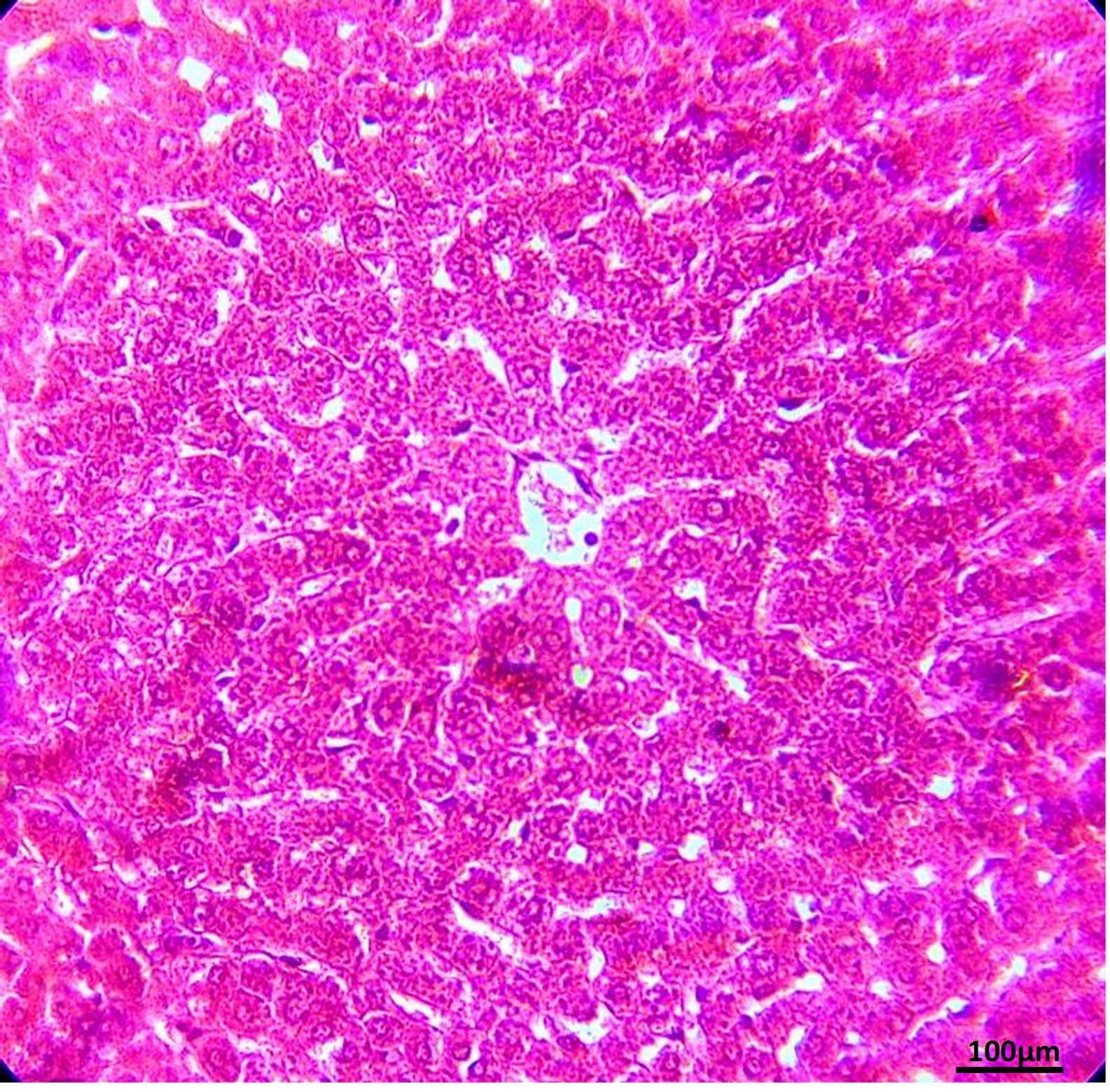
PLATE 1: The histological architecture of livers of rats used as Normal Control](X100) – Haematoxylin and Eosin stain
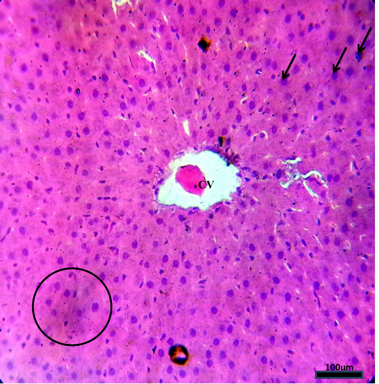
Plate 2: The effect of pre-treatment with Silymarin on the histological architecture of livers of rats administered with a single over-dose of acetaminophen, (X100) – Haematoxylin and Eosin stain. CV = central vein
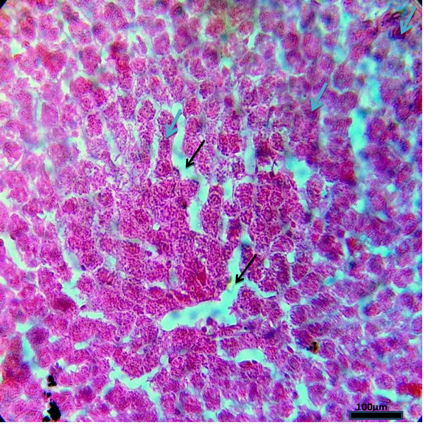
PLATE 3: The effect of a single over-dose of acetaminophen the histological architecture of livers of rats (Negative Control), (X100) – Haematoxylin and Eosin stain
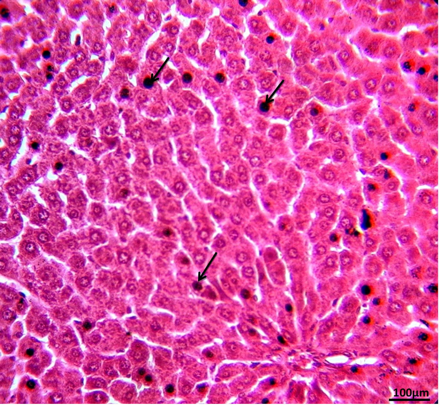
PLATE 4: The effect of pre-treatment with aqueous extract of amaranthusviridis, (Green Amaranth), leaf on the histological architecture of livers of rats administered with a single over-dose of acetaminophen, (X100) – Haematoxylin and Eosin stain
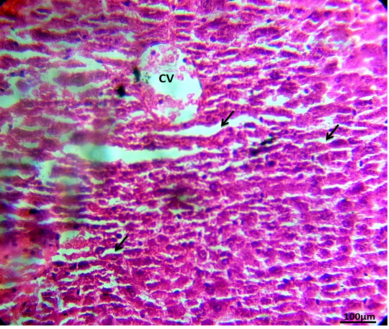
PLATE 5: The effect of pre-treatment with ethanol extract of Amaranthusviridis, (Green Amaranth), leaf on the histological architecture of livers of rats administered with a single over-dose of acetaminophen (X100) – Haematoxylin and Eosin stain.C.V = central vein.
DISCUSSION
The proximate composition of the leaf of Amaranthus viridis reveal that the vegetable is rich in protein, fibre and ash. The Phytochemical screening of the leaf extracts of Amaranthus viridis showed the presence of Phenols, Tannins, Terpenoids and Cardiac glycosides both aqueous and ethanol extracts.Polyphenols and triterpenoids has been shown to possess antioxidant capacity which can ameliorate the effect of oxidative stress on liver cells (Asogwa et al., 2020). The aqueous extract contain higher quantities of tannin and saponin while ethanol leaf extract contained more Phenol and alkaloids. This may be attributed to the different solubility of the phytochemicals in the different solvents used. The oral administration of 2g/kgbw single dose of acetaminophen to the rats (negative control), that were not pre-treated with either the aqueous or ethanol leaf extracts of Amaranthus viridis, induced some level of liver injury. This was evidenced by the significant (p<0.05) increase of the serum activities of the Aspartate aminotransferase (AST), Alanine aminotransferase (ALT) and Alkaline phosphatase (ALP) of the rats in the negative control group (fig 1). Al-Mamary (2002) reported that the aminotransferase are abundant in the liver and are released into the bloodstream following hepatocellular damage, making them sensitive markers of liver damage. Therefore, a marked increase in the serum ALT and AST activities is indicative of liver damage. Serum levels of aminotransferases are used as an indicator of damage to the liver structural integrity because these enzymes are cytoplasmic in location and are released into the circulating blood only after structural damage (Okediran et al, 2014). A marked rise in the serum activity of ALT, reduction in total serum protein and abnormal increase in serum bilirubin had been reported in hepatotoxicity, (Olamide and Mattew, 2013; Olorunnisola et al. 2011; Martin and Friedman, 1992). A decrease in total protein and albumin shows that the ability of the liver to synthesize/metabolize protein (example albumin), may have been impaired, hence indicative of liver damage. Histological profile of the livers of the rats in the negative control group showed poorly preserved hepatic lobular architecture, sharply demarcated hepatocyte, necrosis and some exhibiting peri-portal sinusoidal congestion, marked sinusoidal dilatation which is indicative of liver injury.
The pre-treatment of the animals (rats), with 400mg/kgbw of either aqueous or ethanol leaf extracts of Amaranthus viridis, gave protection against toxicity and oxidative stress arising from a 2g/kgbw single dose of the drug over nine hours (9hrs). The protection may be due to the antioxidant properties of these plants, which stem from their phytochemical components. Researchers have found that phytochemicals have the potential to reduce oxidative damage to cells. Polyphenols and flavonoids compounds have been said to play a major role in various antioxidant effects shown by plant extracts (Asogwa et al., 2020). Triterpenoids is one of the phytochemicals present in Amaranthus and it has been shown to possess hepato-protective activity according to the report by (Xu et al., 2018). However, it is not known whether the health benefits are the result of individual phytochemicals, the interaction of various phytochemicals, the fibre content of plant foods, or the interaction of phytochemicals and the vitamins and minerals found in the same foods. It was found in this study that Amaranthus viridis is a rich source of phytochemicals.
Generally, the administration of aqueous and ethanol leaf extracts of Amaranthus viridis led to decrease of the serum activities of ALT and ALP of the animals pre-treated with the extracts, but markedly increased the serum activity of AST. However, the decreases in the activities of ALT and ALP in the serum of the rats pre-treated with these extracts were not as much as that caused by Silymarin. The extracts also markedly increased the serum albumin and total protein of the pre-treated rats with the preservation of the hepatic lobular architecture.
It can therefore, be suggested that Amaranthus viridis produced effects which may be attributed to its earlier repoted antioxidant activities which may have ameliorated the oxidative damage in the acetaminophen-intoxicated ratsr (Ekor, et al., 2006). From the foregoing, it could be suggested that the plant under study possess great deal of hepato-protective potentials which can account for the low activities of ALT and ALP in the serum of the pre-treated rats and the preservation of the livers’ lobular architecture, compared to the control group.
Conclusion:
The present study provides evidence that Amaranthus viridis offered protection to the hepatic cells. Therefore, it can be a good source of raw material for the production of drugs used for the prevention and treatment of liver diseases.
REFERENCES
- Aja, P.M., Okaka, A.N.C., Onu P.N., Ibiam, U and Urako, A.J (2010).hytochemical Composition of Talinum triangulare (Water Leaf) Leaves.Pakistan Journal of Nutrition 9 (6): 527-530
- Alamgir,H. M. Sheikh Arman Mahbub, Muniruddin Ahmedand Shahidulla Kayser, M.d. (2016). Phytochemical and Pharmacological Investigation ofLagenaria siceraria, Cucumis sativus andCucurbita maxima.European Journal of Medicinal Plants.12(3): 1-13
- Asogwa K. K., Udedi S. C., Ani O. N. and Ekwualor K. U. (2020) Evaluation of micronutrient composition and antioxidant effects of the extracts of the leaf of Justicia carnea. Sky Journal of Biochemistry Research 7(2):021–031.
- AOAC (2006) Official methods of analysis, 21st edition. Association of Analytical Chemist, Washington D.C. pp 74-175
- Bancroft J. D., Suvarna K. S and Christopher, L. (2013):Bancroft’s Theory and Practice of Histological Techniques.Seventh ed. Churchill Livingstone-Elsevier.
- Chevellier, A. (1996). The Encyclopedia of medical plants. London Dorling Kindersley Ltd. (online), http://www.chelibrary.org/plant. (accessed20th August, 2016).
- Dahanukar, A., Foster, K., Van der Goes, Van Naters W.M. and Carlson J.R., (2001). A Gr eceptor is required for response to the sugar trehalose in taste neurons of Drosophila. Nat Neurosci 4: 1182–1186.
- Edeoga, H. O., Okwu, D. E. and Mbaebie, B. O. (2005). Phytochemical constituents of some Nigerian medicinal plants. African Journal of Biotechnology,4(7): 685 – 688.
- Ekor, M., Adekpoju G. K. A. and Epoyun, A. A. (2006). Protective Effect of Methanolic Leaf extracts of Persea Americana (Avocado) Against Paracetamol-Induced Acute Hepatotoxicity in Rats. International Journal of Pharmacology 2 (4): 416-420.
- Enemali, M. O. and S. C. Udedi (2018) Comparative evaluation of the protective effect of leaf extracts of Vernonia amygdalina (bitter leaf) and Ocimum canum (curry) on acetaminophen induced acute liver toxicity. Journal of Pharmacognosy and Phytotherapy 10 (7): 116 – 125. http://www.academicjournals.org/JPP – DOI: 10.5897/JPP2018.0497
- Enemali, M. O., T. O. Bamidele and M. A. Ubana (2018). Protective effect of ethanolic extract of Cucurbita maxima (PUMPKIN) leaf on acetaminophen-induced acute liver toxicity. Journal of Pharmacognosy and Phytotherapy 10(8):142-148. http://www.academicjournals.org/JPP DOI: 10.5897/JPP2018.0497
- Enemali, M.O., Udedi, S C., Ubaoji, K. I., Haruna, G. S. and Augustine, E. (2019). Modulatory Effect of Leaf Extracts of Pterocarpus santalinoides Hook F. (Red Sandal Wood) on Acetaminophen-Induced Hepatotoxicity in Albino Rats. FUW Trends in Science & Technology Journal 4(2); 366 – 371 www.ftstjournal.com.
- Erica Dimitropoulos and Emily M. Ambizas.(2014). Acetaminophen Toxicity: What Pharmacists Need to Know. US Pharmacology. 39(3):HS2-HS8
- Ezekwe, C.I., Uzomba Chidinma R and Ugwu Okechukwu P.C. (2013).The Effect of Methanol Extract of Talinum Triangulare (Water Leaf) on the Hematology and Some Liver of Experimental Rats.Global Journal of Biotechnology & Biochemistry8 (2): 51-60
- Feher, J. and Lengyey, G. (2017).Silymarin: A Potent Antioxidant, Liver Protector, and Anti-Cancer Agent. Smart publications incorporated. Smart Publications, Petaluma, CA, pp 1-2. Křena, V and Walterovab, D (2005).Silybin and Silymarin – New Effects and Applications.Biomedical Papers 149(1): 29–41
- Halliwell, B and Gutteridge, J. M. C.(1999).Free Radicals in Biology and Medicine. 3rd ed., Oxford University Press, New York, pp. 617–783.
- Imaga, N. O. A and Bamigbetan, D. O. (2013).In vivo Biochemical Assessment of Aqueous Extracts of Vernonia amygdalina (Bitter leaf) International Journal of Nutrition and Metabolism.5 (2): 22-27.
- James, L.P, Letzig, L., Simpson, P.M., Capparelli, E., Roberts, D.W., Hinson, J.A. and Lee, W.M. (2009). Pharmacokinetics of acetaminophen-protein adducts in adults with acetaminophen overdose and acute liver failure. Drug Metabolism andDispososal.37: 1779–1784.
- King, E. J. (1965a).The hydrolases-acid and alkalinephosphatases. In Van D (ed.): Practical Clinical Enzymology, Nostrand Co., London, pp.199–208.
- King, E. J. (1965b). The transaminases: alanine and aspartate transaminases. In Van D (ed.): Practical Clinical Enzymology, Nostrand Co., London, 363–395.
- Ladipo, M.K., Doherty, V.Fand Kanife U.C (2010). Phytochemical Screening and Antibacterial Investigation of the Extract of Ocimum Gratissimum (Scent Leaf) on Selected Enterobacteriaceae.patnsukjournal.net/currentissue some Indian herbal formulations as compared to Silimarin, Fitoterapia 62:
- Lowry, O. H., Rosebrough, N. J., Farr, A. L and Randal, R. J. (1951). Protein Measurement with Follin’s Phenol reagent. Biological chemistry.193: 265-275.
- Malloy, H. T and Evlyn E. A. (1937). The determination of bilirubin with photoelectric calorimeter. Journal of Biological Chemistry, 119:481-485.
- Mbang, A., Owolabi, S., Jaja, O and Opeyemi, J.O. (2008). Evaluation of the Antioxidant activity and lipid peroxidation of the leaves of Talinum triangulare Journal of Complementary and Integrated Medicine,5:
- Mitchell, J. R., Jollow, D. , Potter, W. Z., Gillette, J. R and Brodie, B. B. (1973). Acetaminophen-induced hepatic necrosis iv, Protective role of glutathione. Journal of Pharmacological Experiment,187: 211-217
- Morton J.F. (1981). Atlas of medicinal plants of Middle America, Bahama to Yucatan.Charles C, Thomas, Springfield USA pp 1448-1563
- Nilsson, J., Stegmark, R and Akesson, B. (2004). Total antioxidantcapacity in different pea (Pisum sativum) varieties after blanching and freezing. Food Chemistry, 86: 501–507. AOAC (2006).Official methods of analysis, 21st edition. Association of Analytical Chemist, Washington D.C. pp 74-175
- Okediran, B.S., Olurotimi, A.E., Rahman, S.A., Michael, O.G and Olukunle, J.O. (2014). Alterations in the lipid profile and liver enzymes of rats treated with monosodium glutamate. SokotoJournal of Veterinary Sciences 12(3): 42-46.
- Olamide E. Adebiyiand Mathew O. A. (2013). Protective Effects of Enantia chlorantha Stem Bark Extracts on Acetaminophen Induced Liver Damage in Rats. Jordan Journal of Biological Sciences 6 (4): 284-290
- Olorunnisola, O. S., Bradley, G. and Afolayan, A. J. (2011). Antioxidant properties and Cytotoxicity evaluation of methanolic extract of dried and fresh rhizomes ofTulbaghia violacea. African Journal of Pharmacy and Pharmacology.5:2390-2497.
- Sofowora, A. (1993). Screening plants for bioactive agents. In:Medicinal plants and traditional medicine in Africa.2nd Spectrum Books Limited, Ibadan, pp. 134-156.
- Sudha Medapa, Geetha R. J. Singh and Vaishnavi Ravikumar (2011). The phytochemical and antioxidant screening ofJusticia wynaadensis. African Journal of Plant Science.5(9): 489-492.
- Townsend C.C. (1985). Amaranthaceae in polhill RM, (ed), Flora of tropical east Africa Rotterdam, Netherland.1-2: 20-36.
- Udochukwu, U., Omeje, F.I., Uloma,I. S and Oseiwe.F. D. (2015). Phytochemical analysis of Vernonia amygdalina and Ocimum gratissimum extracts and their antibacterial activity on some drug resistant bacteria. American Journal of Research Communication,3(5): 225-235
- Upholf TCTH (1968). Dictionary of economic plants lehre: verlag von j crammer.Vasil’chenko I.T. (1936). Amaranthaceae in: komarov O.I (chief ed). 4:281.
- USDA (2008). United States Department of Agriculture; Natural Resources Conservation Service.Classification for Kingdom Plantae Down to Genus.usda.gov/classification (cited on April 2, 2016).
- Xu, G., Xiao, Y., Zhang, Q., Zhou, M. and Liao, S. (2018). Hepatoprotective natural triterpenoids. European Journal of Medical Chemistry 145(10): 691-716.