
Effect of Tick-Borne Haemoparasitic Diseases on Haematological Parameters of Small Ruminants Managed Under Semi-intensive System in Wukari Town Taraba State Nigeria
- NYIFI, Ambi Silas
- BILBONGA, Garleya
- 457-465
- Dec 18, 2023
- Agriculture
Effect of Tick-Borne Haemoparasitic Diseases on Haematological Parameters of Small Ruminants Managed Under Semi-intensive System in Wukari Town Taraba State Nigeria
*NYIFI, Ambi Silas and BILBONGA, Garleya
Department of Animal Production and Health, Federal University Wukari Taraba State Nigeria
*Corresponding Author
DOI: https://doi.org/10.51244/IJRSI.2023.1011038
Received: 20 November 2023; Accepted: 30 November 2023; Published: 18 December 2023
ABSTRACT
This study was conducted to determine the effect of tick-borne haemo-parasitic diseases on haematological parameters of small ruminants managed under semi-intensive system in Wukari town Taraba State, Nigeria. A total of 100 blood samples were collected from sheep (n = 50) and goats (n = 50) from February to March, 2023. Giemsa stained blood smears were prepared and examined under light microscope, to screen for haemo-parasites. Packed cell volume (PCV) was determined by microhaematocrit centrifugation technique while haemoglobin (Hb) concentration was determined by Sahli’s method. The total white blood cell (WBC) was estimated with Neubauer haemocytometer. The results showed 13% overall prevalence of tick-borne haemo-parasitic diseases. Prevalence based on specie of animal was higher in goats (16%) than in sheep (10%). Prevalence was observed higher in adults than the young, and there was significant difference between age and prevalence of the haemo-parasites. Sex-wise, prevalence of haemo parasites in both goats and sheep was significantly higher in females than males. Anaplasma infection was more prevalent than Babesia infection in both species of the animals examined. The mean values of PCV, Hb concentration of infected sheep were significantly (P<0.05) lower than the uninfected sheep. Similarly, the mean values of PCV and Hb concentration were significantly (p<0.05) lower in infected goats. This study has confirmed that tick-borne haemo-parasitic diseases cause anaemia in infected goats and sheep kept in the study area. We recommend tick control using suitable acaricides, periodic screening and treatment of small ruminants in the study area.
Keywords: Giemsa, Haemo-parasites, Haematological parameters, Taraba, Ticks, Wukari.
INTRODUCTION
Small ruminants production in Nigeria contribute to a great extent to the socio-economic livelihood of many household; they are good source of income generation, household consumption and as security in crop failure (Lebbie,2004).The productivity of small ruminants in Nigeria is threatened by many factors including diseases. Parasitic diseases have been associated with great economic losses in livestock (Singh et al.,2017).Issues related to the health of livestock caused by ecto-parasitic infestation may result to decrease in some blood parameters and other biochemical parameters (Aetish et al.,2007).Ticks are blood sucking ecto-parasites affecting wide range of animals including domestic ruminants such as goats and sheep. As with other species, ticks can limit the production system of small ruminants causing direct and indirect losses (Habela et al.,2003).The feeding habit of ticks cause stress in animals affected by bites, blood losses that can lead to anaemia and even death (Manzano-Roman et al.,2012).Animals that are severely affected by ticks or that do not have immunity against them or the infectious agents they transmit decline in their capacity to produce meat, milk, eggs or leather. Haemo-parasitic diseases in small ruminants transmitted by ticks have been reported in the northern, southern and western regions of Nigeria (Thornton,(2010),Jatau et al.,(2011),Nwoha et al.,(2013), Ukwueze and Ekenma, (2015).Haemo-parasitic diseases such as Babesiosis, Anaplasmosis, Theileriosis and Trypanosomosis are considered as major impediments to ruminant production including small ruminants (Lako et al.,2007).They have generally shown to be associated with destruction of red blood cells resulting in anaemia, anorexia, high morbidity and mortality, infertility, jaundice and weight loss (Akande et al.,2010).Adejinmi et al.,(2004) also confirmed that anaemia is a reliable indicator for the severity of haemo-parasitic infections.
Haematological and serobiochemical alterations are indicators of severity of disease and are considered to be good tools for the diagnosis for effective therapy (Pfaffle et al.,2009).Blood is an important and reliable medium for assessing the health status of individual animals. Examining blood for its constituents is used to monitor and evaluate the health and nutritional status of animal (Gupta et al.,(2017).A reduction in haematological values have been reported in tick infested animals compared to those animals which were free from tick infestation (Biswal et al.,2018).An increase in number of eosinophils and lymphocytes in ticks infested animals have also been reported (William et al.,2017).Pandy et al.,(2017) revealed that some diseases transmitted by ticks such as babesiosis cause anaemia in animals due to intravascular haemolysis of red blood cells. Another report by Ibrahim et al.,(2009) attributed normocytic normochromic anaemia in tick infested animal to theileriosis which have suppressing effect on the bone marrow and interfere with the process of erythropoeisis. Despite several reported cases of haemo-parasitic infections in Nigeria, infection with haemo-parasites remains persisting as a major challenge to livestock production (Onoja et al.,2013,Mans et al.,2015).A proper understanding of the epidemiology of haemo-parasitic diseases is a prerequisite to having a rational design for the effective control and preventive strategies against these dreadful diseases (Opara et al.,2016).
Several in depth studies on the prevalence of these infections have been conducted in various parts of the Sub-Saharan Africa, including Nigeria.Despite the economic significant consequences of haemo-parasites to ruminant, there is still paucity of information on the prevalence and magnitude of haemo parasites in small ruminants in Wukari, Taraba State north-eastern Nigeria.Therefore, information on prevalence, distribution, and potential risk factors of haemo-parasites of small ruminants is significant because the outcome could be used to make objective decisions on control strategies. Hence, the present study was aimed to determine the effect of haemo-parasitic diseases on haematological parameters of small ruminants.
MATERIALS AND METHODS
Study Area
The study was conducted in some selected homes in Wukari town. Wukari is the headquarters of Wukari Local Government Area of Taraba State. It’s situated in the North Eastern Nigeria which lies within latitude 70.52’; 48’N to 70 .87’ N and a longitude 90 .43’ 38’’E to 90 .77’’E at an elevation of 189 meters above sea level (Elechi et al.,2013).The town has an estimated population of 241,546 in 2006 population census (NPC,2006).Taraba State has a tropical climate, marked with two distinct seasons; wet season which starts in March and ends in October and dry season which starts in November and ends in March or April (Taraba State Diary, 2018).
Study Animal /Population
The study animals were local breeds of goats and sheep managed under semi-intensive system.A total of 100 small ruminants were examined for haemo-parasites and their effect on blood parameters (PCV, Hb, and WBCs).All the study animals were de-wormed with anti-helminthic (albendazole) against gastrointestinal parasites before sampling after 7days.
Sample Size, Sampling Technique and Data Collection
Since there is no reasonable research done on this topic in the study area so far, the sample size was determined using Thrus field formula (N= Z2P (1-P)/d2) by considering 50% expected prevalence(P), 95% confidence interval (CI) (Z =1.96) with 5% desired absolute precision (d) (Thrustfield,2007).Although, the required sample size obtained by this formula was relatively larger, but only 100 small ruminants were randomly sampled for this investigation. All procedures involving the handling and collection of blood samples from the animals were approved by the ethical committee for animal research of Federal University Wukari Taraba State (Ref No: CAR/FUW/AR/030).Records on sex, age, specie, presence or absence of ticks were recorded accordingly.Sex differentiation was made based on the appearance of external genitalia that is presence or absence of testis and udder. ages of the sampled animals were considered in two age groups; young (≤ 1 year) and adults (> 1 year).The age was determined by dentition as described by al-qudda (2009).The study animals were randomly selected and examined thoroughly for the presence or absence of ticks on different parts of the body such as head, face, neck, brisket and tail.
Sample Collection
About 4ml of blood samples were aseptically collected from apparently healthy goats and sheep via jugular vein puncture using a sterile hypodermic needle and syringe.The blood samples collected were gently transferred into sample bottles containing Ethylene Diamine Tetra Acetic Acid (EDTA) and labeled appropriately. The whole blood samples were transported on iced pack to Parasitological and Microbiological Laboratory of College of Agriculture, Science and Technology,Jalingo Taraba State for processing and examination.
Parasitological Analysis
A drop of blood (about 20 μl) was placed on a clean glass slide, and covered with clean cover slip. Thin blood smear was prepared using standard method as described by Chees brough,(2003). A drop of blood was placed on one end of a clean, grease-free glass slide and made into thin film with aid of a spreader (a clean glass slide). This was done by allowing the spreader to touch the blood at an angle of 45º, and then spread gently but firmly along the surface of the horizontal slide so that the blood is dragged behind the spreader to form the film with a feathered edge. The prepared thin film was then air-dried, fixed in methanol for 2-5 minutes and stained in freshly prepared 10% Giemsa stain at pH 7.2 (10 ml Giemsa solution and 90 ml buffer solution) for 25-30 minutes. Afterwards, the stained blood smear was rinsed in buffered water and allowed to dry. The smears were then examined at X100 objective magnification (oil immersion) on an Olympus Microscope for the presence of haemoparasites. Parasites were identified using the key standard characteristics of the parasites described by Brar et al.,(2011).
Determination of Haematological Parameters
Packed cell volume (PCV) was determined using microhaematocrit centrifugation technique as described by Brar et al.,(2011).Blood was introduced into microhaematocrit tubes by capillary action and one end of each capillary tube was sealed with plasticin. The tubes were spun in a microhaematocrit centrifuge(Hawksley, England) at 13000 g for 5 min. PCV was measured with a hematocrit reader (Hawksley,England), and recorded appropriately (Kamani et al.,2010). Haemoglobin count was determined using the Acid Hematin method as described by Brar et al. (2011) using Sahli’s instruments. Total white blood cell counts (WBC) were determined by hemo cytometer methods described by Coles (1974).
Data Analysis
The data of positive samples were expressed in simple percentages and presented on tables. The prevalence was calculated by dividing the number of positive animals by the total number of animals examined and times 100.Data analyses were carried out using the Statistical Package for the Social Sciences (SPSS, Chicago, Illinois, USA) for Windows version 22.0. Chi-square (χ2) was used to determine the association of haemo-parasites with risk factors (age and sex) at 95% confidence interval and P < 0.05 was considered as significant. Student’s T-test was performed to determine the difference between mean haematological parameters of infected and uninfected sheep and goats. Significant differences were considered at P< 0.05.
RESULTS AND DISCUSSION
Result
Out of the 100 goats and sheep examined for tick-borne haemoparasitic diseases,13(13%)were positive for haemoparasites. Anaplasma ovis and Babesia ovis were the species of haemoparasites identified in this study. Amongst the 50 goats examined, 6 (12%) were positive for Anaplasma infection while 2(4%) had Babesia infection. In sheep,4(8%) and 1(2%) were recorded for anaplasmosis and Babesiosis respectively. Prevalence based on specie of animal was higher in goats (16%) than in sheep (10%). Anaplasma infection was more prevalent than Babesia infection in both species of the animals examined (Table 1).
Prevalence based on age revealed that adult goats and sheep had prevalence of 10% and 8% respectively while young goats and sheep had prevalence of 6% and 2% respectively. Prevalence was significantly (P<0.05) observed higher in adults than the young.Sex-wise prevalence of haemo-parasites in both goats and sheep was also significantly (P<0.05) higher in female than male (Table 2).
The mean values of Packed Cell Volume (PCV) and Haemoglobin concentration (Hb) of infected goats were significantly below the normal range of values (P< 0.05).The un-infected goats had mean values of PCV and Hb within the normal values and was statistically significant. Total White Blood Cells (WBCs) in infected goats was significantly (P<0.05) above normal range of values and within the normal range in the uninfected (Table 3).
There was a significant reduction in the mean values of PCV and Hb in infected sheep below the normal range of values. Un-infected sheep had PCV and Hb within the normal range of values. WBC count was also significantly higher than normal range of values in infected sheep, but within the normal range of values in the uninfected ones (Table 4).
Table I: Prevalence of Tick-Borne Haemo-Parasites in Goats and Sheep
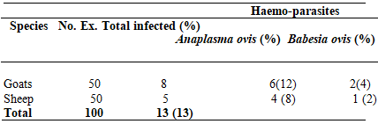
Table II: Prevalence of Tick-Borne Haemo-Parasites Based on Risk Factors
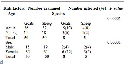
*Significance = P < 0.05
Table III: Mean ± SEM Haematological Parameters of Infected and Un-infected Goats
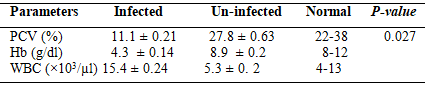
*Significance = P < 0.05
(Normal values, Khan, 2005)
Table IV: Mean ± SEM Haematological Parameters of Infected and Un-infected Sheep

*Significance = P < 0.05
(Normal values, Khan, 2005)
DISCUSSION
The results obtained from this present study revealed that tick-borne haemo-parasites are prevalent in both sheep and goats examined in the study area. An overall prevalence of 13% was obtained in this study.This findings are lower than some reports from previous studies in Nigeria such as 20.38% reported from Kano State (Jatau et al.,2011), 21% from Bauchi State (Adamu and Balarabe,2012), 56.32% from Karu, Nassarawa State (Anyanwu et al.,2016).The reason for the variation in the reported prevalence rates may be due to difference in sample size, breed variation and period of sample collection. Two species of haemo-parasites (Anaplasma ovis and Babesia ovis) were identified from this study. Anaplasma ovis was more prevalent (8%) than Babesia ovis (3%). The observed higher prevalence of Anaplasma ovis compared to Babesia ovis agrees with previous reports of other researchers in Nigeria such as Adamu and Balarabe, (2012), Jatau et al.,(2011), Opara et al.,(2016) and Takeet et al.,(2009). In contrast to our findings, Anyanwu et al.,(2016) reported higher prevalence of Babesia spp than Anaplasma spp in a similar study. The low prevalence of Babesiosis compared to Anaplasmosis in this study could be due to the fact that small ruminants have been reported to be unstable for babesia parasites, those that may have been infected and recovered from Babesiosis become immuned to re-infection as observed by Adamu and Balarabe,(2012),and Jatau et al.,(2011).Age-wise prevalence showed that haemo-parasitic infections were found to be higher in both adult goats (10%) and sheep (8%) compared to the infection rates in young goats (6%) and sheep (2%) examined.There was significant difference between haemo-parasitic diseases and age groups of both species of small ruminants examined. Sex based prevalence of haemo-parasites were found to be significantly higher in the females of both goats (12%) and sheep (6%) when compared to the young goats (4%) and sheep (4%) examined. The higher prevalence in females than males in this study agrees with the reports of Abenga et al.,(2008),Kamani et al.,(2010), and Nwoha et al.,(2013) who have also reported more prevalent haemo-parasitaemia in female animals compared to their males.The discrepancies in the respective findings of haemo-parasitic infections in females against the male animals in this study could be attributed to the proportion of the female animals sampled in both species of the animals examined. Most livestock farmers keep more female animals than males for the purpose of breeding while the males are usually sold out for cash in terms of needs, which may affect the proportion of the sex infection. It has also been reported that the female animals are generally more prone to infection by haemo-parasites due to their extended breeding for economic purposes such as parturition and milk production (Ukwueze and Kalu,2015) as well as the stress of breeding, milking and cyclical hormonal changes associated with gestation, parturition and calving processes. However, the result of the present study is contradictory to previous findings of Anyanwu et al.,(2016) and Paul et al.,(2016) who reported higher prevalence in young animals than the adults. In this study, we recorded a significant decrease (p<0.05) in the mean packed cell volume (PCV), and haemoglobin concentration of goats and sheep infected with haemo-parasites compared to the uninfected ones. The decrease in PCV and Hb concentration of infected goats and sheep below the normal range of values in this study signifies anaemia. The anaemia observed in this study is in line with the findings of Adejinmi et al.,(2004), Okaiyeto et al.,(2008), Okorafor and Nzeako,(2014), Josiah et al., (2015) and Anyanwu et al., (2016) who have also reported that haemo-parasitic infections result in anaemia. Rymaszewska and Grande (2008) also attested that progressive anaemia usually develops during Babesiosis and Anaplasmosis. Kamani et al.,(2010) also reported that infection with Babesia spp, Anaplasma, spp, theileria spp and trypanosomes spp either singly or in combination caused significant reduction in mean PCV of animals. However, our findings contradicts the finding of Ademola and Onyiche, (2013) who reported no significant difference (P>0.05) in the mean packed cell volume (PCV) of animals with mixed or single haemo-parasitic infection in a similar study. The difference in the PCV and Hb concentration of haemo-parasites infected animals from various reports might be as a result of differences in nutritional and health status of the infected animals. Well-nourished and healthy animals may not show significant changes in their blood parameters especially during subclinical infection with blood parasites (Adejinmi et al.,2004; Anyanwu et al., 2016). However, the effects of haemolytic activities of the haemo-parasites might be the cause of anaemia in the infected animals in this present study which supports the fact that haemo-parasites are capable of inducing erythrocytolysis and erythrophagocytosis as previously reported by Okaiyeto et al.,(2008) and Jatau et al.,(2011).
CONCLUSION AND RECOMMENDATIONS
It’s concluded in this study that tick-borne haemo- parasitic diseases such as anaplasmosis and babesiosis can result to anaemia in infected animals. The patho-physiological effects of these diseases might result to significant losses in animal production. The losses could be in form of stunted or decreased growth rate, decrease in milk production, progressive emaciation, late maturity, weight loss, and increased susceptibility to other infectious diseases. To minimize economic losses associated with these diseases, there is need for ticks control using effective acaricides and periodic screening and treatment of sheep and goats with suitable anti-protozoan drugs.
REFERENCES
- Aatish, H.U., Singhu, Z, Iqbal, A, and Jabbar, A. (2007).Prevalence of sheep mange indistrict Dera Ghazi Khan (Pakistan) and associated haematological and biochemical disturbances. International Journal of Agriculture and Biology, 9(6):917-920
- Abenga, J. N, Fajinmi, A, Idowu, T, Kalgo, A, Lawani, F and Samdi, S. (2008). Seasonal variation of trypanosomosis rates in small ruminants at Kaduna abattoir, Nigeria.African Journal of Biomedical Research, 11(2): 229 – 2s
- Adamu, B.S and Balarabe, L.M. (2012). Prevalence of Haemoparasites of Sheep and Goats Slaughtered in Bauchi Abattoir. International Journal of Applied Biological Research, 4(1): 128 –133.
- Adejinmi, J.O, Sadiq, N.A, Fashanu, S.O, Lasisi, O.T and Ekundayo, S. (2004).Studies on the Blood parasites of sheep in Ibadan, Nigeria. African Journal of Biomedical Research, 7(2):41– 43.
- Ademola, I. O and Onyiche T. E (2013). Haemoparasites and Haematological Parameters of Slaughtered Ruminants and Pigs at Bodija Abattoir, Ibadan,Nigeria.African Journal of Biomedical Research, 16(2):101-105
- Akande, F.A., Takeet, M.I., and Makanju, O.A.(2010). Haemoparasites of cattle in Abeokuta, South West Nigeria. Scientific World Journal, 5(4): 9– 21.
- Al-Qudda, A.A., and Awawdeh, L.A.(2009). Root and canal morphology of mandibular first And and second molar teeth in Jordanian population International Endodontic Journal, 442(9):775-84
- Anyanwu, N. C. J., Iheanacho, C. N., and Adogo, L.Y. (2016). Parasitological Screening of Haemo-Parasites of Small Ruminants in Karu Local Government Area of Nassarawa State, Nigeria. British Microbiology Research Journal, 11(6): 1 – 8.
- Biswal, B., Misra, S.C., and Bisol, P.C .(2018).Effect of tick infestation on haematological parameters of cattle. Journal of Veterinary Parasitology 2(1):9-13
- Brar, R.S., Sandhu, H.S., and Singh, A. (2011).Veterinary Clinical Diagnosis by Laboratory methods. First Edition. India, Kalyani Publishers., 28: 216– 217.
- Cheesbrough, M. (2003). District Laboratory Practice in tropical Countries, Part 2 University Press Cambridge United Kingdom, p. 266-342
- Coles EH (1974). Veterinary clinical pathology, 1Ed. U.S: WB Saunders Company, pp.67-92
- Elechi I.M., Tikyaa EV and Isikwe B.C (2013).Dynamics of daily rainfall and temperature in Nigeria. International Journal of Science and Research,4(7):493 – 494
- Gupta R, Gupta A, and Andrew P (2017).Study of haematological parameters in sepsis patients and its prognostic implication. International Journal of Research in Medical sciences, 7(3):828
- Habela, M, Fruto, J.M, Moreno, A, and Gragera-slikker, A (2003). Infestacion por garrapatas. Repercucionesy planes de lucha y control enlas exployes clones de pequenos ruminantes. Mundo Ganadero,156(3):44-50
- Ibrahim, A.K., El Behairy, A.M., Mahran, K.A, and Awad, W. (2009). Clinical and laboratory diagnosis of piroplasmid in naturally infected cattle in Egypt. Journal of Egyptian Veterinary and Medical Association, 69(2):105-203
- Jatau, I.D., Abdulganiyu, A, Lawal, A.I., Okubanjo, O.O., and Yusuf, K.H.(2011).Gastrointestinal and haemo parasitism of sheep and goats at slaughter in Kano, Northern-Nigeria. Sokoto Journal of Veterinary Sciences, 9(1): 7 – 11
- Josiah, G.J., Omalu, I.C.J, Makun, H.J., Chiezey, N.P., and Abah, O.I (2015).Haemonchosis And Haemoparasites of small Ruminants Reared in North Western, Nigeria. Animal Research International, 12(3): 2284 – 2291
- Kamani J, Sannusi A, and Egwu O.K (2010). Prevalence and Significance of Haemoparasitic Infections of Cattle in North-Central, Nigeria. Veterinary World,3(9):44
- Khan, T.A., and Zafar, F. (2005). Haematological Study in Response to Varying Doses of Estrogen in Broiler Chicken. International Journal of Poultry Science, 4(10):748-751.
- Lako, N.J., Tchoumboue, J, Payne, V.K., Njiokou, F, Abdoulmoumini, M, and Awah-Ndukum, J.(2007).Prevalence of trypanosomosis and babesiosis among domestic ruminants in the western Highlands of Cameroon: Proceedings of the 12th International conference of the Association of Institutions of Tropical Veterinary Medicine, Montpellier, France 20 –22 August, 2007, pp. 405 – 410.
- Lebbie (2004).Goats under household conditions. Small Ruminants Res.,51(2), 131-136 Mans, B. J., Pienaar, R. and Abdula Latif, A. (2015). A review of Theileria diagnostics and epidemiology. International Journal for Parasitology,4 (2): 104 – 118.
- Manzano-Roman R, Diaz-martin V, Perez-sanchez, R .(2012). Characteristics anatonicals, epidemiologicals sonidad animals sito argent no de produccion animal, 1-8
- Mosqueda, J, Olvera-Ramirez, A, Aguilar-Tipacamu, G, and Canto G.J. (2012).Current advances in detection and treatment of babesiosi Current Medicinal Chemistry, 19(10):1504-18
- NPC, (2006). Nigerian National Population Census Report. National Population Commission, Pp 109
- Nwoha, R.I.O., Onyeabor, A, Igwe, K.C., Daniel-Igwe, G, Onuekwusi, G.C.O., and, Okah U. (2013). Prevalence of Haemoparasites in Livestock in Ikwuano Local Government Area of Abia State. Journal of Fisheries and Livestock Production, 2(1): 109-111
- Okaiyeto, S.O., Tekdek, L.B., Sackey, A.K.B., and Ajanusi, O.J. (2008). Prevalence of haemo and gastrointestinal parasites in sheep and goats kept by the Normadic Fulanis in some Northern States of Nigeria. Research Journal of Animal Sciences,2(2): 31 – 33.
- Okorafor, U.P., Nzeako, S.O.(2014). Prevalence of Haemoparasites of Cattle from Three Abattoirs in Ibadan Metropolis, Oyo State, Nigeria. International Journal of Science Research and Environmental Sciences ,2(7): 244 – 249.
- Onoja, I.I., Malachy, P, Mshelia,W.P., Okaiyeto, S.O., Danbirni ,S, and Kwanashie, G. (2013).Prevalence of Babesiosis in cattle and goats at Zaria Abattoir, Nigeria. Journal of Veterinary Advances,3(3): 211 – 214.
- Opara, M.N., Santali, A, Mohammed, B.R., and Jegede, O.C (2016). Prevalence of Haemoparasites of Small Ruminants in Lafia Nassarawa State: A Guinea Savannah Zone of Nigeria. Journal of Veterinary Advances, 6(6): 1251 – 1257.
- Paul, B.T., Bello, A.M., Ngari, O, Mana, H.P., Gadzama, M.A., Abba, A, Malgwi, K.D., Balami, S.Y., Dauda J, and Abdullahi, A.M. (2016). Risk factors of haemoparasites and Some haematological parameters of slaughtered trade cattle in Maiduguri, Nigeria. Journal of Veterinary Medicine and Animal Health ,8(8): 83 – 88.
- Pandy, N, Misras, S. (2017).Haematological and biochemical response to haemolytic anaemia of clinical Babesiosis in cattle and therapy. Indian Veterinary Journal, 64:882-886
- Pfaffle, M, Petney, T, Elgas, M, and Skubella, J. (2009).Tick induced blood-loss leads to regenerative anaemia in the European hedgehog. European Parasitology, 136(4):443-45
- Rymaszewska, A, and Grenda, S. (2008). Bacteria of the genus Anaplasma– characteristics of Anaplasma and their vectors: A Review. Veterinary Medicine, 53(11):573-584.
- Singh, E, Kaur, P, Singla, L.D., Bal, M.S.(2017)a. Prevalence of gastrointestinal parasitism in small ruminants in western zone of Punjab India. Veterinary World 10(1):61-66.
- Soulsby, E.J.L, (1982). Helminths, Arthropods and protozoa of domesticated animals,7thedition. The ELBS and Baillieve, tindal cais ltd, London,1982-766
- Taraba state Government Diary (2018).Taraba official Diary, Government printer Jalingo, Nigeria. Pp 25-29
- Takeet, M.I,, Akande, F.A., and Abakpa, S.A.V. (2009). Haemoprotozoan Parasites of Sheep in Abeokuta, Nigeria. Nigeria Journal of Parasitology, 30(20): 142 – 146.
- Thornton, P.K.(2010). Livestock production: recent trends, future prospects. Philosophical Transactions of the Royal Society B: Biological Sciences, 365: 2853 – 2867
- Thrusfield, M.V.(2007).Veterinary epidemiology (3rd edn.).Blackwell Science Oxford, London, UK.,pp. 483.
- Ukwueze C.S., Ekenma (2015). Prevalence of haemoparasites in red sokoto goats slaughtered at Ahiaeke Market, Umuahia, Abia State, Nigeria. Journal of Veterinary Advance., 5(2):826-830
- Ukwueze, C.S., and Kalu, E.J (2015). Prevalence of haemoparasites in red Sokoto goats slaughtered at Ahiaeke Market, Umuahia, Abia State, Nigeria. Journal of Veterinary Advances,5(2): 826– 830.
- William, R.E., Hair, J.A., and Bulknor, R.G (2017).Effect of gulf coast tick on blood composition and weight of dry lot Hereford. Journal of Economics Entomology, 70(2):229-233