
Molecular Isolation of Theileria and Babesia Species and the Risk Factors Associated with Their Infection in Cattle at Makurdi and Abinsi in Benue State, Nigeria
- Veronica Ene Egwu.
- Terwase Febian Ikpa.
- Okwoli Amali
- 347-357
- Jul 30, 2024
- Zoology
Molecular Isolation of Theileria and Babesia Species and the Risk Factors Associated with their Infection in Cattle at Makurdi and Abinsi in Benue State, Nigeria
*Veronica Ene Egwu., Terwase Febian Ikpa., Okwoli Amali
Department of Zoology, Joseph Sarwuan Tarka University makurdi, PMB 2773 Makurdi, Benue State, Nigeria
*Corresponding Author
DOI: https://doi.org/10.51244/IJRSI.2024.1107025
Received: 31 May 2024; Accepted: 25 June 2024; Published: 30 July 2024
ABSTRACT
The study was conducted to investigate the Molecular Isolation of Theileria and Babesia Species and the Risk Factors associated with their infection in cattle at Makurdi and Abinsi in Benue State, Nigeria. Blood samples were collected randomly from 160 cows in three locations in Makurdi (Wadata, Wurukum, North bank and one location in Abinsi and examined for Theileria and Babesia infection using molecular methods. A questionnaire was used to assess the risk factors associated with the infections. The result of the PCR reaction indicated the presence of Theileria orientalis and Babesia bovis at 335 and 278 base pair respectively. Chi-Square statistical analysis was used to determine the significance of the prevalences of the infections among cattle. The results showed that Theileria orientalis was more prevalent than Babesia bovis with the prevalences of 11.25 and 7.50 respectively. The results of multivariate logistic regression analysis showed that management practices, acaricidal control on the farm, and the presence of other animals are statistically significant risk factors associated with an increased likelihood of infections in cattle. However, age and location was not statistically significant and sex showed marginal significance. In conclusion therefore, there was high prevalences of Babesia and Theileria species in cattle in Makurdi and Abinsi. Based on Molecular description it is therefore recommended that farmers should implement more effective tick control measures in the Abinsi and Makurdi area of Benue State, Nigeria to reduce the vectors responsible for transmitting Theileria and Babesia species furthermore, farmers should be educated to know the importance of regular tick control practices against the infection.
INTRODUCTION
The prevalence of haemoparasites of cattle in Nigeria is generally considered to be very high due to the preponderance of their arthropod vectors (Musa et al., 2014). The prevalence of various genera of haemoparasites of cattle (Trypanosomes, Babesia, Anaplasma, and Theileria) has been reported in different parts of the country (Okorafor and Nzeako, 2014 and Qadeer et al., 2015) and elsewhere in the world (Alim et al., 2012; Velusamy et al., 2014). Babesia and Theileria species are listed among the most economically important genera of haemoparasites in Nigeria (Velusamy et al., 2014) and their impact on cattle production and productivity accounts for heavy economic losses to livestock producers in the tropics and subtropics (FAO, 2017). They are responsible for destruction of erythrocytes leading to anaemia, jaundice, anorexia, weight loss and infertility in livestock (Okorafor and Nzeako, 2014).
Babesia and Theileria species infect a wide range of both domestic and wild animals (Gupta et al., 2017). Babesia divergens, Babesia bigemina, Babesia bovis, and Babesia major have been associated with Babesiosis in cattle (Jane and Adam, 2014; Abdela et al., 2018). For theileriosis, Theileria annulata, Theileria orientalis and Theilria parva are responsible for bovine theileriosis
The diagnosis of these important blood protozoa are mainly based on clinical symptoms and microscopic examination of Giemsa-stained blood smears. Several researchers identified bovine piroplasms using microscopic examination for epidemiological investigation (Al Mahmud et al., 2015). In some studies serological examination has been used to confirm the tentative diagnosis of the infections (Ali et al., 2016 and Rahman et al., 2015). However, these studies are only reliable for the diagnosis of acute cases but have limited value in subclinical cases due to low parasitaemia (Hosen et al., 2020). Compared to serological testing and microscopic identification of piroplasms, molecular detection of piroplasms infection by polymerase chain reaction (PCR) has proven to be more sensitive, accurate, and the gold standard test particularly in detecting Babesia and Theileria in carrier cattle (Belotindos et al., 2014).
In recent years, molecular studies have been performed to detect piroplasmosis in cattle in many part of the world (Roy et al., 2018; Le Huy et al., 2020). However, these studies were on a few tick-borne diseases with inadequate information on their genotypes or even knowledge on their molecular epidemiology, which is critical for the control and prevention of these diseases.
Molecular identification, with or without clinical indications of bovine piroplasmosis, provides an alternative method for the direct detection of piroplasms in carrier animals to overcome economic losses (Moumouni et al., 2015). Therefore, the present study, a molecular survey of Babesia and Theileria species based on PCR amplification was conducted in the cattle population of the Abinsi and Makurdi of Benue State. Cattle contribute significantly to food security and value chain of the Nigerian economy (Lawal-Adebowale, 2011) but their productivity is threatened by ticks and associated haemoparasitic diseases, especially Babesiosis and Theileriasis (Jatau et al., 2011). The diseases condition is clinically manifested as fever, anorexia, anaemia, emaciation, threatened abortion and death in the acute form of the infections (Maharana et al. 2016). The effect of haemoparasitic disease of cattle causes huge loss of income to the herders, such as still birth, stunted growth and low birth rate.
The present work initialized the Prevalence of Theileria and Babesia species and the risk factor associated with Their Infection in Cattle in Makurdi and Abinsi in Benue State, Nigeria.
MATERIALS AND METHODS
Study Area
The study was conducted in Makurdi and Abinsi, Benue State. Which are located in north central Nigeria along the Benue River. They samples were collected in Northbank, Wadata, and Wurukum in Makurdi and Abinsi
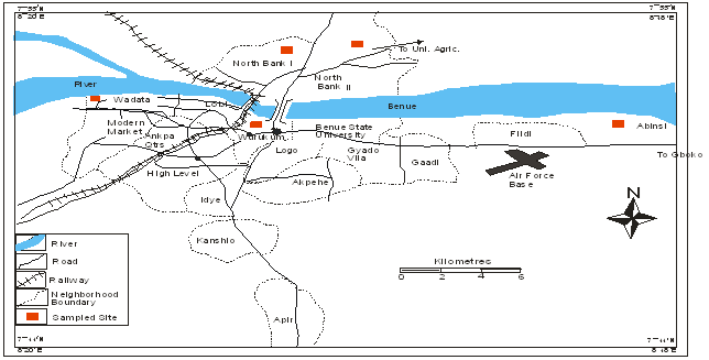
Map of the Study Area Showing Sample Sites for the Collection of Babesia and Theileria Species in Cattle
Sources: Ministry of Land, Survey and Solid Minerals, Makurdi
Sampling Techniques
The samples were collected from four different locations; Wadata, Wurukum, North bank, and Abinsi. One hundred and sixty (160) blood samples were collected from cattle of all ages and both sexes. The sample size was determined from previous prevalence of 15.3 % (Opara et al., 2016). The animals were restrained with rope and 5mls of blood was collected immediately from the severed jugular vein into vacutainer tubes, containing 1 mg of ethylene diaminetetra-acetic acid (EDTA) and were labelled appropriately. The age and sex of each animal were also identified based on morphometric characteristics and recorded in a case book. Cattle less than three (3) year old were categorized as young while older ones were considered as adult. The samples were transported on icepacks at 4°C to the biological laboratory, Joseph Sawuan Tarkaa University Makurdi for parasitological examinations.
A close-ended questionnaire was developed and filled in at flock level by interviewing the herders during sampling in order to assess potential risk factors for Theileriosis and Babesiosis. Those included flock size, type of management (extensive: free ranging without supplementary feed; semi-intensive: supplementary feed provided), source of grazing/browsing place.
LABORATORY ANALYSIS
Haematological Examination
Thin blood smears were prepared using the method described by Ademola and Onyiche (2013). The smears were examined at ×100 magnification (oil immersion) on an Olympus microscope for the presence and identification of blood parasites according to keys and descriptions as given by Soulsby (1982) and Taylor et al. (2016).
Sample Preparation for Polymerase Chain Reaction (PCR)
Blood samples collected were transported to the Molecular Biology Laboratory for PCR diagnosis. Genomic DNA was isolated using ZYMO DNA MINI KIT USA according to the manufacturer’s instructions for solid tissue samples.
Collected samples were treated with lysis buffer and Proteinase K to remove potential inhibitors of PCR present in the blood. For each sample, 500 μl of blood was aliquoted into a microfuge tube filled with 1ml lysis buffer (0.22 % NaCl, 0.015 % saponin, 1 mM EDTA, pH 7.5).
Tube contents were mixed and centrifuged at 9,300Xg for 5 minutes and the resulting supernatant discarded: pellets were resuspended in 750 μl of lysis buffer, centrifuged and supernatants discarded; this process was repeated until the pellet was clear of haemoglobin. The final pellet was resuspended in 100 μl of 50 mM KCl, 10 mM tris-HCl pH 8.0, 0.5 % Tween 20 and 100 μg proteinase K per ml. The tubes were then incubated in a water bath at 56˚C for 2 hours after which they were immediately stored at -20˚C. 1μl of the lysate was used as a template for PCR reactions.
PCR Assay for Diagnosis of Tick-borne Pathogens and Validation by Sequencing
A semi-nested PCR was used for the detection of TBP based on methodology and primer sets previously developed and validated with species specific DNA samples. A primary reaction was carried out using universal primer sets designed against the 18S rRNA gene for T. annulata, T. orientalis, B.bigemina and B. bovis. (Simuunza 2011 and Ghaemi 2012). The set of primer for primary reaction of Theileria and Babesia showing the forward and reverse reaction are shown below:
| PRIMER SET 1 | |||
| Theileria/Babesia universal | F: 5/-GACACAGGGAGGTAGTGACAAG-3/
R: 5/CTAAGAATTTCACCTCTGACAGT3/ |
385 to 429 | Simuunza et al. (2011) |
The primer use for species identification are:
| T. orientalis | F: 5/-ACATTTCTCTTGTTTGAGT-3/ | 335 | Ghaemi et al. (2012) |
| B. bovis | R: 5/-GCTCAATTATACAGGCGAAACCTGC-3/ | 278 | Simuunza et al. (2011) |
Each PCR reaction was carried out in a 25 μl reaction volume containing 10 pmol/μl of each primer, 1 μl of template (DNA in sample lysate) and 12.5 μl of 2X GoTaq1 Green Master Mix (Promega Corporation). Negative controls consisted of water or E. coli DNA. Thermal cycler conditions for the primary universal reactions were: 35 cycles of 30 sec at 94˚C, 30 sec at 57˚C. Species specific semi-nested PCR, primary reaction samples generating positive PCR products were diluted 1:100 and 2 μl of diluted primary reaction mix was used as a template for the semi-nested secondary reaction.
Thermocycler conditions for the nested PCR reactions were similar to primary reaction except the annealing temperature for the Theileria annulata and Theileria orientalis primer sets were 48˚C for 90 sec and 55˚C for 90 sec, respectively. PCR products were separated by gel electrophoresis on a 1.5 % agarose gel and visualized by ethidium bromide stain under UV using a Gel Doc (Syn-gene) apparatus. The primers used in the studies were tested for specificity previously using combinations of field isolates and standards but as an additional confirmation, positive PCR reactions were validated for amplification of the species-specific target gene by direct sequencing of two representative amplicons for each species-specific primer set. Amplified DNA samples were sent for direct sequencing (Eurofins Scientific India Pvt. Ltd.) and sequences were validated by NCBI BLAST searches (https://blast.ncbi.nlm.nih.gov/Blast.cgi) to validate identity to the target species as the top hit.
Data Analysis
The prevalence rates among the different categories were expressed as percentage of the total animals sampled. The Chi-square test multivariate logistic regression analysis was used to test for significant differences in risk factors associated with Theileriosis and Babesiosis among cattle in the study area at 0.05 level of significance. All analysis was done using SPSS, version.
RESULTS
PCR Screening Results for Different Parasites in Four Different Locations
The species T. orientalis had a high prevalence among the examined cases. There were 18 cases of T. orientalis infection identified, which accounts for 11.25 of the total number of cases. (Table 1) In contrast, B.bovis had a low prevalence. There were 12 cases of B. bovis infection, which represents 7.5 of the total cases. This means that approximately 7.5 of the sampled cattle were found to be infected with B. bovis
According to the result in Plate 1: The PCR detection of surveyed Theileria and Babesia species of cattle in Northbank, Wadata, Wurukum and Abinsi showed the presence of both genera found in the first primary reaction. The expected size of the PCR fragment was between 385-439bp. This result indicates that there was either a deletion or alteration of the gene during the PCR amplification.
Plate 2 shows the secondary PCR reaction surveyed to detect the specific species presence in either Theileria or Babesia. The results indicated the presence of Theileria orientalis with 335pb fragment.
Plate 3 shows that another secondary PCR reaction surveyed to detect the specific species present in Babesia. The result indicated the presence of Babesia bovis with 278bp fragment.
Table 1: PCR screening results for different parasites in four different Locations
| Location | Number Examined | Positive | B. bovis | T. orientalis |
| Wadata | 40 | 5(3.12) | 2(1.25) | 3(1.88) |
| Northbank | 40 | 9(5.63) | 4(2.50) | 5(3.13) |
| Wurukum | 40 | 7(4.38) | 4(2.50) | 3(1.88) |
| Abinsi | 40 | 9(5.63) | 2(1.25) | 7(4.38) |
| Total | 160 | 30(18.75) | 12(7.50) | 18 (11.25) |
Prevalences of Theileria and Babesia Species in Relation to Sex of Cattle in Makurdi and Abinsi
The result in Table 2 indicate that out of 160 samples, an overall prevalence of 30 (18.75) was recorded. The highest prevalence was observed among male cattle. Out of the male cattle sampled, 16(23.2) were found to be positive with Theileria and Babesia species. The prevalences among the female cattle in the sample shows 14(15.4) these means there was no significant difference between the prevalences in male and female cattle (p>0.05). This indicates that, the observed differences in prevalences between male and female cattle are not statistically significant. In other words, the variation in prevalence between the two groups could have occurred by chance and may not be a meaningful difference.
Prevalences of Theileria orientalis and Babesia bovis species in Makurdi and Abinsi
The results in Table 3 reveal that Theileria species had the highest infection rate among the cattle samples. Specifically, 14 cases of Theileria infection were identified, which amounts to 8.8 of the total number of cattle samples examined. The second highest infection rate was recorded for Babesia species. In this case, 12 cases of Babesia infection were identified, accounting for 7.8 of the total cattle samples. In addition to single infections with either Babesia or Theileria species, the study also noted cases of mixed infections. These occurred in 4 cases, which represent 2.5 of the total cattle samples. A mixed infection means that the same cattle were found to be infected with both Babesia and Theileria species simultaneously.
Table 2: Prevalences of Theileria and Babesia Species in Relation to Sex of Cattle in Makurdi and Abinsi.
| Gender | Number Examined | Positive | Prevalence (%) |
| Male | 69 | 16 | 23.2 |
| Female | 91 | 14 | 15.4 |
| Total | 160 | 30 | 18.8 |
X2= 1.57, df= 1, p= 0.21
Table 3: Prevalences of Theileria orientalis and Babesia bovis Species in Makurdi and Abinsi
| Organism | Number Examined | Positive | Prevalence (%) |
| Theileria orientalis | 160 | 14 | 8.8 |
| Babesia bovis | 12 | 7.8 | |
| Mixed infection | 4 | 2.5 |
X2 = 5.97, df= 2, p= 0.05
Classification of Cattle as Babesiosis Positive or Negative in Relation to Different Risk Factors
The result in Table 8 shows the risk factors associated with the occurrence of babesiosis in cattle. The odds ratio for age was 1.06, indicating that for every one-unit increase in age, the odds of cattle contracting babesiosis increase by a factor of 1.06. However, the p-value for age was 0.784, which is not statistically significant. The 95 % confidence interval for age ranged from 0.68 to 1.65. Cattle of a male sex had an odds ratio of 1.81, suggesting that they had a higher odds of contracting babesiosis compared to the female sex. The p-value for sex was 0.06, which is marginally significant. The 95 % confidence interval for sex ranged from 0.99 to 3.30. Cattle subjected to free range management practices had an odds ratio of 5.18, signifying that cattle in free range practices were associated with significantly higher odds of babesiosis. The p-value for management practices was 0.001, indicating strong statistical significance. The 95 % confidence interval for management practices ranged from 2.74 to 9.79. Cattle in farms without acaricidal control measures had an odds ratio of 4.34, indicating a substantial increase in the odds of babesiosis. The p-value for not applying acaricidal control on the farm was 0.001, signifying statistical significance. The 95 % confidence interval for acaricidal control ranged from 2.34 to 8.04.
The presence of other animals was associated with an odds ratio of 1.74, suggesting that it increased the odds of babesiosis. The p-value for the presence of other animals was 0.001, indicating strong statistical significance. The 95 % confidence interval for the presence of other animals ranged from 1.25 to 2.44. The odds ratio for location was 1.12, implying that the odds of babesiosis varied by location. However, the p-value for location was 0.09, which is not statistically significant. The 95 % confidence interval for location ranged from 0.04 to 1.28.
Table 4: Classification of cattle as Babesiosis Positive or Negative in Relation to Different Risk Factors
| Risk factor | Category | Babesiosis | OR | CI | P | |
| +ve (n=18) | -ve (n=142) | |||||
| Age | 1-2 | 4 | 30 | 1.06 | 0.68–1.65 | 0.784 |
| 3-4 | 6 | 42 | ||||
| 5> | 8 | 70 | ||||
| Sex | Male | 10 | 59 | 1.81 | 0.99–3.30 | 0.06 |
| Female | 8 | 83 | ||||
| Management practices | Free range | 14 | 106 | 5.18 | 2.74–9.79 | 0.001 |
| Intensive | 4 | 36 | ||||
| Acaricidal control in the farm | ||||||
| Yes | 4 | 36 | 4.34 | 2.34–8.04 | 0.001 | |
| No | 14 | 106 | ||||
| Presence of other animal | ||||||
| Yes | 14 | 106 | 1.75 | 1.25–2.44 | 0.001 | |
| No | 4 | 36 | ||||
| Location | ||||||
| Wadata | 3 | 37 | 1.12 | 0.048-1.28 | 0.096 | |
| Northbank | 5 | 35 | ||||
| Wurukum | 3 | 37 | ||||
| Abinsi | 7 | 33 | ||||
Abbreviations: CI, confidence interval at 95 %; OR odds ratio; P, P value
Classification of Cattle as Theileriosis Positive or Negative in Relation to Different Risk Factors
The result in Table 9 shows the risk factors associated with the occurrence of Theileriosis in cattle. The odds ratio for age was 1.06, indicating that for every one-unit increase in age, the odds of cattle contracting babesiosis increase by a factor of 0.64. However, the p-value for age was 0.22, which is not statistically significant. The 95 % confidence interval for age ranged from 0.31 to 1.30. Cattle of a male sex had an odds ratio of 067, suggesting that they had a higher odds of contracting Theileriosis. Compared to the female sex. The p-value for sex was 0.72, which is marginally significant. The 95 % confidence interval for sex ranged from 0.45 to 1.00. Cattle subjected to free range management practices had an odds ratio of 6.19, signifying that cattle in free range practices were associated with significantly higher odds of Theileriosis. The p-value for management practices was 0.001, indicating strong statistical significance. The 95 % confidence interval for management practices ranged from 3.13to 12.24. Cattle in farms without acaricidal control measures had an odds ratio of 7.97, indicating a substantial increase in the odds of Theileriosis. The p-value for not applying acaricidal control on the farm was 0.001, signifying statistical significance. The 95 % confidence interval for acaricidal control ranged from 3.96 to 16.03. The presence of other animals was associated with an odds ratio of 3.83, suggesting that it increased the odds of Theileriosis. The p-value for the presence of other animals was 0.001, indicating strong statistical significance. The 95 % confidence interval for the presence of other animals ranged from 2.62 to 5.60. The odds ratio for location was 383, implying that the odds of babesiosis varied by location. However, the p-value for location was 0.112, which is not statistically significant. The 95 % confidence interval for location ranged from 2.62 to 5.60.
Table 5. Classification of Cattle as Theileriosis Positive or Negative in Relation to Different Risk Factors
| Risk factor | Category | Theileriosis | OR | CI | P | |
| +ve (n=12) | -ve (n=148) | |||||
| Age | 1-2 | 3 | 31 | 0.64 | 0.31–1.30 | 0.22 |
| 3-4 | 5 | 43 | ||||
| 5> | 4 | 74 | ||||
| Sex | Male | 6 | 63 | 0.67 | 0.45–1.00 | 0.72 |
| Female | 6 | 85 | ||||
| Management practices | Free range | 11 | 109 | 6.19 | 3.13-12.24 | 0.001 |
| Intensive | 1 | 39 | ||||
| Acaricidal control in the farm | ||||||
| Yes | 1 | 39 | 7.97 | 3.96-16.03 | 0.001 | |
| No | 11 | 109 | ||||
| Presence of other animal | ||||||
| Yes | 11 | 109 | 3.83 | 2.62–5.60 | 0.001 | |
| No | 1 | 39 | ||||
| Location | ||||||
| Wadata | 2 | 38 | 3.83 | 2.62–5.60 | 0.112 | |
| Northbank | 4 | 36 | ||||
| Wurukum | 4 | 36 | ||||
| Abinsi | 2 | 38 | ||||
Abbreviations: CI, confidence interval at 95 %; OR odds ratio; P, P value
DISCUSSION
The overall prevalence of Theileria and Babesia was 30 (18.8 %) out of 160 samples examined. This implies that Theileria and Babesia species are prevalent in the area. The prevalence of 18.8 % suggests that nearly one-fifth of the cattle in the study area were carrying these parasites. These parasites can have various effects on cattle, including causing diseases and affecting their overall health. The result indicates that there is a noteworthy presence of Theileria and Babesia in this area, which may require attention in terms of prevention, management or potential treatments for affected cattle. However, the overall prevalence of (18.8 %) gotten in this study is lower than that recorded by Kamani et al., (2010) who reported the overall higher prevalence of 25.7 % for Theileria and Babesia in North-central, Nigeria and also Zawua et al., (2015) who recorded an overall prevalence of 28.9 % in slaughtered cattle from Gboko, Benue State. The difference in the prevalence rate observed in this area may be attributed to differences in geographical location and the husbandry systems. However, Okorafor and Nzeako (2014) and Ademola and Onyiche (2013) reported lower prevalences of Theileria and Babesia species, 6.7 % and 5 in Oyo State. This high disparity in prevalence values could be attributed to local differences in prevalence of Theileria and Babesia due to variations in geographical location (Velusamy et al., 2014).
The study also recorded a higher prevalence of Theileria and Babesia species in male cattle 16(23.2 %), than female 14(15.4 %), However, there was no significant difference between the prevalence in male and female cattle (p>0.05). In other words, the variation in prevalence between the two groups could have occurred by chance and may not be a meaningful difference. The high prevalence in male may be attributed to their type of grazing. Most of the male cattle were allowed to graze freely in the field. While the female cattle were restricted in a semi-intensive raring especially during lactation period. Also the results highlight a significant gender-based difference in parasite prevalence, with a higher prevalences observed in male cattle compared to female cattle. These differences might be due to various factors, such as variations in immune response, behavior or exposure to the parasites. Understanding these gender-specific differences in parasite prevalences is important for managing the health and well-being of cattle populations, as it can inform targeted interventions and strategies for disease control and prevention in different groups of cattle. This result differs from the study of Yu Chen et al (2021), who reported that the prevalence of Theileria and Babesia species was higher in female (48.9 %) than that in male (45.8 %). This difference may be attributed to the geographic location and the type of husbandry.
In respect to age the result show that most of the infected cattle were 5 years and above 12(15.4 %) followed by those within 3-4 years11 (22.9 %) and 1-2 years 7(20.6 %), although there was no significant difference (p>0.05). The high prevalence in older cattle may be as a result of their ability to graze in free range to a long distance while the younger once are confined in a semi-intensive system. This result agrees with several studied as reported by Kamani et al., (2010); Alim et al., (2012); Ademola and Onyiche, (2013); Okorafor and Nzeako, (2014). In their findings, older cattle had a higher prevalence of Theileria and Babesia species compared to their younger counterparts which is in contrast with Ademola et al., (2013) who observed that prevalence of haemoparasites in ruminants decreased with increasing age. This could be as a result of immunity acquired from previous infection by the adult cattle. Kamani et al., (2010) however reported higher prevalence in older Cattle and stated that this could be due to the fact that adult are readily susceptible to Theileria and Babesia compared to younger ones because of their longer period of exposure to the arthropod vectors.
Result of PCR amplification in this study shows that Theileria orientalis species is (11.25 %) and is nearly similar with results of a study conducted by traditional diagnostic PCR method in middle Delta of Egypt with prevelences of (9.18 %; Nayel et al 2012). However a lower prevalence of (5.30 % and 3.97 %) was documented in west Delta and Upper Egypt (Ibrahim et al 2012). On the other hand in other tropical and subtropical areas, PCR showed a prevalence of 26.7 in Brazil (Costa et al 2006). 18.8 % in Philippines (Yu et al 2013) 29 % in Pakistan (Chaudhry et al 2010) and 78.5 % in Portugal (Silva et al 2009) such fluctuation in the prevalence might be attributed to the differences in animal gender, breed, ecology of the area, management practice and irregular use of antiprotozoal and acaricidal drugs. The higher prevalence of T. orientalis infection than B.bovis detected in this study is in agreement with previous surveys performed in Egypt and other locations in Africa. (lbrahim et al 2009, and Mohmmed 2012). The prevalence of theileria species in this investigation is parallel with those previously recorded in different provinces. In part of Egypt Quena (11.1 %), Gharbia (11.3 %) and menofia (16.05 %) (Nayel et al 2012) and that obtained in Turkey 17 % (Acici 1995). On the contrary, high infection rates (65 %) of theileriosis was detected in Egypt by using Giemsa stained blood smear (EL- fayomy et al 2013). Furthermore, prevalences of theileria species higher than 20 % were identified in Tanzania (Ogden et al 2003). Turkey (Altay et al 2008) and Tunisia (M’gherbi et al 2008). Inconstancy in theileriosis and Babesiosis prevalence might be attributed to the disparities in animal age, ecological area, management practice, irregular use of antiprotozoal and presence of other animal species
Among the risk factors that predisposed cattle to infection with Babesiosis and Theileriosis, management practices significantly influence the risk of Babesiosis and Theileriosis infections. In particular, cattle that are allowed to graze freely in a range are at a higher risk of contracting these diseases. This means that animals with unrestricted movement and access to different areas are more likely to become infected. The use of acaricides, which are chemicals or treatments aimed at controlling ticks and mites, is another significant factor. The statement suggests that farms where acaricidal control measures are not practiced have a higher risk of Babesiosis and Theileriosis infection. In essence, the absence of tick and mite control measures on farms increases the susceptibility of cattle to these parasitic infections. The presence of other animal species in the same farm is also highlighted as a significant factor. Farms that have mixed animals (different species of animals) tend to record higher infection rates of Babesiosis and Theileriosis. This implies that the coexistence of various animals in the same environment can contribute to the spread of these diseases. This result is consistent with the finding of Mohamed et al (2017) whose findings revealed that management practices, acaricidal control and presence of other animals were significant risk factors in the infection of Babesiosis and Theileriosis.
CONCLUSION
The findings revealed the presence of Theileria and Babesia species infection in the study area underscoring the importance of regular surveillance and control measures to manage these infections in the cattle population. Furthermore, the identification of risk factors, such as sex (male or female cattle) species and management practices highlights the need for targeted interventions to mitigate the spread of these parasites.
REFERENCES
- Abdela, N., Ibrahim, N., Begna, F. (2018). Prevalence, risk factors and vectors identification of bovine anaplasmosis and babesiosis in and around Jimma town, Southwestern Ethiopia. Acta Tropical. 177: 9-18.
- Ademola I O, Onyiche T E. Haemoparasites and haematological parameters of slaughtered Ruminants and pigs at Bodija Abattoir, Ibadan, Nigeria. African Journal Biomedical Research. 2014;16 (2):101–5
- Al Mahmud, M. A., Belal, S. S. H., & Hossain, M. A. (2015). Prevalence of theileriosis and babesiosis in cattle in Sirajganj district of Bangladesh. Research in Agriculture Livestock and Fisheries, 2(1), 79–86.
- Ali, A.E.F., Radwan, M.E.I. (2012). Molecular detection of Theileria annulata in Egyptian buffaloes and biochemical changes associated with particular oxidative changes. Advance. Life Science, 1(1): 6–10.
- Alim AM, Roy SDK, Sikder MMS, Hassan MM, Siddiki AZ, Hossain MA (2012). Prevalence of Hemoprotozoan Diseases in Cattle Population of Chittagong Division, Bangladesh. Pakistan Veterinary. Journal. 32(2):221-224.
- Altay K., Aydin M.F., Dumanli N., Aktas M. 2008. Molecular detection of Theileria and Babesia infections in cattle. Veterinary Parasitolology 158, 295–301.
- Belotindos, L. P., Lazaro, J. V., Villanueva, M. A., & Mingala, C. N. (2014). Molecular detection and characterization of Theileria species in the Philippines. Acta Parasitological, 59(3), 448–453.
- Chaudhry Z., Suleman M., Younus M., Aslim A. 2010. Molecular detection of Babesia bigemina and Babesia bovis in crossbred carrier cattle through PCR. Pakistan Journal of Zoology, 42,201–204 Babesia and Theileria infections in cattle in Egypt 803
- El-Fayomy A.O., Ghoneim A.M., Abu-Samak O.A., Khidr A.A.2013. Contribution of Babesia to the illness of cows in PortSaid Governorate, Egypt. Global Veterinaria, 11, 118–222
- FAO (2017). Food and Agricultural Organization, Prevention of loss from tick-borne diseases and ticks in cattle imported by developing countries. In: Ticks and Tick-borne Disease Control. A practical field Manual Vol.11, Food and Agricultural Organization, Rome, Italy. pp. 597-621.
- Ghaemi P., Hoghooghi-Rad N., Shayan P., Eckert B. Detection of Theileria orientalis in Iran by semi-nested PCR. Parasitology. Research. 2012; 110:527–531
- Gupta S., Krause P.J., Gewurz B.E., Hill D., Marty F.M., Vannier E., Foppa I.M., Furman R.R., Neuhaus E., Skowron G., (2017) Persistent and relapsing babesiosis in immunocompromised patients. Clinical. Infectious. Diseases. 2008; 46:370–376.
- Hosen, M., Jewel, A., Chowdhury, M., Uddin, M., Hossain, M., Rahman, M., & Rahman, M. (2020). Prevalence of haemoprotozoan diseases in cattle population in Sylhet District of Bangladesh. Advances in Animal and Veterinary Sciences, 8(7), 748–7
- Ibrahim, H. M., Moumouni, P. F. A., Mohammed-Geba, K., Sheir, S. K., Hashem, I. S., Cao, S., & Suzuki, H. (2013). Molecular and serological prevalence of Babesia bigemina and Babesia bovis in cattle and water buffalos under small-scale dairy farming in Beheira and Faiyum Provinces, Egypt. Veterinary Parasitology, 198(1–2), 187–192.
- Jane, E., S., & Adam, J., B. (2014) Babesiosis: Canine and Feline Infectious Diseases. Elsevier. 1: 727-738.
- Jatau ID, Abdulganiyu A, Lawal AI, Okubanjo OO, Yusuf HK (2011). Gastrointestinal and haemoparasites of sheep and goats at slaughter in Kano, Northern Nigeria. Sokoto Journal Veternary Sciences. 9(1):7-11.
- Kamani, J., Sannusi, A., Egwu, O. K., Dogo, G. I., Tanko, T. J., Kemza, S., Tafarki, A. E., & Gbise, D. S. (2010). Prevalence and Signifcance of Haemoparasitic Infections of Cattle in North-Central, Nigeria. Veterinary World, 3(10), 445–448.
- Lawal-Adebowale AO (2012). Dynamics of ruminant livestock management in the context of Nigerian Agricultural System. In Technology. pp. 61-80.
- Maharana, B. R., Kumar, B., Prasad, A., Patbandha, T. K., Sudhakar, N. R., Joseph, J. P., et al. 2016. Prevalence and assessment of risk factors for haemoprotozoan infections in cattle and buffaloes of South-West Gujarat, India. Indian Journal Animal Research, 50(5): 733-739.
- Moumouni, P. F. A., Aboge, G. O., Terkawi, M. A., Masatani, T., Cao, S., Kamyingkird, K., & Liu, M. (2015). Molecular detection and characterization of Babesia bovis, Babesia bigemina, Theileria species and Anaplasma marginale isolated from cattle in Kenya. Parasites and Vectors, 8(1), 1–14.
- Musa, H. I., Jajere, S. M., Adamu, N. B., Atsanda, N. N., Lawal, J. R., Adamu, S. G. and Lawal, E. K. (2014). Prevalence of tick infestation in different breeds of cattle in Maiduguri, north – eastern Nigeria. Bangladesh Journal of Veterinary Medicine, 12(2): 161-166.
- Nayel M, El-Dakhly KM, Aboulaila M, Elsify A, Hassan H, Ibrahim E, Salama A, Yanai T. The use of different diagnostic tools for Babesia and Theileria parasites in cattle in Menofia, Egypt. Parasitological Research. 2012; 111:1019–1024.
- Okorafor, U.P. and Nzeako, S.O. 2014. Prevalence of haemoparasites of cattle from Three Abattoirs in Ibadan Metropolis, Oyo State, Nigeria. International Journal of Scientific Research in Environmental Sciences, 2(7): 244-249.
- Opara M. N., Santali A., Mohammed B. R. and Jegede O. C. (2016). Prevalence of Haemoparasites of Small Ruminants in Lafia Nassarawa State: A Guinea Savannah Zone of Nigeria. Journal Veternary Advancement 2016, 6(6): 1251-1257
- Qadeer, M. A., Gumel, M. A., Chessed, G., Nganjiwa, J. I., Bernard, K., Vandi, P., Hakim, D., & Fadimatu, U. (2015). A cross sectionnal study on the gastrointestinal and haemoparasites of trade cattle in Girei and Yola North Local Government Areas of Adamawa State, Nigeria. Journal of Agriculture and Veterinary Science, 8(4: I), 03–05
- Rahman, A., Sumon, S., Khan, M., & Islam, M. (2015). Current status of subclinical form of babesiosis and anaplasmosis in cattle at Rangpur district in Bangladesh. Progressive Agriculture, 26(1), 51–59.
- Roy, B., Krücken, J., Ahmed, J., Majumder, S., Baumann, M., Clausen, P. H., & Nijhof, A. (2018). Molecular identification of tick-borne pathogens infecting cattle in Mymensingh district of Bangladesh reveals emerging species of Anaplasma and Babesia. Transboundary and Emerging Diseases, 65(2), 231–242.
- Simuunza, M., Yamada, S., Konnai, S., Imamura, S., Chembensofu, M., Chota, A., Nambota, A., Onuma, M., & Ohashi, K. (2011). Quantitative analysis of cytokine mRNA expression and protozoan DNA load in Theileria parva-infected cattle, Journal. Veternary. Medical. Science., 71, 49-54.
- Soulsby, E. J. L. (1982). Healminths, Arthropods Protozoan of Domesticated Animals. 7th Edition London, U. K. Bailliere Tindal, paper 456–475
- Taylor M.A., Coop R.L., Wall R.L. Veterinary Parasitology. 4th ed. Wiley Blackwell; Oxford, UK: 2016.
- Velusamy, R., Rani, N., Ponnudurai, G., Harikrishan, T.J., Anna, T., Arunachalam, K., Senthivel and Anbarasi, P. 2014. Influence of season, age and breed on prevalence of haemoprotozoan diseases in cattle of Tamil Nadu, India. Veterinary World, 7(8): 574- 578.
- Yu Chen, Ying-Yu Chen, Gang Liu, Chuang Lyu, Yang Hu, Qi An, Hong-Yu Qiu, Quan Zhao, Chun-Ren Wang (2022) Prevalence of Theileria in cattle in China: A systematic review and meta-analysis. Microbial Pathogenesis 7, (1) 142-153
- Zawua TP, Amali O, Amuta EU & Sar TT (2015). Haemoparasites of cattle slaughtered for sale within Gboko Metropolis of Benue State, Nigeria. Nigerian Journal of Parasitology, 36(1): 72-76